U
NIVERSIDADE DE
L
ISBOA
F
ACULDADE DE
C
IÊNCIAS
D
EPARTAMENTO DE
B
IOLOGIA
V
EGETAL
X
ENOLOGY OF
B
ETA
-
LACTAMASES
:
ASSOCIATION OF ITS GENETIC SOURCES
AND PUTATIVE PLEIOTROPISM
Hugo Manuel Horta Pinheiro
M
ESTRADO EM
M
ICROBIOLOGIA
A
PLICADA
U
NIVERSIDADE DE
L
ISBOA
F
ACULDADE DE
C
IÊNCIAS
D
EPARTAMENTO DE
B
IOLOGIA
V
EGETAL
X
ENOLOGY OF
B
ETA
-
LACTAMASES
:
ASSOCIATION OF ITS GENETIC SOURCES
AND PUTATIVE PLEIOTROPISM
Dissertação orientada por Dr. Ricardo Dias (BioFIG-FCUL)
e Prof. Dr. Francisco Dionísio (DBV-FCUL)
Hugo Manuel Horta Pinheiro
M
ESTRADO EM
M
ICROBIOLOGIA
A
PLICADA
X
ENOLOGY OF
B
ETA
-
LACTAMASES
:
ASSOCIATION OF ITS GENETIC SOURCES
AND PUTATIVE PLEIOTROPISM
Hugo Manuel Horta Pinheiro
2013
This dissertation was fully performed at Center for Biodiversity,
Functional and Integrative Genomics (BioFIG-FCUL) under the direct
supervision of Dr. Ricardo Dias in the scope of the Master in Applied
Microbiology
of the Faculty of Sciences of the University of Lisbon.
Prof. Dr. Francisco Dionísio was the internal designated supervisor in
the scope of the Master in Applied Microbiology of the Faculty of
Sciences of the University of Lisbon.
CONTENTS ACKNOWLEDGEMENTS i RESUMO ii ABSTRACT vi LIST OF ABBREVIATIONS 1 INTRODUCTION 3 Classification Schemes 5
Group 1 / Class C β-lactamases 6
Group 2 / Class A β-lactamases 9
Group 2 / Class D β-lactamases 11
Group 3 / Class B Metallo-β-lactamases 12
Genetic Environment 14 Functional Pleiotropy 15 Phylogenetic Inference 16 OBJECTIVES 19 METHODS 20 Data 20 Alignments 21 Phylogenetic Analysis 21 Defining Orthologs 22 Functional Annotation 22
Estimation of Positive Selection 22
RESULTS AND DISCUSSION 23
CONCLUDING REMARKS 41
ACKNOWLEDGEMENTS
I would like to thank Prof. Dr. Rogério Tenreiro for his idea for the theme of this dissertation, which was crucial for the conclusion of this Masters Course.
I would like to thank my direct supervisor, Dr. Ricardo Dias, for his complete availability and for his guidance. I also thank Prof. Dr. Francisco Dionísio for having accepted to be my internal supervisor, and for his important intervention and suggestions in the final stage of this work.
RESUMO
Em bactérias Gram-negativas, a fase final da síntese do peptidoglicano ocorre no lado periplasmático da membrana celular, envolvendo reacções de carboxipeptidação e transpeptidação mediadas por DD-peptidases membranares. Estas enzimas pertencem a uma família de proteínas conhecidas colectivamente como Penicillin-Binding Proteins (PBPs). As PBPs constituem o alvo preferencial dos antibióticos -lactâmicos. A molécula do -lactâmico actua como análogo do substrato enzimático, formando um complexo covalente acil-enzima muito estável, uma reacção que é irreversível e que resulta na inactivação da enzima. Consequentemente, dá-se o bloqueio da biossíntese da parede celular, o que pode resultar em lesões, perda de permeabilidade selectiva, e mesmo na lise celular. As bactérias têm a capacidade de detectar interferências no metabolismo da sua parece celular e traduzir o stress resultante em sinais que induzem respostas defensivas. No decurso da sua evolução, as bactérias desenvolveram várias estratégias para lidar com os efeitos prejudiciais dos antibióticos, como os -lactâmicos. Em Gram-negativas de relevância clínica, o mecanismo mais
importante e frequente de resistência aos-lactâmicos consiste na produção de -lactamases. Estas
enzimas hidrolisam a ligação amida do anel -lactâmico, destruindo o local de ligação às PBPs bacterianas e inactivando assim o seu efeito antimicrobiano. Algumas -lactamases requerem a ligação a iões zinco (Zn2+) para romper o anel -lactâmico, mas a maioria das enzimas são serina-hidrolases. Semelhantemente às PBPs, as serina-enzimas têm um motivo conservado Ser-x-x-Lys, constituindo a serina o resíduo do sítio activo. Apesar de partilharem uma estrutura semelhante ao nível do sítio activo, a ligação da PBP ao substrato -lactâmico (via resíduo de serina) inactiva a enzima, ao passo que o mesmo substrato é hidrolisado pela -lactamase, sendo a enzima liberta. As serina--lactamases cromossomais podem ter evoluído a partir das PBPs, com quem partilham algumas homologia de sequência, ou apresentarem uma evolução paralela associada a pleiotropia funcional. Neste caso, estas enzimas teriam sido seleccionadas pela função secundária, associada ao consumo de antimicrobianos. Esta hipótese é apoiada pelo facto dos genes que codificam as -lactamases serem geneticamente conservados, e a sua expressão se encontrar associada a mecanismos fisiológicos responsáveis pelo arranjo da parede celular. A evolução destes genes pode ter sido assim possível devido a pressões selectivas exercidas por organismos produtores de -lactâmicos presentes no solo, ou pela importância pleiotrópica da sua função primária na fisiologia microbiana. Contudo, o uso clínico actual dos -lactâmicos ser o principal factor selectivo a influenciar o isolamento de β-lactamases em organismos patogénicos.
Actualmente, as β-lactamases são classificadas de acordo com a sua estrutura primária (classificação de Ambler) ou com a sua função (classificação de Bush-Jacoby-Medeiros). A classificação de Ambler divide estas enzimas em quatro classes (A – D), com base em motivos de aminoácidos conservados. As enzimas das classes A, C e D são serina-β-lactamases, enquanto que as enzimas da classe B são conhecidas como Metalo-β-lactamases (MBLs). O esquema de classificação de Bush-Jacoby-Medeiros, por sua vez, baseia-se na similaridade funcional (perfil de substrato e inibição) das enzimas incluídas em cada classe do esquema de classificação de Ambler. Este sistema compreende quatro grupos funcionais principais (1 – 4), com vários subgrupos dentro do grupo 2 (a – f).
As enzimas do grupo 1 pertencem à classe C e hidrolisam preferencialmente cefalosporinas, não sendo normalmente inibidas pelo ácido clavulânico ou por tazobactam. Em bactérias Gram-negativas, as cefalosporinases cromossomais mais relevantes constituem o grupo de enzimas conhecidas colectivamente como β-lactamases AmpC (AmpCs). A informação existente sugere uma ligação estreita entre a indução destas enzimas e a reciclagem do peptidoglicano. Neste caso, a hidrólise de β-lactâmicos teria sido uma resposta evolutiva à pressão de selecção efectuada pelas cefalosporinas, de modo a proteger as bactérias da sua acção antimicrobiana. De facto, as AmpC cromossomais são expressas frequentemente como enzimas induzíveis, em resposta à exposição a β-lactâmicos. Estas enzimas não contribuem significativamente para a resistência clínica a estes antibióticos, uma vez que são normalmente expressas a baixos níveis, mas podem causar complicações terapêuticas severas caso os seus genes sejam translocados para plasmídeos. Recentemente, foram identificadas cefalosporinases de espectro hidrolítico alargado, com susceptibilidade reduzida a todo o tipo de cefalosporinas, incluindo as de quarta-geração (ex. cefepima e cefepiroma). Estas AmpCs de amplo espectro (ESACs) encontram-se relacionadas estruturalmente com as AmpC do grupo 1, como resultado de substituições, inserções e delecções de aminoácidos, e encontram-se reunidas no grupo 1e. A disseminação deste tipo de enzimas pode comprometer a utilidade clínica da maioria dos β-lactâmicos.
O grupo 2 inclui β-lactamases pertencentes às classes A e D. Estas enzimas têm mais afinidade para o ácido clavulânico do que para o tazobactam. Destacam-se as β-lactamases de espectro alargado (ESBLs) do subgrupo 2be, as quais derivam das enzimas do subgrupo 2b por apenas algumas substituições de aminoácidos. As ESBLs pertencem à classe A e, à semelhança de algumas enzimas do grupo 1, são capazes de hidrolisar cefalosporinas de quarta-geração. Contudo, as ESBL são susceptíveis ao ácido clavulânico e ao tazobactam, contrariamente às cefalosporinases do grupo 1. Nesta dissertação, o termo “ESBL” foi extendido às β-lactamases da classe D que se encontram reunidas no subgrupo 2de. Estas enzimas derivam das oxacilinases (OXAs) do subgrupo 2d e conseguem hidrolisar cefalosporinas de quarta-geração, retendo igualmente a capacidade de hidrolisar cloxacilina (ou oxacilina).
As MBLs da classe B encontram-se reunidas no grupo 3, sendo inibidas distintivamente pelo EDTA, mas não pelo ácido clavulânico ou pelo tazobactam. À excepção dos monobactamos (ex. aztreonam), que são hidrolisados pelas serina-enzimas, as metalo-enzimas conseguem hidrolisar todos as classes de β-lactâmicos. As MBLs encontram-se actualmente subdivididas em três subclasses estruturais (B1, B2 e B3), as quais se encontram alinhadas com dois subgrupos funcionais (3a e 3b), com base em similaridades funcionais. As enzimas do subgrupo 3a (subclasses B1 e B3) requerem a ligação de dois iões Zn2+ao seu sítio activo para o desempenho da sua actividade hidrolítica de amplo espectro. Pelo contrário, as enzimas do subgrupo 3b (subclasse B2) são inibidas se ocorrer a ligação a um
segundo ião Zn2+. Estas metalo-enzimas possuem um espectro hidrolítico reduzido, actuando
preferencialmente sobre carbapenemos.
As primeiras MBLs descritas foram identificadas em bactérias Gram-negativas de origem ambiental, sendo na sua maioria enzimas cromossomais e induzíveis, associadas provavelmente a uma outra função metabólica ainda desconhecida. Estas enzimas apresentam distribuição ubiquitária e são
reconhecidas actualmente como o reservatório mais importante destes genes de resistência. No entanto, as bactérias que as produzem são geralmente patogénios oportunistas, não associados frequentemente a infecções nosocomiais graves. De facto, são as metalo-enzimas encontradas em elementos transmissíveis, particularmente em integrões ou plasmídeos, que se encontram disseminadas globalmente em bactérias patogénicas de relevância clínica. Assim sendo, é de extrema importância científica e de Saúde Pública a real compreensão do modelo de evolução destas enzimas, como também a identificação dos reservatórios genéticos e avaliação do seu potencial na emergência de novas enzimas hidrolíticas de β-lactâmicos.
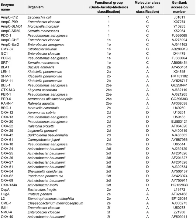
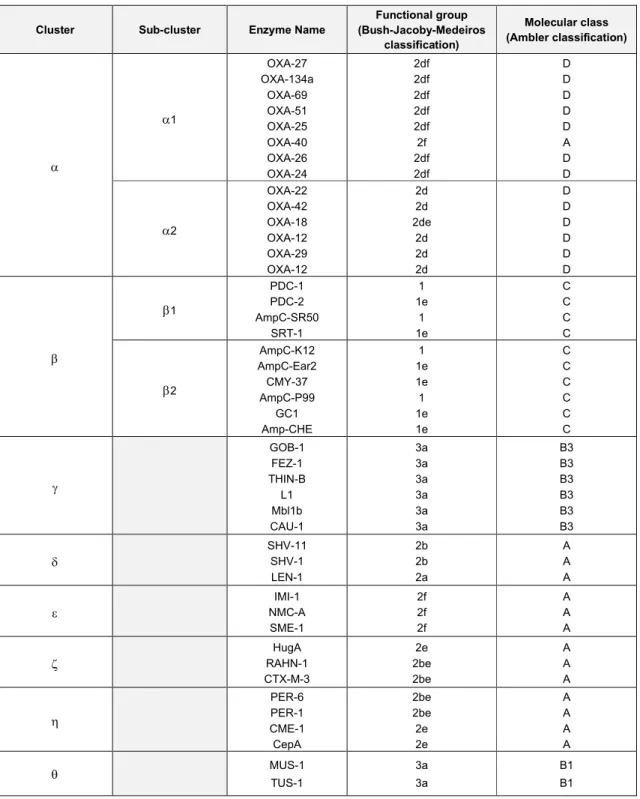
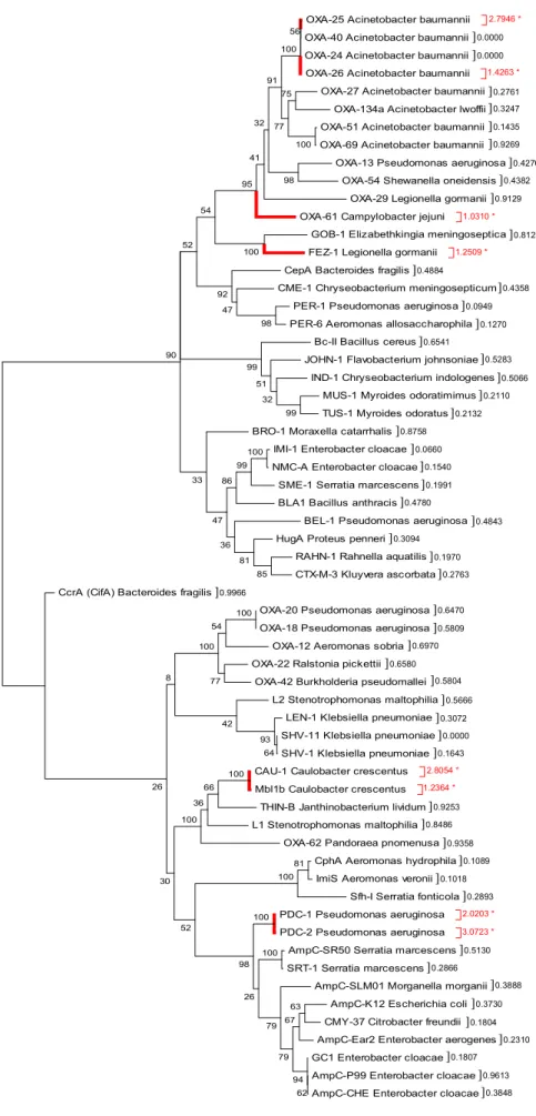
O presente estudo teve por objectivo clarificar as relações evolutivas entre as diferentes classes de β-lactamases, e também procurar compreender como elas se tornaram nas enzimas hidrolíticas de β-lactâmicos dos dias de hoje. Para tal, recorreu-se à análise filogenética e funcional de sequências nucleotídicas e aminoacídicas destas enzimas. Inicialmente, foram alinhadas 61 sequências aminoacídicas de β-lactamases de origem cromossomal, as quais foram posteriormente sujeitas a análise filogenética, recorrendo-se ao método da máxima parcimónia (MP). O critério de selecção destas sequências consistiu em reunir enzimas representativas de cada classe molecular de Ambler e grupo funcional de Bush-Jacoby-Medeiros descritos actualmente, de acordo com os respectivos esquemas de classificação. A partir da árvore filogenética inferida, foi possível organizar as sequências de β-lactamases em 8 grupos (ou clusters) ( – ), os quais revelaram ser consistentes com os agrupamentos de classificação molecular e funcional em vigor para estas enzimas. Enzimas representativas de cada cluster foram então utilizadas para pesquisar proteínas ortólogas nas bases de dados públicas. A filogenia destas sequências foi reconstruída, igualmente, através da abordagem MP. A análise filogenética revelou que a maior parte dos ortólogos putativos surgiu recentemente, juntamente com as β-lactamases com as quais aparentam estar mais relacionados, visto haver pouca divergência em relação ao ancestral comum a partir do qual ocorreu o evento de especiação. Existem também ramos na árvore mais longos, particularmente no grupo das OXAs, os quais representam um grau de divergência mais elevado, resultante de um maior número de substituições de aminoácidos. Estes resultados sugerem que as oxacilinases podem ter surgido na fase inicial da evolução das β-lactamases.
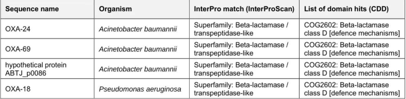
Uma vez que a similaridade por homologia nem sempre é sinónimo de funções idênticas, a inferência de homologia pode não ser suficiente para efectuar previsões funcionais rigorosas. Assim sendo, recorreu-se às ferramentas InterProScan e CDD para a anotação funcional das sequências representativas de cada cluster e dos seus ortólogos putativos. Esta análise funcional revelou que todos os ortólogos putativos partilham alguns domínios conservados com a mesma superfamília de β-lactamases com quem aparentam estar relacionados. Estes domínios funcionais encontram-se relacionados com actividade hidrolítica de β-lactâmicos, o que sugere estarmos perante ortologia genuína. As metalo-enzimas da classe B e os seus ortólogos partilham um domínio funcional específico com outras enzimas da mesma superfamília, as glutationa tiolesterases. Estas enzimas hidrolisam S-D-lactoilglutationa em glutationa e D-ácido láctico, requerendo a ligação a dois iões Zn2+ como cofactor. Estes resultados sugerem a existência de uma possível pleiotropia funcional para as metalo-enzimas.
Por fim, foram estimados os rácios Ka/Ks para os genes codificantes das 61 β-lactamases
cromossomais, visto que a identificação de selecção natural positiva poderia evidenciar alterações adaptativas na função. A filogenia destas sequências foi reconstruída, recorrendo-se ao método da máxima verosimilhança (ML). Um total de 8 genes revelou estar sob selecção positiva (Ka/Ks >1), o
que pode indicar que as proteínas codificadas podem ter alterado a sua função, possivelmente devido à pressão selectiva dos β-lactâmicos. De facto, os genes que codificam as metalo-enzimas CAU-1 e Mbl1b de C. crescentus parecem enquadrar-se neste cenário, tendo sido sugerido que estas enzimas representam hidrolases ancestrais que foram seleccionadas positivamente em resposta à presença de β-lactâmicos produzidos por fungos no solo. Os resultados obtidos fortalecem esta hipótese, bem como a de pleiotropia funcional nas MBLs, evidenciada pelas previsões funcionais. O facto de se terem detectado genes que codificam para enzimas da família OXA sob selecção positiva, suporta a hipótese destas enzimas poderem ter tido uma função importante na fitness bacteriana, como referido anteriormente. Os genes que codificam as cefalosporinases de P. aeruginosa, PDC-1 e PDC-2, parecem partilhar um ancestral comum com as AmpCs. O facto destes genes aparentarem estar sob forte selecção positiva pode significar que a mutação que conferiu a vantagem selectiva na presença do β-lactâmico e, assim, a alteração na função, pode ter-se fixado no seu DNA. Observou-se um
elevado grau de conservação no grupo das AmpCs, sugerido pelos baixos rácios Ka/Ksapresentados,
o qual pode indicar que estes genes são essenciais para as bactérias e, como tal, evoluem mais lentamente do que os genes não-essenciais. De facto, foi sugerida a existência de uma relação estreita entre a indução das AmpCs e a reciclagem do peptidoglicano, antes da pressão selectiva imposta pelas cefalosporinas, o que pode significar que estas enzimas têm um papel crucial na fisiologia bacteriana.
Apesar de por vezes óbvia, nem sempre é possível demonstrar a existência de pleiotropia funcional. Não obstante, esta propriedade parece desempenhar um papel fundamental na evolução dos genes das β-lactamases. De facto, pensa-se que o efeito pleiotrópico pode aumentar quando as bactérias se deparam com novos desafios selectivos, como é o caso dos antibióticos β-lactâmicos. Dependendo de como o novo caracter fenotípico (função) afecta a fitness bacteriana, os genes tornam-se pleiotrópicos ou especializados numa única função. Os resultados apresentados neste estudo contribuíram para o reconhecimento de um certo grau de pleiotropia funcional nas β-lactamases, e para uma maior compreensão de como estas proteínas podem ter evoluído e se tornado em enzimas específicas, responsáveis pela hidrólise de β-lactâmicos. Estudos adicionais combinando análise da sintenia de genes, alinhamentos estruturais, filogenias de genes individuais, interacções proteína-proteína e análises de recombinação e selecção, poderão ajudar a identificar e clarificar os processos evolutivos destas enzimas.
ABSTRACT
In clinically important Gram-negative bacteria, the predominant mechanism for β-lactam resistance is the synthesis of β-lactamases. These enzymes hydrolyze the amide bond in the β-lactam ring of these antibiotics, inactivating their effect. A few β-lactamases are zinc-dependent hydrolases, or Metallo-β-lactamases, requiring at least one zinc ion for disrupting the β-lactam ring. However, most enzymes are serine hydrolases, operating via a serine-ester mechanism. The chromosomal serine β-lactamases may have evolved from the Penicillin-Binding Proteins, with whom they share sequence homologies, or present parallel evolution associated with functional pleiotropy, resulting from the selective pressures performed by β-lactam-producing organisms in the soil. However, the current clinical misuse of β-lactams seems to be the most important factor for the dissemination of β-lactam resistance among pathogenic bacteria.
The present study aimed to clarify the evolutionary relationships between the β-lactamases, and to understand how they became the specific β-lactam-hydrolyzing enzymes nowadays. Phylogenetic and functional analysis from nucleotide and amino acid sequences was used. Phylogenies were reconstructed using Maximum Parsimony. Analysis revealed that most putative orthologs have arisen recently along with some already described β-lactamases, although a higher degree of divergence was evidenced for the oxacillinases, suggesting they may have evolved at the early stages of β-lactamase evolution. Functional predictions revealed that all putative orthologs share conserved domains with the β-lactamase superfamily with whom they seem more closely related. Putative pleiotropism was also evidenced for the metalloenzymes. Finally, Ka/Ksratios were estimated. Eight
β-lactamase genes were found to be under positive selection, suggesting possible adaptive changes in function. Overall, this study has contributed to the acknowledgement of some level of functional pleiotropy within the β-lactamases. Further studies combining analysis of gene synteny, structural alignments, phylogenies of individual genes, protein-protein interactions, and analyses of recombination and selection would help identify and clarify the evolutionary processes of such enzymes.
LIST OF ABBREVIATIONS
In this dissertation, acronyms are expanded on first usage and whenever deemed necessary to improve clarity.
AmpC: Ampicillin Class C
BBLI: β-lactam / β-lactamase Inhibitor
BLAST: Basic Local Alignment Tool
CA: Clavulanic Acid
CDD: Conserved Domain Database
CHDL: Carbapenem-hydrolyzing class D
β-lactamase
D-Ala: D-alanine
DNA: Deoxyribonucleic Acid
EDTA: Ethylenediaminetetraacetic Acid
HGT: Horizontal Gene Transfer
IS: Insertion Sequence
LS: Least Squares Methods
MDR: Multidrug-resistant
ME: Minimum Evolution
MEGA: Molecular Evolutionary Genetics
Analysis
MGE: Mobile Genetic Element
ML: Maximum Likelihood
MP: Maximum Parsimony
MRSA: Methicillin-Resistant Staphylococcus
aureus
MSA: Multiple Sequence Alignment
NaCl: Sodium Chloride
NAG: N-acetylglucosamine
NAM: N-acetylmuramic Acid
NCBI: National Center for Biotechnology Information
NJ: Neighbor-Joining
ORF: Open Reading Frame
PBP: Penicillin-Binding Protein
PSSM: Position-Specific Score Matrix
RBH: Reciprocal Best Hits
TZB: Tazobactam
UPGMA: Unweighted Pair-Group Method Using Arithmetic Averages
β-lactamase Abbreviations
ACT: AmpC-type
ACC: Ambler class C
ADC: Acinetobacterderived
cephalosporinase
ARI: Acinetobacterresistant to imipenem
Bc-II: From Bacillus cereus type II
BEL: Belgium ESBL
BES: Brazil ESBL
BIL: Named after the patient (Bilal) from
whom it was first isolated
BLA: From Bacillus anthracis
BRO: From (Branhamella) Moraxella
catarrhalis
CARB: Active on carbenicillin
CAU: From Caulobacter crescentus
CcrA: Cefoxitin and carbapenem resistant
CepA: Cephalosporinase from Bacteroides
fragilis, class A
CME: From Chryseobacterium
meningosepticum
CMT: Complex mutant derived from TEM-1
CMY: Active on cephamycins
CphA: Carbapenem-hydrolyzing and first (A)
from Aeromonas hydrophila
CTX-M: Active on cefotaxime, first isolated at Munich, Germany
DHA: Discovered at Dhahran Hospital in Saudi Arabia
ESAC: Extended-spectrum AmpC
ESBL: Extended-spectrum β-lactamase
FEC: Faecal Escherichia coli
FEZ: Legionella (Fluoribacter) gormanii endogenous zinc β-lactamase
FOX: Active on cefoxitin
GC1: From Enterobacter cloacae strain
GC1
GES: Guiana ESBL
GIM: German imipenemase
GOB: From Chryseobacterium
meningosepticum, class B
HugA: Hopital Universitaire de Genève,
class A
IMI: Imipenem-hydrolyzing β-lactamase
IMP: Active on imipenem
IND: From Chryseobacterium
(Flavobacterium) indologenes
IRT: Inhibitory resistant TEM
JOHN: From Flavobacterium johnsoniae
KHM: Kyorin Health Science MBL, from
Kyorin University Hospital in Tokyo, Japan
KPC: Klebsiella pneumoniae
carbapenemase
L1 / 2: Labile enzyme(s) from
Stenotrophomonas maltophilia
LAT: Active on latamofex
LEN-1: From K. pneumoniae strain LEN-1
MBL: Metallo-β-lactamase
MIR: Discovered at Miriam Hospital in
Providence, Rhode Island, USA
MOX: Active on moxalactam
MUS: From Myroides odoratimimus
NMC: Not metalloenzyme carbapenemase
NDM: New-Delhi MBL
NPS: From a national Pseudomonas
survey
OXA: Active on oxacillin
P99: From Enterobacter cloacae strain
P99
PaO1: From Pseudomonas aeruginosa
strain PaO1
PC1: From Staphylococcus aureus strain
PC1
PDC: Pseudomonas-derived
cephalosporinase
PER: Pseudomonasextended resistant
PSE: Pseudomonas-specific enzyme
RAHN: From Rahnella aquatilis
RTG: Enzyme with RTG (arginine,
threonine, glycine) triad in conserved box VII
Sfh: Serratia fonticolacarbapenem hydrolase
SFO: From Serratia fonticola
SHV: Sulfhydryl reagent variable
SIM: Seoul imipenemase
SLM01: From Morganella morganii strain SLM01
SME: Serratia marcescensenzyme
SPM: São Paulo MBL
SRT: From Serratia resistant to β-lactam
T-5575
TEM: Named after patient (Temoniera) from
whom it was first isolated
THIN-B: From Janthinobacterium lividum, class B
TLA: Named after the Tlahuicas Indian
tribe
TLE: TEM-like enzyme
TUS: From Myroides odoratus
VEB: Vietnam ESBL
VIM: Verona integron-encoded MBL, first
INTRODUCTION
Peptidoglycan is a complex made of glycan chains of alternating N-acetylglucosamine (NAG) and
N-acetylmuramic acid (NAM), cross-linked by short stem peptides attached to the NAM (160, 209,
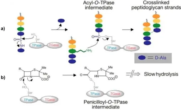
211). It constitutes a mesh-like layer outside the plasma membrane of bacteria and it is an essential component of the cell wall (100), preventing the cell from osmotic lysis and determinating its shape (100, 160, 211). In Gram-negative bacteria, the final stages of peptidoglycan biosynthesis take place at the periplasmic side of the membrane (209), involving distinct carboxypeptidation and transpeptidation reactions catalyzed by membrane-bound DD-peptidases (Figure 1a). These enzymes belong to a group of proteins known collectively as Penicillin-Binding Proteins (PBPs) (39, 104, 160, 192, 211). The term PBPs is a misnomer, since they are produced to complete the cross-linking of the peptide chains that confers rigidity to the peptidoglycan and viability to the bacterial cell, not to bind to penicillins (22). Inhibition of PBPs has harmful consequences in the cell wall structure, such as elongation, lesions, loss of selective permeability, and eventual cell death and lysis (77, 222).
PBPs are the primary targets of β-lactam antibiotics (39, 100, 209), a broad class of antibacterial agents which typically contain a β-lactam ring in their molecular structure and that include all penicillins, cephems (cephalosporins and cephamycins), monobactams and carbapenems (Holten and Onusko, 2000). The PBPs interact with their peptide substrates and β-lactam antibiotics through a similar mechanism (160), by forming a covalent acyl-enzyme complex with the antibiotic molecule via the serine residue at the enzyme’s active-site (39, 209, 211). This is an irreversible reaction which inactivates the enzyme (160), blocking carboxypeptidation and transpeptidation reactions, and consequently impairing the later stages of cell wall biosynthesis (209, 222, 224) (Figure 1b).
Fig. 1: Schematic representation of the biosynthesis of the cell wall (adapted from 236): a)
a) In the final step, peptide chains from adjacent glycan strains are cross-linked to each other by a peptide
bond exchange (transpeptidation) between the free amine of the amino acid in the third position of the pentapeptide and the D-alanine (D-Ala) at the fourth position of the other peptide chain, releasing the terminal D-alanine of the precursor. Transpeptidation and carboxypeptidation reactions are mediated by DD-peptidases (PBPs).
b) β-lactams (e.g. penicillin) bind to the substrate-anchoring site of the PBP, acting as a substrate analog of
D-alanyl-D-alanine, which is target of transpeptidation. The β-lactam-PBP intermediate is very stable, and it inactivates the enzyme. This interference with the normal transpeptidation reactions in the cell wall results in cellular lysis.
Perturbations in cell wall metabolism are sensed by bacteria and the resulting stress is translated into signals that induce defensive responses (100). In the course of evolution, bacteria have developed several strategies to deal with the severe effects caused by antibiotics such as β-lactams (218). Mechanisms frequently found in Gram-positive bacteria involve alteration of PBPs, which results in low affinity to β-lactams (191, 223), or acquisition of new PBPs (211), both responsible for β-lactam resistance. Alterations in PBPs of Streptococcus pneumoniae (219) and Listeria monocytogenes (182) confer high-level penicillin and imipenem resistance, respectively. High-level of resistance to methicillin and all other β-lactams in Gram-positive Staphylococcus aureus (MRSA) is due to acquisition of the mecA gene, which encodes the new PBP 2a with reduced affinity for β-lactams (127).
In clinically important Gram-negative bacteria, the predominant mechanism for resistance to β-lactam antibiotics is the synthesis of β-lactamases (12, 24, 34, 37, 38, 84, 86, 100, 123, 130, 132, 141, 205, 218), enzymes that catalyze the hydrolysis of the amide bond in the β-lactam ring of these
antibacterial agents, inactivating their antimicrobial effect (72, 84, 86, 104, 123, 141). A few β-lactamases require a zinc ion to disrupt the β-lactam ring, but most enzymes are serine hydrolases
(102, 130, 168, 190). The serine β-lactamases operate via a serine-ester mechanism (38, 130); following noncovalent binding of the enzyme to the antibiotic, the β-lactam ring is attacked by the free hydroxyl group on the side chain of the active-site serine residue, yielding a covalent acyl-enzyme ester intermediate. This intermediate is then deacylated from the serine by hydrolysis and the active enzyme is freed, as well as the hydrolyzed, inactive, drug (39, 130). Although the PBPs are also able to react with β-lactam molecules to give serine esters, these do not hydrolyze readily, unlike the
similar esters produced by the β-lactamases (39, 77). Hydrolytic deacylation of the serine β-lactamases is possible due the presence of a glutamate residue located on a small peptide strand,
the Ω-loop, which is a conserved structural motif that transverses the active-site of the enzyme (25, 39).
The serine β-lactamases are evolutionary related (39, 132, 224) and belong to a superfamily of penicillin-recognizing enzymes that also includes the DD-peptidases, and a variety of other PBPs (86, 104, 192); not only they bind to similar ligands, but also operate by a common acyl-enzyme mechanism (39, 77, 104, 160, 192). In addition, all these enzymes contain a conserved Ser-x-x-Lys motif, where the serine is the active-site residue (19, 104, 160, 192). Although the active-sites of the PBPs and β-lactamases are similar, the covalent binding of the PBPs to its β-lactam substrate
inactivates the enzyme, while the same substrate is hydrolyzed by the β-lactamases (90, 160). Sequence homologies suggests that the chromosomal β-lactamases may have evolved from the PBPs (30, 84, 108, 244), with whom they share several highly conserved amino acid sequences (141), or present a parallel evolution associated to functional pleiotropy. The class A and C serine enzymes apparently arose from a common PBP ancestor, an actinomycete DD-carboxypeptidase involved in bacterial wall peptidoglycan metabolism (90, 108, 141). The evolution of β-lactamase genes was probably a result of the selective pressure carried out by β-lactam-producing soil organisms in the natural environment (30, 84, 108, 130; 141). However, the current clinical misuse and overprescription of β-lactam antibiotics seems to be the most important factor for the selection and dissemination of β-lactam resistance in pathogenic bacteria (10, 30, 51, 84, 86, 141, 176, 190).
Classification Schemes
β-lactamases are currently classified according to their primary structure (namely, Ambler classification) (8) or their function (namely, Bush-Jacoby-Medeiros classification) (34, 35, 36, 37, 38). The Ambler classification scheme (8) divides β-lactamases into four major molecular classes, designated A to D, based on conserved and distinguishing amino acid motifs (8, 24, 33, 34, 35, 36, 37, 38, 84, 130, 168, 132, 176, 183, 193). Classes A, C and D are serine β-lactamases (38, 86, 132,176, 190), enzymes that hydrolyze their substrates through the serine-ester mechanism mentioned earlier. Class B enzymes, known as Metallo-β-lactamases (MBLs), utilize at least one active-zinc ion linked to a histidine or cysteine residue to react with the carbonyl group of the amide bond of most carbapenems, cephalosporins and penicillins, facilitating its hydrolysis (37, 38, 39, 84, 193, 239). Contrarily to the serine enzymes, MBLs lack the ability to hydrolyze monobactams and are not susceptible to clavulanic acid or tazobactam (38, 39, 132, 193, 239).
A functional classification is more important in Microbiology than classification based on amino acid homology, because it helps to correlate the properties of a specific enzyme with the observed resistance profile for a particular clinical isolate (38, 39, 130, 176). The Bush-Jacoby-Medeiros classification scheme (37), updated in 2010 (38), groups β-lactamases within each class of the Ambler system according to functional similarities, namely relative hydrolysis rates of a variety of substrates (substrate specificity) and sensitivity to various enzyme inhibitors (inhibitor susceptibility). This system comprises four main functional groups (1 – 4), with multiple subgroups under group 2 (a – f) (34, 37, 38, 84, 141, 176, 193).
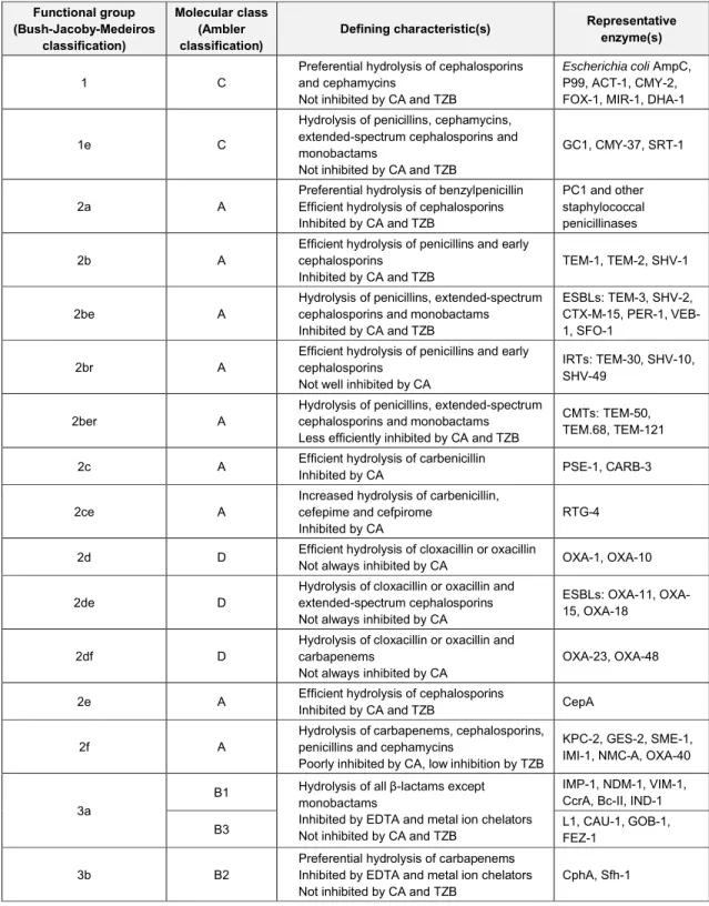
Table 1 aligns functional groupings with molecular assignments as closely as possible, based on the available public information. A summary of functional characteristics, such as ability to hydrolyze specific β-lactam classes and susceptibility to the β-lactamase inhibitors clavulanic acid (CA) and tazobactam (TZB), is also included. A description of each of the functional groups and molecular classes follows. For the purpose of this dissertation, the term “class” will be employed to describe the molecular classes of the Ambler classification scheme. Likewise, the terms “group” and “subgroup” will refer to the functional clusterings within the Bush-Jacoby-Medeiros classification scheme.
Table 1: Classification schemes for bacterial β-lactamases (adapted from 38). Functional group (Bush-Jacoby-Medeiros classification) Molecular class (Ambler classification)
Defining characteristic(s) Representative
enzyme(s)
1 C
Preferential hydrolysis of cephalosporins and cephamycins
Not inhibited by CA and TZB
Escherichia coli AmpC, P99, ACT-1, CMY-2, FOX-1, MIR-1, DHA-1
1e C
Hydrolysis of penicillins, cephamycins, extended-spectrum cephalosporins and monobactams
Not inhibited by CA and TZB
GC1, CMY-37, SRT-1
2a A
Preferential hydrolysis of benzylpenicillin Efficient hydrolysis of cephalosporins Inhibited by CA and TZB
PC1 and other staphylococcal penicillinases
2b A
Efficient hydrolysis of penicillins and early cephalosporins
Inhibited by CA and TZB
TEM-1, TEM-2, SHV-1
2be A
Hydrolysis of penicillins, extended-spectrum cephalosporins and monobactams
Inhibited by CA and TZB
ESBLs: TEM-3, SHV-2, CTX-M-15, PER-1, VEB-1, SFO-1
2br A
Efficient hydrolysis of penicillins and early cephalosporins
Not well inhibited by CA
IRTs: TEM-30, SHV-10, SHV-49
2ber A
Hydrolysis of penicillins, extended-spectrum cephalosporins and monobactams
Less efficiently inhibited by CA and TZB
CMTs: TEM-50, TEM.68, TEM-121
2c A Efficient hydrolysis of carbenicillin
Inhibited by CA PSE-1, CARB-3
2ce A
Increased hydrolysis of carbenicillin, cefepime and cefpirome
Inhibited by CA
RTG-4 2d D Efficient hydrolysis of cloxacillin or oxacillinNot always inhibited by CA OXA-1, OXA-10
2de D
Hydrolysis of cloxacillin or oxacillin and extended-spectrum cephalosporins Not always inhibited by CA
ESBLs: 11, OXA-15, OXA-18
2df D
Hydrolysis of cloxacillin or oxacillin and carbapenems
Not always inhibited by CA
OXA-23, OXA-48 2e A Efficient hydrolysis of cephalosporinsInhibited by CA and TZB CepA
2f A
Hydrolysis of carbapenems, cephalosporins, penicillins and cephamycins
Poorly inhibited by CA, low inhibition by TZB
KPC-2, GES-2, SME-1, IMI-1, NMC-A, OXA-40
3a
B1 Hydrolysis of all β-lactams except monobactams
Inhibited by EDTA and metal ion chelators Not inhibited by CA and TZB
IMP-1, NDM-1, VIM-1, CcrA, Bc-II, IND-1
B3 L1, CAU-1, GOB-1, FEZ-1
3b B2
Preferential hydrolysis of carbapenems Inhibited by EDTA and metal ion chelators Not inhibited by CA and TZB
CphA, Sfh-1
Group 1 / Class C β-lactamases
Group 1 β-lactamases belong to class C and are generally encoded on the chromosomes of many
Enterobacteriaceae and a few other organisms (38, 235). These enzymes hydrolyze preferentially
for aztreonam (34, 35, 37, 38, 39). Representative enzymes include CMY-1 (14), CMY-2 (13), FOX-1 (122), MIR-1 (174), MOX-1 (95) and LAT-1 (233), all from Klebsiella pneumoniae, P99 from
Enterobacter cloacae (75), PDC-1 from Pseudomonas aeruginosa 202) and BIL-1 from Escherichia
coli(72). In Gram-negative bacteria, one of the most important chromosomal cephalosporinases is the
group of enzymes collectively known as AmpC β-lactamases (19, 123). Data suggests that the AmpC enzymes have evolved to deal with cephalosporins rather than for some other cellular function (33, 101), although there is evidence suggesting a close relationship between the induction of these enzymes and peptidoglycan recycling (33, 90, 100, 101, 123, 130, 252). This may imply an alternative metabolic function of the class C β-lactamases at the early stages of its evolution, possibly some physiological role in cell wall metabolism (130, 141). In this context, the hydrolysis of β-lactams should be considered an evolutionary response to the challenge by cephalosporins, in order to defend bacteria against these antimicrobial agents. The β-lactam substrate would have provided the selective pressure that drove the evolution of the class C enzymes (33).
Chromosome-mediated AmpC β-lactamases are often expressed as inducible enzymes in response to β-lactam exposure (19, 38, 100, 101, 123, 235). In Gram-positive bacteria, the presence of a β-lactam molecule at the external face of the cytoplasmic membrane is sensed by the cell, which transmits this signal to the intracellular regulatory components that control β-lactamase expression (19). In Gram-negative bacteria, the induction system is more complex, involving four regulatory genes,
ampR, ampD, ampG and ampE, which constitute the amp operon, together with the structural gene,
ampC(19, 100, 101, 123, 252). When the β-lactam inducer is present, it interacts with the PBPs of the
bacterial cell, preventing the cross-linking step of cell wall biosynthesis (19, 223). This interaction causes peptidoglycan breakdown products (muropeptides) to accumulate in the periplasm (19, 252), which are captured and transported by the transmembrane permease AmpG to the cytoplasm, serving as the induction signal to activate the transcriptional regulator, AmpR. When on its active form, AmpR acts as an activator for ampC transcription, stimulating β-lactamase synthesis (19, 100, 101, 123, 141, 252). AmpD is a cytosolic amidase that down-regulates β-lactamase expression, maintaining AmpR in its repressor form (100, 101, 141, 252), possibly assisted by the cytoplasmic membrane protein, AmpE (19, 123, 252).
Most members of the Enterobacteriaceae (e.g. Citrobacter freundii, E. cloacae, Serratia marcescens,
Morganella morganii, Proteus mirabilis, Yersinia enterocolitica) and P. aeruginosa possess a
naturally-occurring, chromosome-mediated β-lactamase (11, 15, 29, 30, 38, 64, 84, 119, 123, 202, 203, 235, 245, 252). Such chromosomal ampC genes are usually expressed at low level, regulated by induction of β-lactams (19, 38, 100, 101, 123, 134) or by mutation of regulatory genes (123, 134). In other organisms, one or more components of the induction system are missing (38); E. coli, Acinetobacter
baumannii, Shigella dysentery and Shigella flexneri lacks an ampR gene, so that their AmpC enzymes
are noninducible (101, 123, 252). Chromosome-mediated AmpC β-lactamases can also be expressed at continuously high level (123); spontaneous mutations, deletions or insertions in regulatory genes, most frequently in ampD (134), results in constitutive hyperproduction of the enzyme and in increased resistance to third-generation cephalosporins (12, 64, 134, 202, 203). Such derepressed phenotypes can also be achieved by less common mutations in ampR locus (12, 101), which results in the
production of more active transcriptional regulator AmpR (12, 100). In addition, mutations in ampC gene can expand the substrate specificity of the AmpC β-lactamases, contributing as well to naturally-occurring resistance to extended-spectrum cephalosporins (12, 134). Another form of chromosomal β-lactamase expression is at continuously low level (123), due to least common mutations in ampG (100, 101). Inactivation of AmpG also impairs the peptidoglycan recycling process, because in addition to modified β-lactamase regulation, such mutants have lost the ability to recycle muropeptides (100). Most class C β-lactamases are produced in Gram-negative bacteria as chromosomal enzymes (30, 168). Such enzymes may not contribute to clinical β-lactam resistance, since they are often expressed at low levels, but may cause severe therapeutic problems if their genes are translocated onto plasmids (119, 245). The ability of plasmids harbouring resistance genes to spread between bacterial cells by conjugation (167, 169) greatly enhances the nosocomial dissemination of resistant strains, which represents a serious challenge to the treatment of infections (39, 123, 217).
All plasmid-mediated β-lactamases, including the AmpC enzymes, are likely to have chromosomal origins (101, 123, 130, 134). Plasmid-encoded AmpC enzymes have been described worldwide since 1989 (38, 101, 123, 235), mainly in bacteria without or with incomplete chromosomal ampC gene, such as Klebsiella spp., E. coli, P. mirabilis, Salmonella spp. and some Shigella spp., but also in
bacteria with complete chromosomal ampC, such as E. cloacae, Acinetobacter spp. and
P. aeruginosa (123). Generally, plasmid-encoded ampC genes lack regulatory gene ampR, and
expression seems to be at continuously high level and noninducible (123). The exceptions are the DHA-1 enzyme from Salmonella enteritidis (11), as well as DHA-2 (71) and ACT-1 (29) enzymes from
K. pneumoniae, which are inducibly expressed because their ampC genes are linked to ampR genes (101, 123). In addition to resistance to cephamycins, a combination of high level production of the ACT-1 β-lactamase with the loss of a major outer membrane porin can confer imipenem resistance in
K. pneumoniae (29, 176, 241). This development is of particular concern for the Public Health, since that carbapenems, such as imipenem and meropenem, have been the drugs of last-resort for Gram-negative bacteria (123).
Most AmpC-type β-lactamases, with the exception of plasmid-borne ACC-1 from K. pneumoniae (15), confer resistance to cephamycins (cefoxitin and cefotetan) and to a lesser extent, to oxyimino- β-lactams (or oxyimino-cephalosporins), such as cefotaxime, ceftazidime, cefuroxime and ceftriaxone, and to the monobactam aztreonam (119, 134, 202, 203). AmpC-overproducing strains, additionally, have demonstrated increased resistance to extended-spectrum cephalosporins, particularly oxyimino-cephalosporins (134). Recently, cephalosporinases with broadened substrate activity have been reported (101, 134, 202, 203); these enzymes confer reduced susceptibility to all cephalosporins, including fourth-generation cephalosporins cefepime and cefpirome (128), having been termed Extended-Spectrum AmpC (ESAC) β-lactamases (12, 38, 39, 101, 134, 202, 203). ESACs are structurally related to the cephalosporinases from group 1, as a result of amino acid substitutions, insertions or deletions (38, 101, 134), and were assigned into group 1e (38). They are very likely chromosomally-encoded enzymes (134), and include enzymes such as SRT-1 from S. marcescens (140), GC1 from E. cloacae (168), PDC-2 from P. aeruginosa (202) and ADC-33 from A. baumannii (203). Plasmid-mediated ESACs, such as CMY-10 from Enterobacter aerogenes (119), CMY-19 from
K. pneumoniae (235) and CMY-37 from C. freundii (3), have also been described. Along with other
resistance mechanisms, such as AmpC overproduction, increased drug efflux and decrease or loss of outer membrane permeability, ESACs were described as also contributing to carbapenem resistance in P. aeruginosa isolates (101, 202). Spread of such an extended-substrate activity phenotype may potentially compromise the clinical utility of most β-lactams (134).
Group 2 / Class A β-lactamases
Group 2 enzymes, which include classes A and D, represent the largest group of β-lactamases (37, 38). Contrarily to group 1 cephalosporinases, group 2 enzymes tend to have more affinity to clavulanic acid than for aztreonam (34, 35). Subgroup 2a penicillinases are a small cluster of β-lactamases that belong to class A and are predominant in Gram-positive bacteria (34, 35, 38). These enzymes have a relatively narrow-spectrum hydrolytic activity, acting preferentially in benzylpenicillin and other penicillin derivates, over cephalosporins, carbapenems or monobactams (38). Cloxacillin is not an effective enzyme inhibitor (34, 35), as are clavulanic acid and tazobactam (37, 38, 39). Subgroup 2a penicillinases are mainly chromosomal (37, 38), such as BLA-1 from Bacillus anthracis (46) and LEN-1 from K. pneumoniae (9), but there are some plasmid-encoded enzymes (38), such as PC1 from
S. aureus(132, 251) and NPS-1 from P. aeruginosa (129, 173).
Subgroup 2b β-lactamases are broad-spectrum enzymes belonging to class A (34, 35, 37, 38, 39, 130). They readily hydrolyze penicillins and first-generation cephalosporins, such as cephalothin, cefazolin and cephaloridine (30, 38, 39, 84), presenting low hydrolysis rates for extended-spectrum cephalosporins, aztreonam and imipenem (34, 35, 130, 176). These enzymes are strongly inhibited by clavulanic acid and tazobactam (34, 35, 37, 38, 39). The most common and widely distributed enzymes TEM-1, TEM-2 and SHV-1 are included in this subgroup (34, 35, 37, 38, 39). TEM-1 is the most frequently found β-lactamase in Gram-negative bacteria (30, 130, 132, 176), mainly in E. coli,
K. pneumoniaeand P. mirabilis (84). This enzyme is transposon- (81, 175) and plasmid-mediated (7,
30, 84, 116), which explains its widespread dissemination among bacteria (30). For instance, transposons carrying TEM-1 genes have been responsible for the plasmid-mediated ampicillin and penicillin resistance reported in Haemophilus influenzae (30, 62, 132) and Neisseria gonorrhoeae (30,
63, 132), respectively. The SHV-1 β-lactamase is the most prevalent chromosomal β-lactamase in
K. pneumoniae(30, 84, 130, 176, 234), although is plasmid-mediated in other species (130), such as
E. coli(30, 84).
Substitutions in one or more amino acids in classic subgroup 2b β-lactamases resulted in enzymes with an extended-spectrum phenotype (30, 130, 176, 234). These enzymes, known as Extended-Spectrum β-lactamases (ESBLs), are capable of hydrolyzing latest-generation oxyimino-cephalosporins, as well as the monobactam aztreonam (24, 30, 37, 38, 39, 130, 176, 177), at rates usually >10% that for benzylpenicillin (34, 35, 37, 38, 176), and were assigned into group 2be (24, 30, 37, 38, 39, 176). Being mostly mutants of subgroup 2b TEM-1, TEM-2 and SHV-1 (30, 38, 130, 177), subgroup 2be ESBLs are class A enzymes that retain hydrolytic activity against penicillins and early cephalosporins (30, 37, 38, 39, 176), while remaining susceptible to clavulanic acid and tazobactam (24, 30, 34, 35, 37, 38, 39, 130, 176). This is the main feature differentiating ESBLs and group 1
AmpC-type enzymes, which are also capable to hydrolyze oxyimino-cephalosporins, but are not susceptible to such β-lactamase inhibitors (176).
Additionally to the TEM- and SHV-type ESBLs, CTX-M-type enzymes have been described (24, 30, 38, 84, 176). These plasmid-mediated enzymes have apparently originated from chromosomal β-lactamase genes of environmental Kluyvera spp. (24, 30, 38, 106, 176, 201, 244) that have been captured by mobile genetic elements (MGEs) (177), such as insertion sequence ISEcp1 (106, 177, 218). CTX-M β-lactamases are actually the most prominent class A / subgroup 2be ESBLs (24, 86, 176, 177, 199, 218), given its high dissemination among bacteria and the number of outbreaks reported worldwide (24, 30, 84, 106, 176, 177). They hydrolyze preferentially cefotaxime over ceftazidime (24, 30, 38, 84, 106, 176, 201), being so known as cefotaximases (24, 201). Another distinctive property of these enzymes is that unlike TEM or SHV ESBLs, they are better inhibited by tazobactam than by clavulanic acid or sulbactam (24, 30, 38, 84, 176). CTX-M-15 is the most widely distributed CTX-M ESBL, which was first reported in E. coli isolates from India (47, 84, 106, 177, 217). Multidrug-resistant (MDR) CTX-M-15-producing E. coli, particularly strain ST131 O25:H4, is now emerging globally in community and hospital settings, constituting an important public health threat (39, 47, 177). Other examples of class A / 2be ESBLs, not closely related to TEM, SHV or CTX-M, include plasmid-mediated and integron-associated enzymes (24, 30, 38, 39, 84, 176), such as VEB-1 (156, 183), GES-1 (184), TLA-1 (214), SFO-1 (138), BES-1 (23), as well as the chromosomally-encoded enzymes PER-1 (164), PER-6 (80), RAHN-1 (18) and BEL-1 (189).
The “ESBL” terminology was originally associated with class A / subgroup 2be β-lactamases, mainly TEM and SHV derivatives, capable of hydrolyzing oxyimino-cephalosporins and susceptible to β-lactamase inhibitors, such as clavulanic acid (24, 30, 37, 38, 84, 176). This term was expanded to include not only the CTX-M and VEB enzymes, but also unusual class C / group 1e AmpC cephalosporinases with increased activity against cefepime, and a subset of class D / subgroup 2de OXA enzymes (described later), both with extended-spectrum phenotypes, but not meeting the definition of subgroup 2be, since they are not inhibited by clavulanic acid (38, 176). For the purpose of this dissertation, the term “ESBL” will refer to class A / subgroup 2be and class D / subgroup 2de enzymes, as previously suggested (176).
Clinical introduction of β-lactam / β-lactamase inhibitor (BBLI) combinations could result in the emergence of subgroup 2b mutants with acquired resistance to clavulanic acid and sulbactam, while retaining the broad-spectrum of activity of their parent enzymes (30, 38, 130, 132), but that remain susceptible to tazobactam (30). Such variants belong to subgroup 2br (37, 38) and were originally given the designation Inhibitory-Resistant TEM (IRT) β-lactamases, given that the first mutants were TEM-1 and TEM-2 derivatives (30, 130), such as TEM-30 (IRT-2) from E. coli (40). However, there are also inhibitory-resistant variants of SHV-1 (30), such as SHV-49 from K. pneumoniae (57). IRT β-lactamases represent an adaptive resistance mechanism specifically developed by bacteria to overcome the activity of β-lactamase inhibitors (161). The point mutations that lead to the inhibitory-resistant phenotype take place at a few specific amino acid residues (30, 109, 132) and are distinct from those that lead to the ESBL phenotype (30). However, concurrence of amino acid substitutions in
clavulanic acid with an extended-spectrum phenotype. They are known as Complex Mutant TEM (CMT) β-lactamases of subgroup 2ber (30, 38, 39, 41, 199), and include enzymes such as TEM-50 (CMT-1) (217) and TEM-158 (CMT-9) (200) from E. coli (217), TEM-68 (CMT-2) from K. pneumoniae (67) and TEM-121 (CMT-4) from E. aerogenes (187).
The remaining class A β-lactamases are comprised within subgroups 2c, 2ce, 2e and 2f (38). Subgroup 2c enzymes preferentially hydrolyze carbenicillin and ticarcillin (penicillins), at a rate >60% that for benzylpenicillin, with cloxacillin or oxacillin being hydrolyzed at a rate <50% that for benzylpenicillin (36, 37, 38). They are well inhibited by clavulanic acid and tazobactam (38), and include the chromosomal enzyme from Moraxella catarrhalis BRO-1 (26) and the plasmid-mediated enzymes from P. aeruginosa, PSE-1 (CARB-2) (98) and CARB-3 (115). Recently, a carbenicillinase with extended activity against the latest-generation cephalosporins cefepime and cefpirome, RTG-4 (CARB-10), was described in A. baumannii. This enzyme was assigned into subgroup 2ce (38). Subgroup 2e β-lactamases are cephalosporinases, which can lead to be confused with group 1 AmpC enzymes. However, contrarily to group 1 cephalosporinases, subgroup 2e enzymes are inhibited by clavulanic acid and have low affinity for aztreonam (34, 36, 37, 38, 39, 176). Representative enzymes include chromosomal CepA from Bacteroides fragilis (204), L2 from Stenotrophomonas maltophilia (237), CME-1 from Chryseobacterium meningosepticum (205), and HugA from Proteus penneri (125), as well as plasmid-mediated FEC-1 from E. coli (138). Class A β-lactamases are still represented by the serine carbapenemases from subgroup 2f, carbapenem-hydrolyzing enzymes that are less inhibited by clavulanic acid than by tazobactam (37, 38, 39, 176, 193). Bacteria expressing these enzymes, such as K. pneumoniae, E. cloacae and S. marcescens, have reduced susceptibility to imipenem (193). Chromosomal enzymes, such as SME-1 (154), IMI-1 (196), NMC-A (153) and OXA-40 (91) have been described for this subgroup (37, 38, 166, 193), but the plasmid-encoded enzymes, particularly the KPC and GES families, are those that raises more clinical concerns (38, 39, 193). The KPC carbapenemases have been more often associated with nosocomial infections by MDR Gram-negative pathogens, and continue to spread worldwide (39, 193).
Group 2 / Class D β-lactamases
Similarly to class A and C β-lactamases, class D β-lactamases possess an active-site serine (4, 86, 176, 193), representing the most diverse enzymes among the four molecular classes (190). Subgroup 2d β-lactamases belong to class D and includes enzymes that preferentially hydrolyze oxacillin or cloxacillin (4, 30, 36, 37, 38, 39, 84, 86, 176, 190), at a rate >50% that for benzylpenicillin (37, 38, 176), but that may also efficiently hydrolyze carbenicillin (38). These oxacillinases, or OXA-type β-lactamases (OXAs), are poorly inhibited by clavulanic acid (4, 30, 37, 38, 39, 84, 190), but can be inhibited by sodium chloride (NaCl) (36, 38, 190). Many bacteria encode OXA enzymes in their chromosome, but most oxacillinase genes are part of gene cassettes within class 1 integrons (4, 10, 190). These integrons are frequently associated with plasmids or transposons (197), which facilitates the spread of OXA genes among bacteria (4, 193). Chromosomal enzymes include OXA-12 from
Aeromonas jandaei (formerly Aeromonas sobria) (190, 195), OXA-13 (149) and OXA-20 (155) from
from Burkholderia pseudomallei (163) and OXA-61 from Campylobacter jejuni (4). Plasmid-mediated oxacillinases of clinical importance include OXA-1 from E. coli (172), OXA-2 from S. typhimurium (147) and OXA-10 (PSE-2) from P. aeruginosa (97, 197). OXA-type enzymes now comprise the second large family of β-lactamases, preceded by TEM and followed by SHV (38, 39).
Several subgroup 2d / class D β-lactamases have expanded its hydrolytic spectrum towards oxyimino-cephalosporins, while retaining the ability to hydrolyze oxacillin or cloxacillin. These enzymes are regarded as ESBLs (30, 38, 39, 84, 176, 190) and belong to subgroup 2de (38, 39). OXA-type 2de ESBLs are mostly OXA-10 derivatives by few amino acid substitutions and are predominant in
P. aeruginosa (30, 38, 176, 190). They include chromosomally-encoded enzymes, such as OXA-18 (180), but also plasmid-mediated enzymes, such as OXA-11 (85) and OXA-15 (52).
Some OXA-type β-lactamases may also hydrolyze carbapenems (38, 190), but they are not able to combine such carbapenem-hydrolyzing activities with traditional ESBL hydrolytic profiles (190, 193), at least at a significant level (193). Carbapenem-hydrolyzing class D β-lactamases (CHDLs) are allocated in subgroup 2df (38), being more frequent in A. baumannii (38, 190, 193). With exception of the clinically important enzymes from A. baumannii, OXA-23 (ARI-1) (56) and OXA-58 (188), which have been shown to be plasmid-encoded, the CHDLs described so far in this opportunistic pathogen are very likely chromosomally-located (38, 190, 193). Both these acquired enzymes, OXA-23 and OXA-58, have demonstrated to contribute significantly to carbapenem resistance (190, 193). Plasmid-mediated OXA-48 from K. pneumoniae (50, 185) is another clinically relevant CHDL (38, 39), which has demonstrated to be the enzyme with the highest known catalytic efficiency for imipenem (185, 190). Naturally-occurring CHDLs include enzymes such as OXA-24 (28), OXA-25 (2), OXA-26 (2), OXA-27 (2), OXA-51 (OXA-Ab1) (31) and OXA-69 (32), all from A. baumannii, OXA-54 from
Shewanella oneidensis (186), OXA-62 from Pandoraea pnomenusa (212) and OXA-134 from Acinetobacter lwoffii(68).
Group 3 / Class B Metallo-β-lactamases
Group 3 MBLs belong to class B (34, 36, 37, 38, 39). These metalloenzymes are characterized by its ability to hydrolyze carbapenems, but most can also hydrolyze cephalosporins and penicillins (37, 38, 39, 132, 193, 239). They require a bivalent metal ion (Zn2+) at the active-site for optimal enzymatic activity (34, 36, 76, 193, 239), and are distinguishably inhibited by EDTA and metal ion chelators, but not clavulanic acid or tazobactam (34, 36, 37, 38, 39, 132, 166, 176, 193, 239). MBLs are currently subdivided, based either on their structure (subclasses B1, B2, B3) (38, 39, 76, 193, 239) or function (subgroups 3a and 3b) (38, 39). The three structural subclasses are aligned within the subgroups, according to functional similarities (38). Subgroup 3a comprises subclasses B1 and B3 (38, 39), both requiring two bound zinc ions at the active-site for their similar broad-spectrum hydrolytic activity. In contrast, subclass B2 enzymes are inhibited if a second zinc ion is bound and have a narrow-spectrum of activity, preferentially hydrolyzing carbapenems (38, 39, 76, 193, 239). Subclass B2 is associated with subgroup 3b (38, 39).
Some bacteria from environmental niches carry ubiquitously MBLs, mostly chromosomal and inducible (39, 193, 239), which are probably associated with another cellular function that is yet to be entirely
elucidated (239). These enzymes are now recognized as the most important reservoir for these β-lactamase genes (157). Therefore, it is important to clarify the evolutionary relationships between the described β-lactamases, to understand how they evolved and became the specific β-lactam-hydrolyzing enzymes nowadays, and to identify the genetic sources associated with the emergence of putative new β-lactam-hydrolyzing enzymes. Indeed, the first MBLs identified and described were environmental, chromosomal enzymes produced by Gram-negative bacteria (38, 39, 166, 193, 239), such as CphA from Aeromonas hydrophila (subclass B2) (38, 137), L1 from S. maltophilia (subclass B3) (49) and CcrA (CifA) from B. fragilis (subclass B1) (194), and also by some Gram-positive bacteria (39, 193, 239), such as Bc-II from Bacillus cereus (subclass B1) (99, 114, 126). The latter was the first MBL for which an amino acid sequence was determined (99), having been described as a lipoprotein-like, membrane-bound enzyme (26, 99). Membrane-bound hydrophobic forms of Gram-positive β-lactamases were also described for Bacillus licheniformis and S. aureus and have been proposed to be the precursors of the periplasmatic enzymes (26, 162). Chromosomal metalloenzymes also include Sfh-1 from Serratia fonticola (subclass B2) (208), GOB-1 from Chryseobacterium meningosepticum (subclass B3) (16), IND-1 from Chryseobacterium indologenes (subclass B1) (17), FEZ-1 from L.
gormanii (subclass B3) (27), CAU-1 from Caulobacter crescentus (subclass B3) (55), THIN-B from Janthinobacterium lividum (subclass B3) (206), JOHN-1 from Flavobacterium johnsoniae (subclass
B1) (157), TUS-1 and MUS-1 from Myroides spp. (subclass B1) (133). The chromosomally-encoded MBLs were usually found in bacteria that also expressed at least one serine β-lactamase (39, 193, 239). For instance, S. maltophilia can hydrolyze third-generation cephalosporins (and carbapenems) due to coregulated overexpression of the L1 MBL and its chromosomal class A enzyme, L2 (239). With exception of the L1 MBL from S. maltophilia, bacteria producing chromosomal metalloenzymes have not been frequently associated with severe nosocomial infections, as they are generally opportunistic pathogens (193, 239), and also because chromosomal MBL genes are not easily transferred (193).
Contrarily to the chromosomal MBLs, whose presence depends directly on the prevalence of the producing species (193), the metalloenzymes encoded by transferable genes have disseminated globally (39) and are now widely spread among clinical pathogenic bacteria (193, 239). The most common transferable MBLs include the IMP, VIM and GIM families (38, 39, 193, 239). All these metalloenzymes are allocated in subgroup 3a (38, 39) and are found as gene cassettes in a variety of integron structures (193, 239), mostly from class 1 (239). Clinical important transferable MBLs include integron-borne enzymes, such as IMP-1 (117, 242), VIM-1 (118) and GIM-1 (44), and also plasmid-mediated SPM-1 (152, 232), all originally found in clinical isolates of P. aeruginosa (193, 239). Other relevant acquired MBLs includes the SIM-1 (120) enzyme, located within a class 1 integron from
A. baumannii(120, 193), and also the plasmid-mediated KHM-1 enzyme from C. freundii (213).
Recently, a novel MBL was identified in a clinical isolate of K. pneumoniae (39, 145, 250). This metalloenzyme, NDM-1, was found to be carried on a large 180-kb transmissible genetic element encoding other resistance determinants, including the broad-spectrum β-lactamase CMY-4, genes inactivating erythromycin, ciprofloxacin, rifampicin and chloramphenicol, and genes encoding an efflux pump. This genetic element was able to spread rapidly among other enterobacterial strains and also
nonfermentative bacteria, conferring resistance to all antibiotics, except colistin (39, 145, 250). NDM-1 was originally described as being integron-borne (250), but is now carried by a variety of plasmids, which were responsible for its widespread dissemination worldwide (39). An E. coli isolate in Germany was reported as having the blaNDM-1gene probably integrated into the chromosome, which could be
related with the genetic structures surrounding that gene. In addition to the NDM-1 MBL, the same isolate coproduced the CTX-M-15 ESBL, as well as the enzymes TEM-1, OXA-1 and OXA-2 (39, 179).
Genetic Environment
The species definition for prokaryotes has advanced over the last years, following the genomic era (144). The current paradigm is that bacterial species are more accurately described by their pan-genome (142, 144), which is the sum of two components: a core pan-genome consisting of genes ubiquitously found in all strains, and an accessory genome containing partially shared and strain-specific genes (20, 113, 142, 144, 215). The core cluster genes are highly conserved within the species and tend to remain stable throughout evolution, and horizontal gene transfer (HGT) is less likely to be detected (20, 53, 113). These genes typically encode proteins that are essential for the survival of the organism (215), which are mainly associated with housekeeping functions (e.g. transcriptional and translational apparatuses), the cell envelope, regulatory roles and transport of nucleotides and amino acid (144). A considerable fraction of the core genome consists in hypothetical proteins or open reading frames (ORFs) with no known function (53, 54, 215), but whose presence (i.e. conservation) suggests that they may have an important function (53). The components of the core genome can be particularly useful for phylogenetic inference, i.e. for clarifying the evolutionary history of clonal lineages through time (144).
Unlike the core genome, the accessory (or adaptive) genome consists of genes that vary among strains within a species (20, 113, 142, 144, 215). These genes typically encode proteins that are responsible for adaptation to specific niches, hosts or environments, reflecting specific phenotypes that are advantageous under certain selective conditions (113, 144, 215). The bacterial pan-genome evolves mainly through HGT of elements of the accessory genome, along with mutations, deletions and rearrangements within the core genome (113). Since gene acquisition through HGT often results in biological fitness cost, the acquired traits must confer an advantage sufficient to overcome not only inactivation by mutation, but also elimination by segregation (113, 169). The accessory gene pool may include genes encoding virulence factors that promote bacterial persistence in several host species, as well as genes encoding resistance to multiple classes of antibiotics, which are closely associated with MGEs (113, 144, 215). The most common MGEs for HGT between bacteria are plasmids, genomic islands and bacteriophages, which are relatively complex, usually encoding regulatory and structural mechanisms for replication and transfer. MGEs carrying accessory genes can become chromosomally-integrated; they can carry smaller and simpler insertion sequences (IS), transposons and integrons, which can facilitate genome rearrangements, gene duplications and deletions, and capture of new genes (113, 144, 169, 215).
Although bacteria have intrinsic resistance mechanisms encoded within their core genome, such as multidrug efflux pumps and naturally-occurring β-lactamases, is the dissemination of antibiotic