Faculdade de Ciências da Universidade de Lisboa
Departamento de Biologia Vegetal
Validation of neuroprotective effect of blackberries digested
metabolites in a model of neurodegeneration based on mice
neurons primary culture
Inês de Sousa Costa
Dissertação
Universidade de Lisboa
Faculdade de Ciências da Universidade de Lisboa
Departamento de Biologia Vegetal
Validation of neuroprotective effect of blackberries digested
metabolites in a model of neurodegeneration based on mice
neurons primary culture
Inês de Sousa Costa
Dissertação
Mestrado em Biologia Molecular e Genética
Orientadores: Lucélia Rodrigues Tavares, PhD, IBET/ITQB Rui Artur Paiva Loureiro Gomes, PhD, FCUL
ACKNOWLEDGEMENTS
To Dr. Cláudia Nunes dos Santos and Dr. Lucélia Tavares, both my supervisors, for all the guidance, support and teaching. For allowing me this chance and for always believing in me and my skills to accomplish this work. With both of you I could learn how to be part of a unique and functional team, and how to be a good professional, always maintaining the good work, personal values and respect for others. A special thanks to Lucélia Tavares, who has always been there for me, who always demonstrated a huge knowledge, a special ability to teach, an incredible patience to clarify my innumerous doubts and to have a posture that always let me confident at work. Thank you for all the practical work we have shared and done has a team, for the talking and advice! It was a pleasure!
I would like to acknowledge professor Ricardo Boavida Ferreira, for having received me at Disease and stress Biology Laboratory at ITQB.
I would also like to acknowledge Dr. Helena Vieira from CEDOC and all the team members for having received me at Cell Death and Disease Laboratory and having made available all the human and technical resources that allowed me to learn everything about primary cultures of cerebellar granule cells.
Thank you for all DSB team members who helped me during this process, specially Andreia Gomes, Inês Figueira and Carolina Jardim, because at some point during this year, I needed a specific skill from each one of you, which you had to offer with no hesitation and a sense of friendship and teamwork that I had rarely felt before. It was a pleasure getting to know you and working with all of you.
To my internship colleagues, Tânia Silva, Gonçalo Garcia and Vítor Gonçalves for all your companionship, friendship, help, enthusiasm, support, relaxing moments, deep reflections and jokes …This internship and all this experience wouldn’t have been the same without you. With you I learned about friendship. Many thanks!
believing in me and for giving me wise words and strength, courage and determination when those seem to lack. I will be eternally grateful.
“Success is not final, failure is not fatal: it is the courage to continue that counts.” Winston Churchill (1874-1965)
ABSTRACT
Neurodegenerative diseases represent a large and heterogeneous group of neurological disorders with increasing incidence associated with aging. Oxidative stress and glutamate excitotoxicity are thought to be one of the main contributing factors to neurodegeneration. The ingestion of fruits and vegetables have been associated to a decreased risk of neurodegenerative and cardiac diseases amongst others, which justifies the increased interest for the preventive properties of (poly)phenols from berry fruits. Thus, the aim of this work was to validate the neuroprotective effect of blackberries digested metabolites in a model of neurodegeneration based on mice neurons primary culture.
Establishment of cerebellar granule cells in culture was performed from 7 day-old mice. Initially, an optimization of some media components was performed and integrity and functionality of primary neurons was confirmed. To mimic neurodegeneration, glutamate was chosen as the chemical stimulus under 90 µM, as it has given the ideal percentage of death to allow cell response to digested extracts. In order to evaluate the neuroprotective effect of blackberries digested metabolites, different concentrations, close to the physiological range concentrations of phenolic compounds detected in human plasma, were tested, and as no toxic effects were detected, concentrations from 0.25-1 µg GAE mL-1 were chosen for further cytoprotective assays. Significantly similar neuroprotective effects were obtained for the three concentrations tested when compared to cells exposed only to glutamate. Mitochondrial electron transport chain complexes were analyzed at protein level through western blot. Preliminary analysis at DNA level for further quantitative PCR was also initiated.
In conclusion, this work allowed the establishment of primary cultures of cerebellar granule cells and the validation of blackberries digested metabolites neuroprotective effect. It was attempted mitochondrial complexes I, II and IV as the most affected under glutamate induced excitotoxicity, with a partial recovery of complex IV incubations with digested extract.
RESUMO
As doenças neurodegenerativas, como são exemplo as doenças de Alzheimer, Parkinson, Huntington e Esclerose Lateral Amiotrófica, representam um amplo e diversificado grupo de distúrbios neurológicos com características clínicas e patológicas bastante heterogéneas. Esta diversidade é resultado do envolvimento de subconjuntos específicos de neurónios presentes em sistemas funcionais também eles específicos. O stress oxidativo tem sido descrito como um importante fator que condiciona o envelhecimento e as alterações cognitivas associadas a este tipo de doenças, podendo ser provocado por várias razões, incluindo a acumulação de glutamato extracelular.
As células neuronais são altamente vulneráveis ao stress oxidativo não só devido às suas necessidades energéticas elevadas e ao elevado consumo de oxigénio, mas também por outro lado por apresentarem baixos níveis de antioxidantes endógenos, o que justifica a necessidade de sistemas biológicos de defesa (antioxidante) capazes de manter o balanço fisiológico relativamente à formação/eliminação das espécies reativas de oxigénio/azoto (ROS/RNS). O glutamato é o neurotransmissor endógeno mais predominante no cérebro humano, sendo as suas respostas mediadas por dois tipos de recetores pós-sinápticos: os ionotrópicos (N-metil-d-aspartato (NMDA), ácido α-amino-3-hidroxi-5-metil-4-isoxazolepropiónico (AMPA), cainato) e os metabotrópicos. A acumulação excessiva de glutamato extracelular na fenda sináptica por um período prolongado de tempo, que tem por consequência danos neuronais e morte celular, refere-se ao processo designado por excitotoxicidade.
A mitocôndria representa um dos organelos mais críticos para a sobrevivência da célula, pois apresenta o maior consumo de oxigénio celular, contém DNA próprio e sofre alterações contínuas através dos processos de fissão e fusão consoante as necessidades/condições energéticas da célula. A fosforilação oxidativa, processo através do qual a célula consegue produzir energia, ocorre através de cinco complexos multiproteicos e dois transportadores de eletrões que constituem a cadeia transportadora de eletrões. Esta constitui a maior fonte de ROS, que a par das anomalias mitocondriais representam fatores de grande importância no processo de envelhecimento e patogénese das doenças neurodegenerativas.
A manutenção das faculdades cognitivas, que por sua vez é sinónimo de qualidade de vida, é influenciada por inúmeros fatores, sendo a nutrição um dos principais. Nesse sentido, os (poli)fenóis são moléculas que têm vindo a ser bastante investigadas nesta área. Estruturalmente estes contêm um ou mais anéis de benzeno, aos quais pelo menos um grupo hidroxilo está ligado, estando essencialmente presentes em frutas, vegetais, bebidas e algumas ervas, para as quais contribuem ao nível da cor, sabor e aroma. O seu consumo tem sido associado a uma diminuição do risco de doenças degenerativas em geral, de foro neurológico mas também doenças como o cancro, diabetes, osteoporose e doenças
cardíacas, já que as inúmeras propriedades às quais os (poli)fenóis estão associados vão desde anti-inflamatórias e antioxidantes, até antialérgicas, antivirais, anti proliferativas, anti carcinogénicas e neuroprotetoras. No que diz respeito aos (poli)fenóis presentes em pequenos frutos, estes são reconhecidos pela sua atividade química antioxidante e efeitos neuroprotetores. Nesse sentido, este trabalho teve como principal objetivo a validação do efeito neuroprotetor de metabolitos de amoras digeridas num modelo de neurodegeneração estabelecido a partir de culturas primárias de neurónios de ratinho. A abordagem utilizada contempla o estudo destes compostos após sofrerem uma digestão in vitro. Este processo pretende simular as alterações bioquímicas e físico-químicas que ocorrem no trato gastro intestinal superior, permitindo assim uma extrapolação dos metabolitos bioacessíveis ao organismo. No que respeita às amoras, estas são conhecidas por representarem uma rica fonte de (poli)fenóis e a sua popularidade a nível alimentar tem vindo a aumentar, sendo já vários os benefícios documentados resultantes da ingestão deste fruto. No entanto, têm vindo a ser realizadas várias abordagens para análise destes efeitos: in vivo / in vitro; extrato total / extrato digerido; linhas celulares / células primárias, sendo que nem todos os resultados obtidos apresentam o mesmo significado biológico para a nutrição humana e neuroprotecção.
A cultura de células representa uma ferramenta importante para o estudo das respostas celulares sob condições controladas e quando comparadas entre si, as culturas primárias e linhas celulares divergem em bastantes características, fazendo ressaltar a relevância da utilização de culturas primárias. Estas mimetizam o estado fisiológico das células in vivo, o que por sua vez permite a obtenção de dados mais relevantes no que diz respeito aos sistemas vivos. Os neurónios granulares constituem a maior população neuronal do cérebro de mamíferos, sendo por isso frequentemente utilizados como modelos para diferentes estudos. Para além disso, as células cerebelares granulares in vitro desenvolvem as características observadas nas mesmas células maduras in vivo. Por estas razões, as células cerebelares granulares foram adotadas neste estudo.
Neste trabalho, o estabelecimento de culturas de células cerebelares granulares foi realizado a partir de ratinhos com 7 dias, sendo todas as experiências iniciadas ao 7º dia das células em cultura. Inicialmente, procedeu-se à otimização de alguns dos componentes do meio, nos quais o suplemento B-27 foi testado relativamente à sua composição em
debilitados, o neurotransmissor glutamato foi selecionado como o estímulo químico a introduzir nas células, sendo que após diferentes concentrações serem testadas, se considerou que 90 µM induziam uma percentagem de morte que permitiria detetar uma resposta celular aos extratos anteriormente adicionados. Em seguida, e tendo como finalidade avaliar o efeito neuroprotetor dos metabolitos de amora após sofrerem digestão in
vitro, diferentes concentrações de extrato compreendidas entre 0 e 1 µg GAE mL-1 foram testadas, concentrações essas que se encontram próximas do intervalo de concentrações fisiológicas definidas para compostos fenólicos no plasma humano. Tendo em conta que não foram observadas diferenças significativas entre as diferentes concentrações, nem quaisquer efeitos tóxicos paras as células decorrentes das mesmas, foram selecionadas para os estudos subsequentes as concentrações de 0.25, 0.5 e 1 µg GAE mL-1. Uma vez testados os extratos digeridos, foram observados resultados de neuroprotecção significativos e semelhantes para as 3 concentrações testadas, quando comparadas com as células expostas apenas ao estímulo tóxico (90 µM glutamato). Numa análise posterior, embora preliminar, foram analisados os complexos mitocondriais da cadeia transportadora de eletrões a nível proteico através da técnica de “western blot”, utilizando uma mistura de anticorpos para os 5 complexos da cadeia de fosforilação oxidativa. Os resultados obtidos permitiram sugerir um maior envolvimento dos complexos I, II e IV em situações de stress relativamente aos restantes. De forma a inferir sobre a possível biogénese mitocondrial, foi iniciado um estudo acerca do número diferencial de cópias de DNA mitocondrial para as diferentes condições testadas. Foram realizados testes preliminares de extração de DNA, quantificação, avaliação da qualidade do mesmo e amplificação por PCR, no qual os primers a serem utilizados foram testados, para posterior aplicação na reação de PCR quantitativo.
Em conclusão, este trabalho permitiu em primeira instância a aquisição do conhecimento necessário ao estabelecimento de culturas primárias de células cerebelares granulares, o estabelecimento de um modelo de neurodegeneração a partir da aplicação de 90 µM de glutamato às células cerebelares granulares em cultura, e a validação do efeito neuroprotetor de diferentes concentrações (0.25, 0.5 e 1 µg GAE mL-1) de metabolitos de amoras digeridas, anteriormente verificado num modelo de neurodegeneração em linha celular (SK-N-MC) estabelecido através da aplicação de 300 µM de peróxido de hidrogénio. Para além disso, a análise de proteómica mitocondrial sugeriu a possibilidade de os complexos I, II e IV serem os mais afetados numa situação de stress, havendo uma parcial recuperação do complexo IV na presença dos extratos de amora. No que diz respeito à análise ao DNA, algumas condições inerentes à reação de PCR necessitam ainda de ser otimizadas previamente à execução de PCR quantitativo.
Palavras-chave: doenças neurodegenerativas, células granulares do cerebelo, metabolitos
GENERAL INDEX
ACKNOWLEDGEMENTS ... III ABSTRACT ...V RESUMO ... VI GENERAL INDEX ...X FIGURES INDEX ... XII TABLES INDEX ... XIII ABBREVIATIONS ... XIV
1. THEORETICAL FUNDAMENTS ... 1
1.1 NEURODEGENERATION ... 1
1.1.1 OXIDATIVE STRESS AND GLUTAMATE EXCITOTOXICITY ... 1
1.1.2 MITOCHONDRIA FUNCTION AND BIOGENESIS ... 3
1.2 BERRIES AND (POLY)PHENOLS ... 6
1.2.2 BLACKBERRIES DIGESTED METABOLITES - IN VITRO DIGESTION (IVD) ... 7
1.2.3 BLACKBERRIES AND NEUROPROTECTION ... 7
1.3 NEURODEGENERATION CELL MODEL ... 8
2. OBJECTIVES ...10
3. MATERIALS AND METHODS ...11
3.1 PLANT MATERIAL, EXTRACT PREPARATION AND IN VITRO DIGESTION ...11
3.2 CELL CULTURE ...11
3.2.1 CEREBELLAR CELL CULTURE ...11
3.2.2 B-27 SUPPLEMENTATION TEST ...12 3.2.3 IMMUNOFLUORESCENCE ...12 3.3 CELL VIABILITY ...12 3.4 GLUTAMATE OPTIMIZATION ...13 3.5 CYTOTOXICITY EVALUATION ...13 3.6 NEUROPROTECTIVE EVALUATION ...13
3.7 WESTERN BLOT (WB) ANALYSIS ...13
3.7.2 ELECTROPHORESIS AND ELECTROTRANSFER ...14
3.7.3 IMMUNODETECTION ...14
3.8 POLIMERASE CHAIN REACTION (PCR) ...15
3.9 STATISTICAL ANALYSIS ...15
4. RESULTS AND DISCUSSION ...17
4.1 ESTABLISHMENT OF PRIMARY CULTURE OF NEURONS ...17
4.2 ESTABLISHMENT OF NEURODEGENERATION MODEL USING PRIMARY CULTURE OF NEURONS ...20
4.3 EVALUATION OF NEUROPROTECTIVE EFFECT OF BLACKBERRIES DIGESTED METABOLITES...22
4.4 ASSESSMENT OF MITOCHONDRIA BIOGENESIS ...24
4.5 PCR ...26
5. FINAL CONSIDERATIONS AND FUTURE PERSPECTIVES ...28
FIGURES INDEX
Figure 1 - Schematic representation of the glutamate-induced excitotoxicity mechanism23.... 2 Figure 2 - Schematic representation of the mitochondria electron transport chain. Electro leak from the ETC results in the production of superoxide (O2-), which is quickly and efficiently converted to H2O2, which is in turn the principal cellular mediator of oxidative stress because
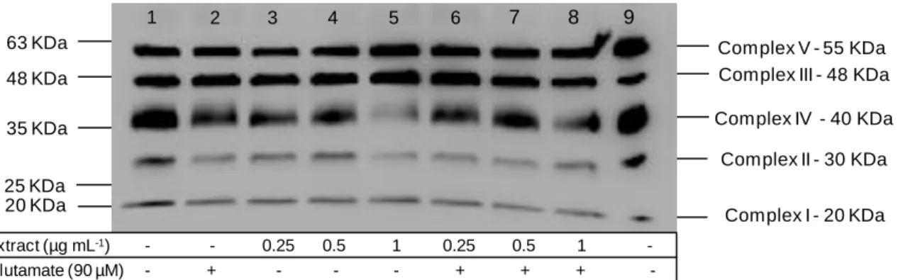
of its relative stability and membrane permeability38. ... 4 Figure 3 - Schematic representation of mitochondrial oxidative damage and its consequences39. ... 5 Figure 4 - Overall flowchart of the main steps involved on cerebellar granule cells culture, occurring during the four tasks proposed. ...16 Figure 5 - Viability of primary neurons monitored from day 1 to 11 in culture, through PI/Hoechst staining. ...18 Figure 6 (continued from previous page) - Morphological aspects of cerebellar granule cells cultured for 11 days in medium with B-27 without antioxidants: (a) day1 in culture; (b) day 2 in culture; (c) day 3 in culture; (d) day 4 in culture; (e) day 5 in culture; (f) day 6 in culture; (g) day 7 in culture. Magnification: 40X. ...19 Figure 7 – Two different aspects of cerebellar granule cells stained with the neuronal specific marker βIII-tubulin (green), the dopaminergic neurons marker tyrosine hydroxylase (red) and the nuclear marker DAPI (blue) after 7 days in culture. Magnification: 40X...20 Figure 8 - Cell viability after 24 h incubation with different glutamate concentrations in order to optimize the most suitable concentration to establish the neurodegeneration model. Values are average ± SD for at least three independent experiences. Statistical differences between treatments in relation to control are expressed as: *p<0.05 and ***p<0.001. ...21 Figure 9 – Morphological differences between cerebellar granule cells at day 9 in culture (a) under control conditions and (b) in the presence of 90 µM of glutamate. ...22 Figure 10 - Cell viability after 24 h incubation with different extract concentrations. Values are average ± SD for at least three independent experiences. ...22 Figure 11 - Cytoprotective effect determination of blackberry digested extracts after 24 h incubation with extracts (0.25, 0.5 and 1 µg GAE mL-1) followed by another 24 h incubation with glutamate (90 µM). Values are average ± SD for at least three independent experiences. Statistical differences between treatments in relation to control are expressed as: ***p<0.001. ...23 Figure 12 - Mitochondrial complexes profile of cerebellar granule cells after 24 h incubation with extracts (0, 0.25, 0.5 and 1 µg GAE mL-1) in the absence of glutamate, depicted on lanes 1, 3, 4 and 5 respectively; or in the presence of 90 µM of glutamate for 24 h, depicted on lanes 2, 6, 7 and 8 respectively. Rat heart mitochondria was used as positive control (lane 9). ...24
Figure 13 - DNA was extracted from 9-day old cerebellar granule cells and submitted to (a) 1% agarose gel electrophoresis for DNA integrity assessment. Lanes: (L) DNA ladder 1Kb; (1) sample 1; (2) sample 2; and (b) 2% agarose gel electrophoresis for primers functionality assessment, after PCR reaction. Lanes: (L) DNA ladder 100bp (1) negative control for GAPDH; (2) empty; (3) DNA amplified with GAPDH primers; (4) negative control for cyt b; (5) empty; (6) DNA amplified with cyt b primers. ...27
TABLES INDEX
Table 1 - Two biological samples of DNA extracted from 9-day old cerebellar granule cells. Further quantification and ratios (nucleic acids/proteins (260/280) and nucleic acids/sugars (260/230) determination was performed. ...26
ABBREVIATIONS
ROS - Reactive Oxygen Species RNS – Reactive Nitrogen Species O2- - Superoxide
OH- - Hydroxyl
H2O2 - Hydrogen peroxide
HOCl - Hypochlorus acid NO - Nitric oxide
ONOO- - Peroxynitrite ON- - Nitroyl
NO2 - Nitrogen dioxide
CNS - Central Nervous System
NMDA - N-methyl-D-aspartate (NMDA)
AMPA - α-amino-3-hydroxy-5-methyl-4-isoxazole PTP - Permeability Transition Pore
ATP – Adenosine triphosphate mtDNA - Mitochondrial DNA
OXPHOS - Oxidative Phosphorylation ETC - Electron Transport Chain
MOMP – Mitochondrial Outer Membrane Permeabilization IVD - In vitro Digestion
BBB – Blood Brain Barrier CVD – Cardiovascular Disease GAE – Gallic Acid Equivalents RT – Room Temperature
PBS – Phosphate-Buffered Saline BSA – Bovine Serum Albumine DAPI - 4’,6-diamidino-2-phenylindole PI – Propidium Iodide
WB – Western Blot
N-PER – Neuronal Protein Extraction Reagent EDTA – Ethylenediaminetetraacetic acid
SDS-PAGE – Sodium Dodecyl Sulphate - Polyacrylamide Gel Electrophoresis PVDF – Polyvinylidene Fluoride
MBA – Membrane Blocking Agent TBS – Tris-Buffered Saline
qPCR – Quantitative Polymerase Chain Reaction ANOVA - Analysis of Variance (ANOVA)
HSD - Honest Significant Difference PCR – Polimerase Chain Reaction Cyt b – Cytochrome b
GAPDH – Glyceraldehyde 3-phosphate dehydrogenase TH – Tyrosine Hydroxylase
1. THEORETICAL FUNDAMENTS
1.1 NEURODEGENERATION
Neurodegeneration corresponds to any pathological condition primarily affecting neurons, which is represented by a large group of neurological disorders with heterogeneous clinical and pathological expressions affecting specific subsets of neurons in specific functional anatomic systems1,2.There are evidences that oxidative stress insult is thought to be one of the main contributing factors to the decrements in cognitive and/or motor performance seen in aging and other neurodegenerative diseases, such as Alzheimer, Parkinson and Amyotrophic Lateral Sclerosis diseases3-6.
1.1.1 OXIDATIVE STRESS AND GLUTAMATE EXCITOTOXICITY
Glutamate excitotoxicity and oxidative stress are mutually related and represent two important hallmarks of neurodegeneration. The adult brain contains about 1011-1012 neurons2, and although it represents only 2% of the body weight, it receives 15% of blood and accounts for 20% of total body oxygen consumption7. This, in addition to its high metabolic demand (dependence on oxidative phosphorylation for energy), oxygen consumption and lipid peroxidation content, as well as its low levels of endogenous antioxidants, make neuronal cells highly sensitive to oxidative stress2,8,9. Oxidative stress can be defined as the condition occurring by chemical imbalance between oxidizing agents (reactive oxygen/nitrogen species (ROS/RNS)) and antioxidants (reducing agents and enzymes)2,10,11. The radicals produced due to this imbalance will typically attack lipids, proteins and DNA, damaging cell membranes, enzymes and genetic material12, leading to many degenerative diseases and aging13. ROS are a family of highly reactive small molecules consisting of oxygen free radicals and associated entities that include, among others, superoxide (O2-), hydroxyl (OH-),
hydrogen peroxide (H2O2), and hypochlorus acid (HOCl)2,10,14. RNS in the other hand refer to
nitric oxide (NO) and derived molecules, such as peroxynitrite (ONOO-), nitroyl (ON-) and nitrogen dioxide (NO2)2,14. These species are formed either during normal metabolism or
through external factors, such as x-rays, ultra-violet radiation and pollution15. In that sense, in order to prevent high levels of ROS/RNS formation, living organisms have complex endogenous antioxidant defense systems (either enzymatic or non-enzymatic)12,16, which are necessary to maintain a physiological balance12,14,17.
Glutamate is the predominant excitatory neurotransmitter, mainly present in the central nervous system (CNS) of the mammalian brain, with up to 40% of all synapses being glutamatergic18-25. Its concentration inside the cells is 5000 to 10000 times greater than outside (at the millimolar level within the vesicle)26,27, is relatively inactive as long as it is intracellular, but extracellularly its action occurs on two cells: presynaptic and postsynaptic
neurons and glial cells inside the brain25. The excitatory responses to this endogenous excitatory amino acid are mediated by a number of pharmacologically and functionally distinct cell-membrane (post-synaptic) receptors: the ionotropic N-methyl-D-aspartate (NMDA), α-amino-3-hydroxy-5-methyl-4-isoxazolepropionic acid (AMPA) and kainate receptors, as well as the metabotropic ones11,19,20,24,25 (fig. 1). Ionotropic receptors are ligand-gated ion channels that open upon the binding of glutamate, and regulate intracellular levels of Na+, K+ and Ca2+, and metabotropic receptors belong to the G-protein coupled receptor superfamily24. Glutamate is both essential and highly toxic at the same time: under physiological conditions, it is released from nerve terminals and is transported primarily to astrocytes, where it is converted to glutamine, which is then stored in glial cells in an inactive form, where glutamate is then regenerated. AMPA receptors are usually co-expressed with NMDA ones at synapses, where they jointly contribute to the process of synaptic plasticity22. Almost every class of glutamate receptors has been implicated in excitotoxic cell death, but it is now accepted that the NMDA receptors play a major role, mainly owing to their Ca2+ permeability. At chemical synapses, glutamate is stored in vesicles, until the Mg2+ cation is removed, as NMDA receptor is blocked whenever bonded to this ion. At that point, this receptor allows the influx of Ca2+ leading to post-synaptic depolarization11,22 (fig.1).
Excitotoxicity refers to the neuronal injury and death that occurs as a consequence of excessive accumulation of extracellular glutamate in the synaptic cleft for a prolonged period, a subsequent overstimulation of glutamatergic receptors on the postsynaptic membrane and the associated excessive influx of ions into the cells18,19,21,22. The increase in cytoplasmic Ca2+ activates a number of Ca2+ dependent enzymes involved in the catabolism of proteins, phospholipids and nucleic acids, increases oxidative stress and may irreversibly damage mitochondria, culminating in neuronal death (fig.1)20,28. Death can occur either by excitotoxic necrosis (involving an uncontrolled influx of Na+ that results in rapid cell swelling and lysis) or
excitotoxic apoptosis (mediated by excessive Ca2+ influx causing the activation of caspases and nuclear chromatin condensation)20. Mitochondrial damage occurs due to the overactivation of NMDA receptors that induce an excessive influx of Ca2+ via these ion channels, which in turn attenuates mitochondrial membrane potential, leading at last to the opening of the permeability transition pore (PTP). Through the disruption of mitochondrial potential, excess Ca2+ can reduce adenosine triphosphate (ATP) synthesis, rendering the cell more vulnerable to death insults, and making mitochondria to be the primary mediators of cell death caused by excitotoxicity24. In general, the common mechanisms of excitotoxicity can be defined as free radical generation and inhibition of antioxidant enzymes, mitochondrial energy enzymes and glutamate transporters19. Therefore, an evident relationship can be established, as excitotoxicity leads to oxidative stress which not only causes neuronal dysfunction and cell death, but also triggers excitotoxicity itself. Excitotoxicity happens by impairing glutamate removal and activating microglia, that contain abundant stores of glutamate, leading at the end to age-related chronic neurodegenerative diseases19,25,29.
1.1.2 MITOCHONDRIA FUNCTION AND BIOGENESIS
As critical organelles in cell survival, by their participation in a number of cellular functions such as modulation of: cellular calcium homeostasis; free radicals; apoptosis; amino acid biosynthesis; fatty acid oxidation; steroid metabolism; and maintenance of cellular energy reserves through ATP production, mitochondria play an essential role in eukaryotic cells2,8,11,30. It has been estimated that about 90% of mammalian oxygen consumption is at mitochondria, which primarily serves to ATP synthesis, making mitochondria particularly important in neurons, owing to their high demands for energy already mentioned2,31. Mitochondria suffer continuously changes on their morphology, length, size, number, function and distribution, through a delicate balance between fission and fusion processes29,30. Fission and fusion are antagonistic processes that predominate under different conditions to adapt mitochondrial morphology and dynamics to the bioenergetic requirements of the cells32. During fission, mitochondrial DNA (mtDNA) and other critical mitochondrial contents are redistributed to the daughter mitochondria by being transformed into isolated, small and round organelles. These fragmented mitochondria are frequently found in resting cells, when high respiratory activity is not required, contributing to the maintenance of bioenergetic capacity33. Fusion on the other hand allows the mixture and exchange of mitochondrial contents (including mtDNA and its encoded proteins), which will then enable individual mitochondrion to rapidly replenish its possible lost components. Fused mitochondria are frequently found in respiratory active cells, when optimal mitochondrial function is needed, either due to the absence of a functional respiratory chain or due to a significant reduction of
Figure 2 - Schematic representation of the mitochondria electron transport chain. Electro leak from the ETC results in the production of superoxide (O2-), which is quickly and efficiently converted to H2O2, which is in turn the principal
cellular mediator of oxidative stress because of its relative stability and membrane permeability38.
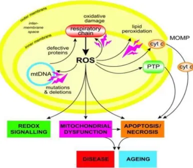
cellular ATP levels, as fusion allows constituents of the respiratory machinery to cooperate most efficiently32,33. However, an altered balance between fission and fusion may affect the removal of damaged mtDNA and thus contributes to the accumulation of mutant mtDNA33. Although the biogenesis of mitochondria depends on an intricate cross talk between mitochondrial and nuclear genomes, mitochondria are the only organelles in animal cells (besides nucleus) that contain their own separate DNA34, which by complementarity with the nuclear genome encodes genes involved in local mitochondrial protein synthesis and protein subunits of the oxidative phosphorylation (OXPHOS) complexes21,35. These multiprotein complexes constitute the mitochondrial electron transport chain (ETC), which is composed of a series of electron carriers that are spatially arranged according to their redox potentials and organized into four complexes (fig.2), named NADH:ubiquinone oxireductase (complex I), succinate:ubiquinone oxireductase (complex II), ubiquinol-cytochrome c reductase (complex III) and cytochrome c oxidase (complex IV), as well as the electron carriers ubiquinone and cytochrome c and the H+-ATP synthase complex30,36.
Knowing that mitochondrial oxidative phosphorylation is the major source of ROS generation7,14,37, this mechanism is believed to be important in the aging process as well as in the pathogenesis of neurodegenerative diseases, considering complex I the major physiologically and pathologically relevant ROS generating site38. The formation of ROS occurs when unpaired electrons escape the electron transport chain and react with molecular
Figure 3 - Schematic representation of mitochondrial oxidative damage and its consequences39.
3). In addition, mitochondrial ROS production leads to induction of the PTP, which renders the inner membrane permeable to small molecules in certain situations39. As mentioned before, mitochondria are essential for neuronal function because the limited glycolytic capacity of neurons makes them highly dependent on aerobic OXPHOS for their energetic needs7. However, oxidative stress directly leads to mitochondrial dysfunction14, which means that any mitochondrial injury can have severe consequences for neuronal function and survival (fig. 3)21. In fact, mitochondrial dysfunction (associated with a functional decline) is becoming appreciated as a unifying characteristic of diverse degenerative pathologies40, contributing to the aging process, in which an activity reduction of any ETC component is detected, and is in turn associated with increased mitochondrial biogenesis.
There is information stating that small, physiological increases in ROS are required for normal increases in mitochondrial biogenesis and moreover that this process may represent an attempt by cells either to increase their aerobic set point, or to maintain a pre-existing aerobic set point in the face of declining mitochondrial function. This relationship is no longer observed either when the decline keeps further to the point that mitochondrial biogenesis can no longer compensate functional declines or by the administration of certain compounds, that may actually serve to disrupt this delicate physiologic feedback loop34. Accordingly, measurement of mitochondrial synthesis is considered to be the correct approach to assess mitochondrial biogenesis, at protein or DNA level (instead of the frequently used assessments of signaling or content)37.
There is no doubt that mitochondrial abnormalities and dysfunction are involved in neurodegenerative diseases, contributing to the aging process37. However, it is still a matter
of debate whether or not such abnormalities are a cause or merely a consequence among many of the diseases21.
1.2 BERRIES AND (POLY)PHENOLS
Quality of life at older age is strongly linked to the preservation of cognitive function that can be influenced by numerous factors and one of the most obvious, but yet under-recognized, being the role of nutrition41.
(Poly)phenols represent a wide variety of compounds essentially found in fruits, vegetables, beverages and some herbs, which are responsible for their colour, flavour, aroma and appearance, and whose primary function is protection of plants against ROS produced during photosynthesis and against herbivores41-44. Characteristically, they have at least one aromatic hydrocarbon ring attached with one or more hydroxyl groups, are mostly found as sugar conjugated41, have ROS scavenging properties and seem to be important in the prevention of different diseases associated with oxidative stress, such as neurodegenerative and cardiovascular diseases and cancer42. Although (poly)phenols are greatly abundant in our diet, they are poorly absorbed from the intestine, highly metabolized and rapidly eliminated, therefore their metabolites are the bioactive compounds within the body42,44. (Poly)phenols can be classified into two main categories, namely flavonoids and non-flavonoids. Flavonoids include flavonols (eg.: quercetin, kaempferol and myricetin), flavones (eg.: apigenin and luteolin), flavan-3-ols (eg.: catechin and condensed tannins (proanthocyanidins), flavanones (eg.: naringenin, eriodictyol and hesperitin), anthocyanins (eg.: callistephin, chrysanthemin and peonidin-3-O-glucoside) and isoflavonoids. Non-flavonoids on the other hand include phenolic acid (eg.: hidroxybenzoates), lignans and stilbenes (resveratrol)13,41,44,45.
Consumption of fruits and vegetables has been associated with a decreased risk for certain degenerative diseases, not only for neurological ones, but also for cancer, stroke, heart disease, diabetes mellitus and osteoporosis, due to the (poly)phenols' anti-inflammatory, antioxidant, antiallergic, antiviral, antiproliferative, anticarcinogenic and neuroprotective and thus "anti-aging" health effects4,46.
Fruits, and specially berries phytochemicals are well known for their chemical antioxidant activity47 and neuroprotective effects48. In addition to their attractive sensorial attributes
choice (different berries) has a substantial impact on the amount of anthocyanins and other (poly)phenols consumed, and consequently, the amount of health benefits they are associated with51.
1.2.2 BLACKBERRIES DIGESTED METABOLITES - IN VITRO DIGESTION (IVD)
When applying fruits and other food components to in vitro studies, it is expected that they have been previously gone through digestion, absorption and metabolization processes. Therefore, in this sort of studies it is used an extrapolation of the bioaccessible metabolites, which refers to the amount of potentially absorbable form of ingredients into the body41.
In vitro digestion models are widely used to study the structural changes, digestibility,
absorption and release of food components under simulated gastrointestinal conditions, mimicking as much as possible the human digestion52, in other words, it is a procedure that simulates the physicochemical and biochemical changes that occur in the upper gastrointestinal tract. This is easily reproducible, although requires adjustments according to the type of food being digested. For the present work, the main purpose of performing in vitro digestion of blackberry fruits is essentially trying to obtain the metabolites that will possibly reach blood stream and consequently the blood brain barrier (BBB) and exert any neuroprotective effect. This process is particularly important, as it was previously demonstrated that the original undigested berry extract (specifically blackberry), although exhibiting a significant chemical antioxidant capacity in vitro, was not able to decrease ROS production in cells submitted to an oxidative stress. On the contrary, digested berry extracts potentiated the ROS decrease3.
1.2.3 BLACKBERRIES AND NEUROPROTECTION
Blackberry fruits are well known to be a rich source of (poly)phenols (mainly anthocyanins and ellagitannins)13 and to exhibit high natural chemical antioxidant capacity, which is one of the major reasons for their increasing popularity in the human diet3,48,53. There are several positive and beneficial documented effects from blackberries ingestion, such as age-related changes and prevention of age-related neurodegenerative diseases13, management of inflammation by suppressing pro-inflammatory cytokines and cancer prevention, as it was shown potential in inhibiting the growth of human colon cancer15.
Dissecting the information obtained from the studies available, its different conditions in terms of type of the assay (in vitro or in vivo), type of berries, way of administration (digested or non digested), and targets (either animals or humans) becomes evident, there is a huge and variable pool of results regarding berries and its benefits. However, not all have the same biological significance for human nutrition and neuroprotection. In vitro studies of neuroblastoma cell line subjected to an H2O2 stress and wild blackberries digested extract
resulted in recovery of membrane potential and cell membrane integrity, ensuring cell viability, constituting by then promising sources of metabolites with neuroprotection capabilities48. In a similar study, having the same neuroblastoma cells subjected to the same stress, blackberry digested metabolites were able to maintain cell membrane integrity, protecting neurons from death, which in that case was not necessarily associated to enhanced antioxidant capacity3. In addition to those, in vivo studies which use animals (usually rodents) as the study group can also be found. Their advantage is that, apart from all the chemical and biochemical analysis made to cells, they allow the observation of differences in motor and cognitive functions. A number of these studies have documented that rodent diet supplementation with berries is effective in reducing not only oxidative stress but also neuronal, behavioral and cognitive defects associated with aging, inflammation and ischemia54. Other similar in vivo studies, with certain extracts supplementation, have demonstrated retarded age-related decrements, improved performance on motor tests, improved working memory performance and increases in several indices of neuronal signaling55. When rats had a long-term intake of blackberry extracts (anthocyanins and ellagitanins), the markers for the oxidative status in some tissues are changed and the expression and activities of antioxidant enzyme are altered when compared with animals that did not receive (poly)phenols in their diet16. A number of other in vivo studies applied not only to animals but also to humans have been made in order to investigate not only the bioavailability of anthocyanins51, but also the berries impact on cardiovascular health49. Amongst other conclusions, volunteers showed a low risk of cardiovascular disease (CVD) during berries enriched diet (by the maintenance of normal vascular function and blood pressure), a decrease on an inflammation marker, and a decrease of lipid peroxidation in smokers.
From all these assays, it becomes evident that berries, and specifically blackberries have a distinguished role on human health, mainly proved in terms of age-related diseases and neurodegeneration. In that sense, the usage of this berry fruit as this study object becomes urgent and relevant.
1.3 NEURODEGENERATION CELL MODEL
survive the disaggregation process, attach to the cell culture support and proliferate, being morphologically similar to the parent tissue and physiologically similar to in vivo cells. Although they have issues of limited cell accessibility and comparability, difficulty with cell isolation/purification, quality assurance and consistency, and contamination risks, primary cells more closely mimic the physiological state of cells in vivo, which in turn allows the generation of more relevant data representing living systems56,57.
When comparing these two cell types in terms of literature, it easily becomes evident that studies and investigations concerning beneficial effects of nutrition, developed on cell lines is more common. In the case of cell lines such as SK-N-MC3,48, SH-SY-5Y (human neuroblastomas)54, HT-29 (human colon tumor)58 and MCF-7 (human breast tumor)53, a plenty of studies have been developed, usually using a stress inducer, like H2O259 that
proceeds a previous treatment with different (poly)phenols extracts. Regarding primary neurons, either hippocampal47, cerebellar60 or others, few studies have been done. Although these cells are used for a plenty of different studies as well, also using a stress inducer21,23,28,29,61-63, they have not often been applied specifically to test berries neurological preventive effects.
With the increasing evidence that neurodegeneration is related to oxidative stress, which in turn can be triggered by excitotoxicity, exposure of cells to glutamate can be used as a valuable model of the mechanisms of oxidative injury in the CNS, and may prove very useful in the elucidation of protective strategies in neurodegenerative diseases associated with oxidative damage or stress. Taking that in consideration, glutamate has been used extensively as an inducer of oxidative stress in different in vitro models28,61-63, namely those from cerebellar granule cells64.
The great majority of cerebellar neurons, both in cerebellum and whole brain, are granular, which constitute the largest homogeneous neuronal population of the mammalian brain, being often used as a model system for the study of neuronal development, function and pathology, including the analysis of activity-dependent survival/apoptosis of neurons and the mechanisms of neuroprotection. In addition, when cultured in vitro, cerebellar granule cells develop characteristics of mature cerebellar granule cells seen in vivo, such as an extensive neuritic network, expression of excitatory amino acid receptors and production and release of glutamate64.
2. OBJECTIVES
Since few years that both scientific and non-scientific societies have been assisting to an increased interest in berries, in particular due to their highly advertised benefits, with great impact on several areas of the human health. In that sense, nutrition can be considered as an emerging preventive solution for several disorders, mainly neurodegenerative ones. Neurodegeneration is in intimate association with both excitotoxicity and oxidative stress. Several preventive approaches have been studied, namely with regards to berries, which have been referred for their highly neuroprotective potential but still, lacking to be tested on primary cultures of neurons. Therefore, this study renders to be necessary, as it will hopefully bring up new information on how all these mentioned factors work together to generate health and/or disease. In that sense, some main goals for this work were settled:
To establish a primary culture from mice granular cerebellar cells and to learn about their behavior and development along the days in culture;
To determine if the neuroprotective effects previously verified on a neuronal cell line, are present in a model of mice primary culture of neurons, which meets the present goals of the assay. So far, in DSB Lab there used to be an implemented neurodegeneration cell model, developed with SK-N-MC, which is a neuroblastoma continuous cell line, established in culture from human metastatic neuroblastoma tissue65. However, for the purpose of this work, a more physiological model based on mice cerebellar neurons was used instead. The main characteristics of this model make it extensively characterized and used for in vitro studies64;
To evaluate mitochondrial population as an indicative of mitochondrial biogenesis;
To understand how the overall expression of mitochondrial complexes occurs within different culture conditions, to realize if any of them is over or underexpressed, and to determine the main complex(es) involved in oxidative stress/excitotoxicity processes and by then, also determine a possible pathway for neuroprotection;
In order to achieve these goals, four main tasks were proposed:
Establishment of primary culture of neurons from seven day-old mice;
Establishment of a neurodegeneration model based on an oxidative stress by testing different times and concentrations of exposure to glutamate;
3. MATERIALS AND METHODS
3.1 PLANT MATERIAL, EXTRACT PREPARATION AND IN VITRO DIGESTION
Fruits of the commercial blackberries cultivar Apache (Rubus L. subgenus Rubus Watson) produced in Fataca experimental field (Odemira, Portugal) were previously obtained, its extract prepared and stored in DSBLab, as described before48. In addition to that, these hydroethanolic extracts went through phytochemical alterations occurring during digestion, which were done in collaboration with James Hutton Institute and mimicked using the IVD model previously described3. Digested samples were dried and stored at -20ºC until use.
3.2 CELL CULTURE
3.2.1 CEREBELLAR CELL CULTURE
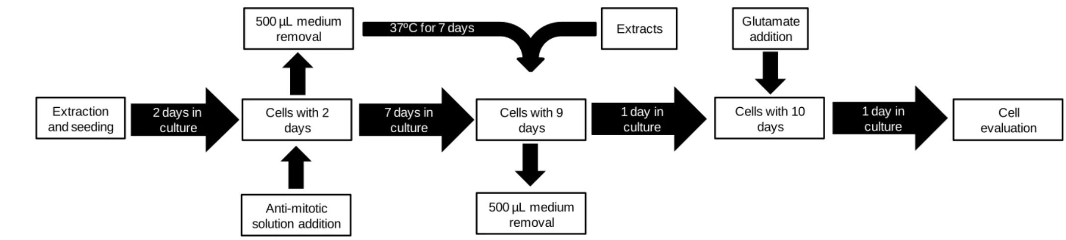
Primary cultures of cerebellar granule cells were prepared according to the already described method60. Briefly, cells were isolated from the cerebella of 7-day-old BALB-C mice and went through a manual disruption process, where cerebella was cut with a razor in parallel and perpendicular movements, after which the tissue was centrifuged 1 min. at 200 g. The following process was a chemical disruption with a mild trypsinization (200 mg L-1, Sigma) of the tissue, followed by trituration in a DNAse solution(10 mg mL-1; Sigma), simultaneously with its constant mixing at a 37ºC water bath. Trypsinization was then inhibited and a new centrifugation for 10 min. at 200 g was made. As the last mechanism to isolate cells from the original tissue, it was minced with a punction needle, and another centrifugation of 5 min. at 200 g was made. After cell counting, cells were ressuspended (0.5 x 10⁶ cells mL-1) and cultured in Neurobasal Medium (Gibco) supplemented with 2% (v/v) B-27 without antioxidants (alpha tocopherol acetate, alpha-tocopherol, catalase, superoxide dismutase and glutathione (Gibco)) and 2% (v/v) KCl (20mM, Sigma), and containing 0.25% (v/v) glutamine (200 mM, Sigma) and 0.48% (v/v) penicillin-streptomycin (5000 U mL-1, Gibco) solution. Cells were lastly cultured in plates or flasks coated with poly-D-lysine (50 µg mL-1, Sigma) and maintained in a humidified atmosphere of 7% CO2 at 37 ºC. Half of the
media (either 500 µL or 5 mL) was removed 48 h after inoculation, maintained at 37ºC, and cytosine arabinoside (20 µM, Sigma) was added to prevent glia cell proliferation, with all the experiments being performed on days 7-11 in culture (fig. 4). The removed media was introduced back in the culture at day 7, and used either for medium renewal or for the introduction of any new compound to the culture. Most of the experiences were performed on 24-well plates covered with coverslips and coated with poly-D-lysine.
3.2.2 B-27 SUPPLEMENTATION TEST
Primary cultures were established with B-27 either with or without antioxidants. Moreover, to analyze if antioxidants from B-27 could interfere with blackberry digested extract effect, different concentrations of digested extract were tested. In order to do that, at day 7, half of the media was removed and the extracts added, which had been previously homogenized in the preserved medium. Concentrations tested were: 0.25, 0.5 and 1 µg gallic acid equivalents (GAE) mL-1. Cells were incubated for 24 h, morphology was monitored by optical microscopy and cell viability was determined.
3.2.3 IMMUNOFLUORESCENCE
The integrity and functionality of primary cultures of cerebellar neurons were confirmed by immunofluorescence of cells cultured for 7 days, according to the protocol previously described66 . Briefly, cells were fixed in a solution of 4% paraformaldehyde plus 4% sucrose for 20 min. at room temperature (RT), washed twice in phosphate-buffered saline (PBS) and then blocked and permeabilized with PBS containing 3% bovine serum albumin (BSA, Sigma) and 0.1% Triton X-100 (Merck) for 20 min. at RT. Cells were incubated with anti-tirosine hydroxilase (rabbit polyclonal, 1:200, Santa Cruz Biotechnology Inc., Nr. Sc-14007) and anti-Beta III tubulin (mouse monoclonal, 1:200, Millipore, Nr. MAB1637) for 2 h at RT. Immunolabeling was visualized by incubation with AlexaFluor 594-conjugated goat anti-rabbit IgG (1:500, Invitrogen) and AlexaFluor 488-conjugated goat anti-mouse IgG (1:500, Abcam). After two washes in PBS, immunolabeled cells were mounted and counsterstained with ProLong® Gold antifade reagent with 4’,6-diamidino-2-phenylindole (DAPI, Invitrogen). After drying, cells were observed and photographed under a computer-driven digital camera (Leica DFC340 FX, Heerbrugg, Germany) on an upright microscope (Leica DMRB microscope, Wetzlar, Germany) using filter cubes with a bandpass of 515-560 nm, 450-490 nm and 355-425 nm of wavelength, for tyrosine hydroxilase, beta III tubulin and DAPI, respectively (fig. 4).
3.3 CELL VIABILITY
Cell viability was assessed in all cases by fluorescence microscopy through Propidium Iodide (PI) and Hoechst 33342 staining, as previously described60. Cells were incubated with
For image analysis of cell viability, 3 micrographs per coverslip were taken at a 40 X magnification, meaning that for each experiment, 9 micrographs per condition were evaluated. Image analysis was performed with ImageJ 1.47. Brightfield images for morphology evaluation were taken at 20 and 40 X magnification, with a computer driven digital camera (Nikon Digital Sight DS-Fi1) on an inverted microscope (Nikon Eclipse TE300).
3.4 GLUTAMATE OPTIMIZATION
To investigate the appropriate glutamate concentrations (that allow a range of death percentage enough to make the cells able to respond to the further stimulus) for the establishment of the neurodegeneration cell model, at day 8 in culture cells were exposed to different concentrations of glutamate solution (Sigma), from 10-100 µM. Controls were cells without glutamate treatment, and processed in parallel. Cells were then incubated for 24 h, morphology was evaluated and viability determined as previously described at 3.3 topic (fig. 4).
3.5 CYTOTOXICITY EVALUATION
To evaluate extracts cytotoxicity, dried digested fractions were ressuspended in cell medium. At day 7 in culture, 500 µL of culture medium was removed and primary neurons were incubated with different concentrations of these fractions, in the range of 0-1 µg GAE mL-1 medium. Cells were then incubated for 24h, morphology was evaluated and viability determined as previously described at 3.3 topic (fig. 4).
3.6 NEUROPROTECTIVE EVALUATION
To determine the neuroprotective effects of blackberries digested metabolites, at day 7 in culture, non-toxic concentrations (0.25, 0.5 and 1 µg GAE mL-1) of blackberry dried fractions were ressuspended in 500 µL of cell medium (previously removed at day 2 and maintained at 37ºC) and added to the cells, after a previous removal of 500 µL of the medium from each well. Cells were then incubated for 24 h and morphology evaluated. At day 8 in culture, 90 µM of glutamate was added to the respective wells. Cells were incubated for 24 h, morphology was evaluated and viability determined as previously described at 3.3 topic (fig. 4).
3.7 WESTERN BLOT (WB) ANALYSIS 3.7.1 PROTEIN EXTRACTION
Neurons cultured on 25 cm2 t-flasks were submitted to protein extraction on day 9 in culture through the Neuronal Protein Extraction Reagent (N-PER, Pierce) supplemented with an
ethylenediaminetetraacetic acid (EDTA)-free protease inhibitor cocktail (1% (v/v), Calbiochem), according to the manufacturer’s instructions. Briefly, cells were lysed with the previous solution on ice for 5 min. following a 10 min. centrifugation at 10.000 g, after which the supernatant was collected and stored at -20ºC until being used. Protein quantification was then processed using modified Lowry’s method.
3.7.2 ELECTROPHORESIS AND ELECTROTRANSFER
Twenty µg of protein, as well as the prestained protein marker VI (Applichem) and the rat heart mitochondria extract as positive control (Mitosciences - Abcam) were subjected to a sodium dodecyl sulphate – polyacrylamide gel electrophoresis (SDS-PAGE) in 10% (w/v) and 1.5 mm thickness acrylamide gel, at 200 V for approximately 40 min. The electrophoresis buffer was composed by 25 mM Tris-base, 190 mM glycine and 0.1% (w/v) SDS67.
The electrotransfer of proteins occurred according to the previously described method68. After SDS-PAGE, gel was soaked in transfer buffer composed by 12 mM Tris-base, 96 mM glycine, 0.1% (w/v) SDS and 20% (v/v) methanol at pH 8.3. A polyvinylidene fluoride (PVDF) membrane (Amersham Biosciences) was also incubated in transfer buffer for 5 min. after a 1 min. pre-incubation in methanol. Both gel and membrane were assembled in the transfer cell (Trans-Blot electrophoretic transfer cell, BioRad) containing transfer buffer, and transference was carried out at approximately 70 V for 2 h at 4ºC.
3.7.3 IMMUNODETECTION
The expression of mitochondrial OXPHOS system components was assessed by using the MitoProfile® Total OXPHOS Rodent WB primary antibody cocktail (MS604, Mitosciences - Abcam) targeting specific subunits from mitochondrial complexes I, II, III, IV and V, according to manufacturer’s instructions and other previously described data69,70.
After transfer, membrane was dried at RT and blocked with 5% (w/v) membrane blocking agent (MBA, GE Healthcare) in Tris-buffered saline (TBS) containing 0.01% (v/v) Tween20® (Sigma) (TBST), for 1 h at RT. Membrane was then incubated with primary antibody (1:250) and left overnight at 4ºC, after which it was washed three times in TBST for 5 min. each, and incubated with secondary antibody (stabilized goat anti-mouse horseradish peroxidase
Rockland Inc.) and membrane images were acquired in the Molecular Imager Chemidoc XRS (Quantity One© software 4.6.6, BioRad).
3.8 POLIMERASE CHAIN REACTION (PCR)
Preliminary tests on PCR with extracted DNA samples were executed, as they represent the necessary analysis preceding a quantitative PCR reaction. In order to do that, genomic DNA was extracted from primary neurons cultured under control conditions (0 µg GAE mL-1 and 0 µM glutamate) using the High Pure PCR Template Preparation Kit (Roche Diagnostics), according to the manufacturer’s instructions, and further stored at -80 ºC until use. For the assessment of DNA quality and yield, samples were analyzed with a NanoDrop 2000c spectrophotometer (Thermo Scientific) and DNA integrity and contamination were determined by DNA visualization in a 1% agarose gel, stained with ethidium bromide.
Potential DNA contamination and primers efficiency were tested through PCR, which was performed using specific forward and reverse primers designed for the mitochondrial cytochrome b (cyt b) gene (forward: 5'-TTC ATG TCG GAC GAG GCT T-3' and reverse: 5’-TCC TCA TGG AAG GAC GTA GG-3’) and for the nuclear glyceraldehyde 3-phosphate dehydrogenase (GAPDH) gene (forward: 5’-CCT TCA TTG ACC TCA ACT ACA T-3’ and reverse: 5’-CCA AAG TTG TCA TGG ATG ACC-3’)71. The PCR reaction contained 2 µL input
(20-100 ng DNA), 0.625 U Taq DNA Polymerase (Fermentas), 1.25 µL of each forward and reverse primer, 2 mM MgCl2, 0.2 mM of each dNTP (Invitrogen), 75 mM Tris-HCl (pH 8.8 at
25 ºC), 20 mM (NH4)2SO4 and 0.01% (v/v) Tween 20. The following program was applied: 1
cycle of 3 min. at 94ºC for denaturation, followed by 41 cycles (45 sec. at 94 ºC for denaturation, 30 sec. at 60 ºC for annealing, 90 sec. at 72 ºC for extension) and a final 10 min. extension at 72 ºC. PCR products previously obtained were visualized in a 2% agarose gel stained with ethidium bromide. Both gel images were acquired in the Molecular Imager Chemidoc XRS (Quantity One© software 4.6.6, BioRad).
3.9 STATISTICAL ANALYSIS
All the results reported are the averages of at least three independent experiments, which data was statistically evaluated either by analysis of variance (ANOVA) – Tukey honest significant difference (HSD) test, or by Paired t-Test, using SigmaStat 3.10 software (Systat Software Inc.).
Cell evaluation Extraction and seeding 2 days in culture Cells with 2 days 7 days in culture Anti-mitotic solution addition 500 µL medium removal Glutamate addition 37ºC for 7 days Cells with 9 days 500 µL medium removal Extracts Cells with 10 days 1 day in culture 1 day in culture
4. RESULTS AND DISCUSSION
4.1 ESTABLISHMENT OF PRIMARY CULTURE OF NEURONS
The initial stage of this work was the establishment of primary culture of neurons from mice cerebellum. The choice of this brain’s region was related to its high content on neuronal cells that increases the probability of getting viable neurons for cell culture. The use of 7 day-old mice is an important issue and using older animals is not recommended, as cells will start to extend axons and form dendrites and synaptic connections, rendering them more susceptible to damage during the purification process64.
B-27 is recommended to be used as a supplement within Neurobasal media. This supplement has a diversified composition, presenting proteins (such as BSA and insulin), vitamins (such as vitamin A and tocopherols), antioxidants (alpha tocopherol acetate, alpha-tocopherol, catalase, superoxide dismutase and glutathione) and other components (such as some hormones and D-galactose), however for our study raises some questions related to the antioxidant and protein contents:
The high content on antioxidants mentioned before (alpha tocopherol acetate, alpha-tocopherol, catalase, superoxide dismutase and glutathione) could interfere and mask any possible effects of blackberries digested metabolites on cells, therefore we have decided to test the commercially available B-27 supplement without antioxidants, and determine whether it could effectively replace B-27 with antioxidants without affecting cell viability. According to the results obtained, for a control situation in which cells were cultured in medium containing B-27 with and without antioxidants in the absence of blackberries digested extracts (0 µg GAE mL-1), no statistical significance was observed on cell viability between both conditions. Moreover, when increasing concentrations of blackberries digested metabolites (0.25, 0.5 and 1 µg GAE mL-1) were added to the culture medium in the presence of both types of B-27, the same statistical indifference was detected between the two medias tested, for each extract concentration (data not shown). Therefore, we decided to culture cells in B-27 without antioxidants supplemented medium;
Concerning protein content of B-27 supplement, and since protein interaction with (poly)phenols further added to the media was a concern, we then considered to test different percentages of B-27 in order to minimize it as much as possible. However, because its percentage within the media is already low, we have considered that the interference between protein and (poly)phenols would be minimal and decided to maintain the original recommended B-27 concentration as 2% of the media.
After the media parameters were defined, cells were monitored in culture for 11 days in order to understand and evaluate their growing state and progression throughout the time in
0% 20% 40% 60% 80% 100% 120% 140% 0 2 4 6 8 10 12 N or m al iz e d V ia bi lit y (% )
Incubation time (day)
Daily monitorization of primary neurons viability
culture. Cell viability was determined for each day in culture, and according to the results obtained, the percentage of viable cells did not change considerably over days (fig. 5), meaning that cells in culture are usable for experiences during that time. Microscopic morphological analysis was also performed and by the results achieved, major differences were evident from day 1 to day 3 in culture, from which they develop their cellular bodies and elongate dendrites, being considered ready to use for experiences after day 7 (fig. 6).
Figure 6 (continued from previous page) - Morphological aspects of cerebellar granule cells cultured for 11 days in medium with B-27 without antioxidants: (a) day1 in culture; (b) day 2 in culture; (c) day 3 in culture; (d) day 4 in culture; (e) day 5 in culture; (f) day 6 in culture; (g) day 7 in culture. Magnification: 40X.
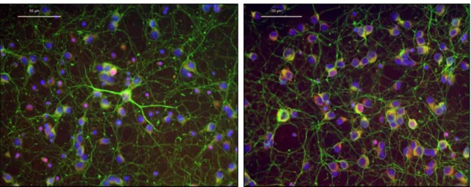
Following the previous analysis, integrity and functionality of primary culture of cerebellar neurons were confirmed by immunostaining for tyrosine hydroxylase (TH) and βIII-tubulin, two cytoplasmic proteins. TH takes part in the first step of the pathway that produces a group of hormones called catecholamines (namely dopamine), by which TH is considered as a marker of dopaminergic neurons28,72. βIII-tubulin on the other hand is a cytoskeleton tubulin protein almost exclusively found in neurons, and has been extensively used as a marker to identify neuronal cells from other brain cells such as glia, which do not express it66. According to the images obtained (fig. 7), under normal conditions, cultured cerebellar granule cells at day 7 in vitro extended long, highly branched dendrites and were heavily connected to each other. The positive staining for both markers indicates that the present cells were effectively neurons and specifically refer to dopaminergic ones.
Figure 7 – Two different aspects of cerebellar granule cells stained with the neuronal specific marker βIII-tubulin (green), the dopaminergic neurons marker tyrosine hydroxylase (red) and the nuclear marker DAPI (blue) after 7 days in culture. Magnification: 40X.
4.2 ESTABLISHMENT OF NEURODEGENERATION MODEL USING PRIMARY CULTURE OF NEURONS In order to mimic a neurodegeneration condition, namely through an excitatory insult, glutamate was used to induce cytotoxicity, although H2O2 is commonly described as the toxic
insult often used in cell models3,48,54. Because the interaction between H2O2 and
(poly)phenols exists, and because the culture media can never be completely removed, the use of glutamate instead of H2O2 eliminates or at least decreases that interference.
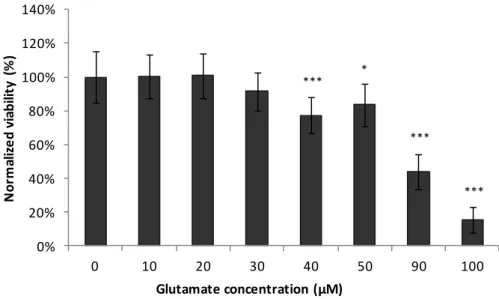
Throughout the literature, there is evidence that the sensitivity of cerebellar granule cells to glutamate depends on the number of days in culture73, as they prevailed insensitive to all of the excitotoxic amino acids at days 3 and 5 in culture, and only became sensitive to toxicity induced by L-glutamine at day 8 in culture74. This information is in accordance to the morphological mature state found on cerebellar granule cells at day 7 in culture. To establish the proposed neurodegeneration cell model with primary neurons, different concentrations of glutamate were tested in a range of 10 to 100 µM, which is within the ranges referred to be found in plasma (30-100 µM) and synaptic cleft (2-1000 µM)27. For this particular work, the selection of the most appropriate glutamate concentration was not only the concentration that caused considerable death in respect to the control condition, but also that enabled cells to have a significant response in the presence of the blackberries digested extracts.
0% 20% 40% 60% 80% 100% 120% 140% 0 10 20 30 40 50 90 100 N or m al iz e d vi abi lit y (% ) Glutamate concentration (µM) *** *** *** *
decrease made both of these concentrations a possible choice for the establishment of the neurodegeneration model.
Nevertheless, accordingly to the similar model established for the neuroblastoma cell line developed in DSB Lab, to which the toxic stimulus (H2O2) is added at a concentration that
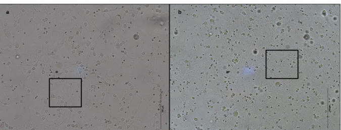
allows an approximate reduction of 50% in cell viability3, 90 µM of glutamate was chosen. Morphological alterations were also observable when comparing primary neurons in culture with and without glutamate exposure. In the absence of glutamate, cerebellar granule cells grew normally in culture, properly extended their axons and formed dendrites and synaptic connections with each other (highlighted in fig. 9a). When, on the other hand, 90 µM of glutamate was present, it’s deleterious effects on neuronal morphology were recognized essentially in dendrites showing an irregular punctuate staining, which is indicative of neuronal damage (highlighted in fig. 9b). Moreover, there is an increase in the number of small rounded and dark cells, which represent dead cells (fig. 9b). As a matter of fact, immunostaining of hippocampal neurons evidences that the length and branching of dendrites in neural networks are severely reduced by the presence of glutamate75. Regarding the cerebellar granule cells that contacted with neurotoxic concentrations of glutamate, it was previously demonstrated that they undergo two different death processes: a subpopulation collapse to acute necrosis during and immediately after the exposure, while the remaining neurons die from delayed apoptosis73.
Figure 8 - Cell viability after 24 h incubation with different glutamate concentrations in order to optimize the most suitable concentration to establish the neurodegeneration model. Values are average ± SD for at least three independent experiences. Statistical differences between treatments in relation to control are expressed as: *p<0.05 and ***p<0.001.
80% 100% 120% ia bi lit y (% ) a b
4.3 EVALUATION OF NEUROPROTECTIVE EFFECT OF BLACKBERRIES DIGESTED METABOLITES Once established the neurodegeneration cell model with cerebellar granule cells, different concentrations of blackberries digested metabolites were tested in a range of 0 to 1 µg GAE mL-1, in order to determine any cytotoxicity effects on the cells. This range corresponds to 0 to 6 µM GAE and is close to the physiological concentrations of certain phenolic components detected in human plasma, which is about 0 to 4 µM3,45. Applying this range of concentrations to the present neurodegeneration cell model, the biological significance will be increased in case of a neuroprotective effect. According to the results obtained, none of the concentrations tested revealed to be cytotoxic for primary neurons in culture, as viability was not significantly different from control conditions, meaning that all of them would be an adequate choice to be used in the neuroprotection study itself (fig. 10). However, because this work is in line with the neuroprotection studies that took place at DSB Lab within the neurodegeneration cell line model present at the time3, and in order to maximize a protective effect, we decided for the highest blackberries digested metabolites non toxic concentrations, which were 0.25, 0.5 and 1 µg GAE mL-1,respectively.
Figure 9 – Morphological differences between cerebellar granule cells at day 9 in culture (a) under control conditions and (b) in the presence of 90 µM of glutamate.