INSTITUTO DE INVESTIGAÇÃO E FORMAÇÃO AVANÇADA
ÉVORA, MAY 2013 Supervisors: Prof. Dr. Maria Amely Zavattieri Prof. Dr. Maria do Rosário Martins Prof. Dr. Ana Teresa Caldeira A
s
Thesis presented to obtain the PhD degree
in Biology by the University of Évora
Thesis presented to obtain the PhD degree
in Biology by the University of Évora
Carla Aparecida Ragonezi Gomes Lopes
In vitro plantlet regeneration from
mature zygotic embryos of Pinus pinea
L.: overcoming the rooting problems
Funding
Contactos:
Universidade de Évora
Instituto de Investigação e Formação Avançada - IIFA
Palácio do Vimioso | Largo Marquês de Marialva, Apart. 94 7002-554 Évora | Portugal Tel: (+351) 266 706 581 Fax: (+351) 266 744 677 email: [email protected]
Ragonezi, C. (2012): In vitro plantlet regeneration from mature zygotic embryos of Pinus pinea L.: overcoming the rooting problems. Doctoral Dissertation presented at University of Évora, Portugal.
Keywords: Adventitious rooting, biotization, ectomycorrhiza, in vitro mycorrhization, micropropagation, Pinus pinea L.
This thesis was financed by the Portuguese Foundation for Science and Technology (FCT) SFRH / BD / 44119 / 2008
I hear and I forget I see and I remember I do and I understand
VI
Como nenhuma grande realização da nossa vida é alcançada sozinha, esta parte da minha tese destina-se àquelas pessoas que fizeram com que este sonho longínquo e aparentemente intangível fosse certeza. Foram muitos anos, muito trabalho, muitas pessoas, distintos laboratórios, inúmeros equipamentos, muitas conversas, telefonemas e favores, por isso espero que aqueles que são citados tenham paciência de ler, e peço desculpas adiantadas para aqueles que me esqueci (não foi por mal, mas estava na fase crítica da entrega da tese).
Primeiro, gostaria de agradecer às instituições que forneceram apoio financeiro, instalações e equipamentos, permitindo que este trabalho fosse possível. Pelo apoio financeiro através da bolsa de doutoramento agradeço à Fundação para a Ciência e Tecnologia (FCT). Este estudo foi realizado na Universidade de Évora, principalmente no Laboratório de Melhoramento e Biotecnologia Vegetal do ICAAM - Instituto de Ciências Agrárias e Ambientais Mediterrânicas (instituição de acolhimento), no Laboratório de Biotecnologia do Departamento de Química e no Laboratório HERCULES, nos quais eu gostaria de agradecer por fornecerem as condições necessárias de trabalho.
Nada seria possível sem o apoio incondicional da minha querida orientadora e amiga (creio que posso utilizar este termo) Prof. Dr. Amely Zavattieri. Esta tese foi construída com uma receita equilibrada entre orientação, autonomia e muito trabalho. Obrigada pelos grandes momentos vividos e por todo o apoio (dentro e fora do Lab.).
Nada mais importante para cooperar com a orientadora do que uma co-orientadora. No meu caso tive sorte pois consegui duas. Prof. Dr. Maria do Rosário Martins e Prof. Dr. Ana Teresa Caldeira, agradeço imenso por todo o auxílio, todas as horas despendidas no laboratório e todos os conselhos que foram oferecidos. Com vocês as componentes moleculares e bioquímicas ficaram mais divertidas.
Dedico um parágrafo à parte para a conselheira científica do nosso grupo Dr. Krystyna Klimaszewska. Obrigada por ser uma excelente pessoa e investigadora, sempre muito dedicada e disponível para colaborar com conselhos extremamente importantes.
Para complementar todo o trabalho despendido na tese, agradeço a toda equipe de profissionais que trabalhei na Universidade de Évora, que foram cruciais para a conclusão deste trabalho. Principalmente Prof. Dr. Dora Teixeira, Prof. Dr. Luís Dias,
VII
Às técnicas de laboratório que comemoraram comigo nos dias felizes, me ajudaram nos momentos de desespero (como quando o meio de cultura não solidificava, ou quando eu não tinha como repicar meus fungos) e me consolaram nos dias chuvosos, tristes e que tinha saudades do Brasil. Sem vocês nenhuma tese de doutoramento é realizada e agradeço imenso toda a assistência. Aqui cito principalmente as técnicas Virgínia Sobral, Elsa Ganhão, Otília Miralto e as técnicas do Departamento de Química
Á minha família em geral (que estão aqui e os que já não estão), principalmente aos meus pais e minha irmã. Minha mãe e meu pai que recebem comigo os frutos deste árduo trabalho, uma vez que sempre investiram em mim apostando na minha formação académica e como pessoa. Obrigada por não me tirarem da “Cultura Inglesa” . Uma gratidão especial vai para a Luana, pois sem ela, não estaria em Portugal finalizando esta etapa tão importante da minha vida. Ao Samuel, meu marido de fé, companheiro incansável que me deu todas as forças para continuar quando a energia já estava acabando (e às vezes a paciência).
A todos os meus amigos e colegas (deste lado do Atlântico e do lado de lá) que me apoiaram, que aguentaram as reclamações e que ouviram com uma tolerância incrível a todos os meus problemas. Obrigada ainda pelos momentos felizes, pelas risadas exageradas e pelos ótimos momentos que tive nestes últimos anos.
Por último, mas não menos importante agradeço a todas as pessoas que de uma forma ou outra, direta ou indiretamente fizeram parte desta fase da minha vida e que contribuíram para a conclusão de um trabalho tão gratificante.
VIII
No great accomplishments are made in life without the help of others, and I would like to thank those who have helped bring this seemingly impossible task to fruition. Many years of hard work have gone into this project, involving many people. I apologize in advance to anyone I may have forgotten.
First of all, I would like to thank the institutions that have provided financial support, facilities and equipment, making this work possible. For Ph.D. scholarship funding I thank the Foundation for Science and Technology (FCT). This study was conducted at the University of Évora, mainly at the Laboratory of Breeding and Plant Biotechnology of the ICAAM - Institute for Mediterranean Agricultural and Environmental Sciences (host institution), the Laboratory of Biotechnology at the Chemistry Department and the HERCULES Laboratory, which I would like to thank for providing support for my work.
Nothing would have been possible without the unconditional support of my dear friend (I think I can call her that) and supervisor Prof. Dr. Amely Zavattieri. This thesis was written thanks to a combination of guidance, autonomy and hard work: many thanks for the great moments and for all the support you gave (both inside and outside the lab).
Nothing is more important than cooperating with the supervisor than collaborating with a co-supervisor. In my case I got lucky because I managed to get both: to Prof. Dr. Maria do Rosario Martins and Prof. Dr. Ana Teresa Caldeira, thank you very much for all the help you gave, all the hours spent in the laboratory and all the advice that you provided. You made the molecular and biochemical components more fun.
To the group scientific advisor, Dr. Krystyna Klimaszewska, thanks for being a great person and researcher, extremely dedicated and always willing to help and provide great advice.
In order to complement all the work spent on the thesis, I thank the entire team of researchers who I worked with at the University of Évora, who were crucial to the successful completion of this work, especially Prof. Dr. Dora Teixeira, Prof. Dr. Luís Dias, Prof. Dr. Augusto Vieira Peixe, Prof. Dr. Celeste Santos-Silva, Prof. Dr. Paulo Oliveira, Mário Castro, M.Sc. and Cátia Salvador, B.Sc..
Laboratory technicians that cheered with me the good days, helped me in times of despair (as when the medium is not solidified, or when I had to cultivate my fungi) and comforted me on rainy and sad days that I missed Brazil. Without you no doctoral thesis
IX
To my family (those who are with us and those who are no longer with us), especially my parents and my sister. My mother and father share with me the fruits of all the hard work I have done, since they have always supported me in my academic training and personal development. Thank you for sending me to the “Cultura Inglesa". My special thanks go to Luana, because without her, I wouldn't now be in Portugal in the last stage of a process which is so important in my life. To Samuel, my husband, a tireless companion who has given me the strength to continue when I was lacking in energy (and sometimes patience), thank you.
To all my friends and colleagues (on both sides of the Atlantic) who have supported me, endured my complaints and listened with incredible tolerance to all my problems. Thank you also for the happy moments, the great laughs and the great moments we've shared throughout all these years.
Last but not least, I would like to thank all those who in one way or another directly or indirectly figures in this stage of my life and who contributed to the completion of such a rewarding piece of work.
X
Regeneração in vitro de plântulas de embriões zigóticos maduros de Pinus pinea L.: superando os problemas de enraizamento
Na natureza, as plantas e os microrganismos estabelecem associações de várias ordens. Nas culturas in vitro de plantas, mesmo as associações favoráveis com microrganismos foram, durante muitos anos, consideradas como contaminantes. Só mais tarde, as vantagens da inoculação in vitro (co-cultura) foram demonstradas e as técnicas de biotização (bacterização ou micorrização) usadas com o objetivo de melhorar as condições de crescimento in vitro.
As dificuldades do enraizamento in vitro de uma das espécies mais importantes da floresta mediterrânica portuguesa, Pinus pinea L., conduziu à escolha deste sistema biológico, como matéria de estudo para a tese. Neste estudo, foram utilizados fungos ectomicorrízicos para otimizar a fase de enraizamento de plantas de Pinus pinea L. micropropagadas via organogénese. A introdução de ectomicorrízas no processo de micropropagação reativou o crescimento das raízes e induziu a melhoria dos vários parâmetros do sistema radicular adventício conduzindo a uma menor perda de plantas durante a aclimatização. Com efeito, a micorrização melhorou a funcionalidade das raízes, facilitando a absorção de nutrientes e de água.
Neste trabalho, efetuou-se também uma extensiva caracterização morfológica e molecular das ectomicorrízas associadas a P. pinea. Das várias co-culturas testadas, selecionou-se a interação Pisolithus arhizus/P. pinea para estudar os sinais bioquímicos pré-simbióticos estabelecidos durante as etapas iniciais da co-cultura in vitro. Para possibilitar este estudo foi desenvolvido um novo sistema de co-cultura, o qual já está patenteado. Os resultados indicam que a presença de compostos fenólicos, nomeadamente o ácido o-coumarico, poderão ser importantes mediadores na interação fungo/planta.
XI
In nature, plants and microorganisms establish symbiotic associations of various orders. However, for many years such associations were deemed unnecessary in in
vitro cultures because the culture medium provides ample amounts of nutrients and
plant growth regulators to a growing plant. Only recently, the benefits of biotization (bacterization or mycorrhization) of plants regenerated in vitro were demonstrated by improvements in their growth and vigor.
Pinus pinea L., which is of one of the most important species of Portuguese
Mediterranean forests, can be regenerated in vitro from embryo cotyledons but the growth of adventitious roots induced in shoots ceases shortly after their formation. Overcoming this particular biological impediment was the study subject of the thesis. In this study, ectomycorrhizal fungi were used to improve adventitious rooting of Pinus
pinea L. plants micropropagated through organogenesis. The introduction of
ectomycorrhizae during the micropropagation process reactivated the root growth and improved several root characteristics leading to a reduced loss of plants during acclimatization. In fact, the mycorrhization enhanced root functionality facilitating the absorption of nutrients and water.
In this work, an extensive characterization of morphological and molecular ectomycorrhizae associated with P. pinea was also undertaken. Of the several fungus species tested, the interaction of Pisolithus arhizus/P. pinea was selected for studying the pre-symbiotic biochemical signals established during the initial stages of co-culture
in vitro. To facilitate this study, a novel co-culture system was developed which has
been patented. The results indicate that the phenolic compounds, in particular the o-coumaric acid ester might be important mediators in the interaction between the fungus and stone pine.
XII
AC - activated charcoal AM - arbuscular mycorrhizal ARF - adventitious root formation BAF - biotin-aneurin-folic acid agar BAP - benzylamino purine
CW - cool white light
DCR medium - Gupta and Durzan (1985) DM - direct measurement;
DNA - deoxyribonucleic acid ECM - ectomycorrhiza
GD medium - Gresshoff and Doy (1972) GL - gro-lux
HEPES buffer - N-2 Hydroxyenthyl piperazine-N´-2-ethane sulfonic acid HPLC-UV - high-performance liquid chromatography
IAA - indole-3-acetic acid IM - indirect measurement ITS - internal transcribed spacer
LC-DAD-MS Liquid chromatography - Diode array detector - Mass spectrometry LP - medium Quorin and Lepoivre (1977)
LSU - large subunit
MMN - modified Melin-Norkrans medium (Marx 1969) MS medium - Murashige and Skoog (1962)
NAA - naphthalene acetic acid
NCBI - National Center for Biotechnology Information PBS - phosphate buffer saline
PCR - polymerase chain reaction PGRs - plant growth regulators PPF - photosynthetic photon flux rDNA - ribosomal genes
rRNA - ribosomal RNA RT- retention time RTD - root tissue density
SH medium - Schenk and Hildebrandt (1972) SRA - specific root area
XIII
UC - unknown compounds
UPGMA - unweighted pair group method with arithmetic average WS - work solutions
WPM - woody plant medium - Lloyd and McCown (1981) WPMRI - woody plant medium root induction
WPMRE - woody plant medium root expression WPMS - woody plant medium solid phase WPML - woody plant medium liquid phase
XIV
Chapter 1 - General introduction
Fig. 1 - A schematic representation of the Pinophyta phylogeny (from Quinn and Price 2003).
Fig. 2 - Taxonomic hierarchy of Pinus pinea L. (from Integrated Taxonomic Information System (ITIS), 2012).
Fig. 3 - The distribution map of Pinus pinea L. along the Mediterranean Basin and adjacent zones (from Euforgen.org).
Fig. 4 - Pinus pinea L. trees in a typical pine stand in Alcácer do Sal district, Portugal.
Fig. 5 - Mature cone of a P. pinea tree (a) and seed without the seed-coat (b). Fig. 6 - Morphological changes in roots as they become mycorrhizal and the effects of those changes on the development of a mycorrhizosphere. A - Generalized non-mycorrhizal root with root hairs and indicated sources of organic materials available as substrates for rhizosphere microorganisms. B - Endomycorrhiza with indicated morphological changes such as reduced tissue sloughing, lack of root hairs, presence of external hyphae, thick-walled spores, and associated soil aggregates; no obvious change in surface area. C - Ectomycorrhiza indicating dramatic morphological changes such as development of a fungal mantle plus extensive external hyphae and rhizomorphs and associated soil aggregates, loss of root hairs, and greatly increased branching and surface area (from Linderman 1988).
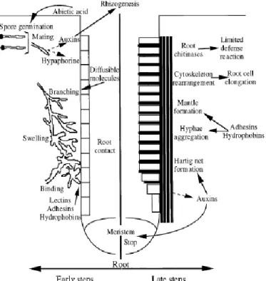
Fig. 7 - Schematic representation of ectomycorrhizal development. Morphological events taking place during early (left) and late (right) stages of ectomycorrhizal formation are indicated (from Baker et al. 1998).
Fig. 8 - Commonly used primers for amplifying parts or the entirety of the ITS region. a) Relative position of the primers, design of the subsets and number of sequences in each subset. b) Primer sequences, references and position of the primer sequence according to a reference sequence of Serpula himantioides (AM946630) stretching the entire nrDNA repeat (from Bellemain 2010).
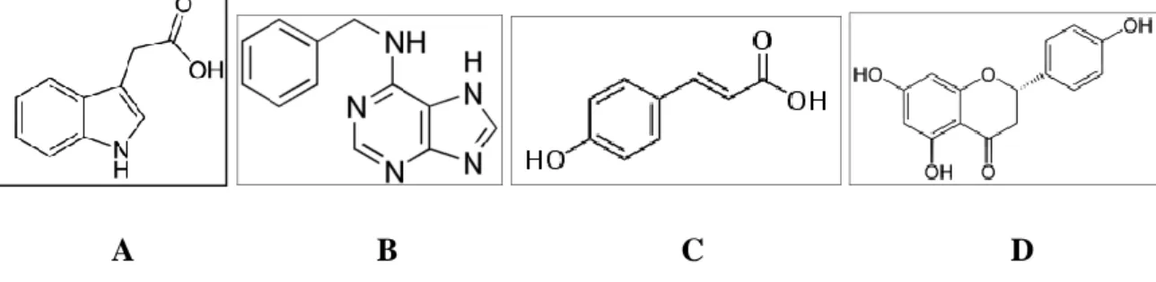
Fig. 9 - Chemical structures of signalling compounds secreted into the rhizosphere by the plant host. A: Indole-3-acetic acid, B: 6-Benzylaminopurine, C: Coumaric acid and D: Naringenin.
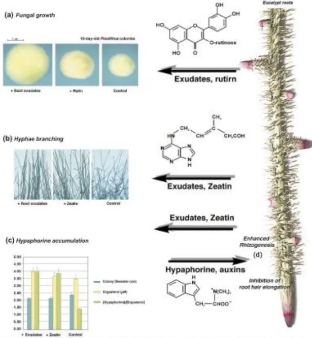
Fig. 10 - The molecular cross-talk taking place in the rhizosphere of Eucalyptus
globulus colonized by Pisolithus. Root exudates alter the morphology of the
mycelium of Pisolithus. These morphological changes are induced by the flavonol rutin, which stimulates fungal growth, expressed as colony diameter (a). Low concentrations of zeatin modify the hyphae branching angle (b) and the accumulation of the tryptophan betaine, hypaphorine (c). On the other hand,
2 3 5 4 12 14 17 21 20 3
XV
in the root system (d). (Béguiristain and Lapeyrie 1997) (from Martin et al. 2001).
Chapter 3 - Influence of light quality and intensity on adventitious root formation in microshoots of Pinus pinea L.
Fig. 1 - Spectra of Sylvania Gro-lux lamps (white) and Philips Cool-white lamps (black).
Fig. 2 - Rooting of P. pinea microshoots under different light quality and intensity. See significance in the Duncan Range test above.
Fig. 3 - Number of roots produced per week considering all microshoots for each
light treatment under Cool-white and Gro-lux lamps at 60 μmol m-2 s-1. Chapter 4 - Mycorrhiza-like structures in rooted microshoots of Pinus pinea
L.
Fig. 1 - (a) After more than one month without transferring to new medium, some genotypes developed coralloid mycorrhizal-like structures. (b) Coralloid structures also appeared as a consequence of NAA in the rooting induction medium. The arrow indicates a normal root type induced by auxin treatment.
Fig. 2 - (a) Differential responses in the in vitro adventitious root formation in co-culture system. Root with dichotomous branching. (b) Normal in vitro growth of the root system. Mycelium could be seen growing on the surface of the culture medium in both culture vessels.
Fig. 3 - Left: Rooted plants in the acclimation phase. Plants after 4 months in the acclimation phase. (a) Inoculated pine plant, identified as DD03, shows a good development of the aerial part and (b) also compact and dichotomous branched root system. (c) Control plant, not inoculated with less aerial part development and a linear root system 23 cm long.
Fig. 4 - (a) Different anatomical structures of the roots in plants derived from co-culture after 4 month in the acclimation phase (pine clones/different fungi isolates). Extensively dichotomous branching without hairs, (b) dichotomous short root with hairs and (c) ectomycorrhizal-like structure.
Fig. 5 - (a) Short root covered with mycelia. (b) Longitudinal section shows the intracellular hyphae.
Fig. 6 - Dichotomous branching observed in the liquid axenic root cultures after one month of growth.
Chapter 5 - Molecular approach to characterize ectomycorrhizae fungi from Mediterranean pine stands in Portugal
65 65 65 65 65 66 67 67 73 73 74 74 74 75
XVI
(b). Secondary mycelia. Each interval 2.5 µm (c).
Fig. 2 - Lactarius deliciosus sporocarps collected in a pine stand selected for
Pinus pinea-ECM associations study (a). The mycelia cultured in Hagen medium
(b). Secondary mycelia. Each interval 2.5 µm (c).
Fig. 3 - In vitro co-culture of Pinus pinea and P. arhizus mycelia (arrows). Fig. 4 - Cryostat transversal root section colonized by P. arhizus pine root showing the mantle hyphae (M) (100x); Scale bar 20 µm (a). Details of the transversal section showing well-differentiated Hartig-net (HN) in cortical cells (1250x); Scale bar 7.5 µm (b).
Fig. 5 - Phylogeny tree based on the ITS sequence.
Fig. 6 - Fingerprinting patterns obtained by amplification of genomic DNA. Lanes: 1 and 11 DNA molecular ladder 100 bp plus (Fermentas®), 2: Pisolithus arhizus culture, 3: Pisolithus arhizus sporocarps, 4: Lactarius deliciosus sporocarps, 5:
Rhizopogon roseolus sporocarps, 6: Penicillium brevicompactum sporocarps, 7: Aspergillus niger sporocarps 8: Cladosporium sp., 9: Fusarium oxysporum
sporocarps, 10: Control.
Fig. 7 - Phylogenetic tree analysis based on the PCR fingerprinting patterns for different species of Basidiomycetes and Ascomycetes. To evaluate the reproducibility of the assay, each sample has been analyzed in at least three independent PCR reactions. The distance values between branches are reported as percentage of similarity (0-100%).
Chapter 6 - Pisolithus arhizus (Scop.) Rauschert improves growth of adventitious roots and acclimatization on in vitro regenerated plantlets of
Pinus pinea L.
Fig.1 - In vitro co-culture of Pinus pinea and the mycelium of Pisolithus arhizus. A) in a double layer WPM medium. Arrow indicates place of inoculation with the mycelium, B) Bottom of a culture vessel showing colour mark (arrow) made to measure the weekly root growth with a ruler, C) Details of a dichotomous ectomycorrhizal structure covered with yellowish-brown mycelium collected from a plant 30 days after inoculation, D and E) Cryostat transversal root section colonized by P. arhizus showing the mantle (M) developed at the root apex of a short root and internal Hartig-net hyphae (HN) (200 ×) (Scale bar 20 µm), D). Details of the transversal section showing well-differentiated Hartig-net (HN) in cortical cells (2000 ×) (Scale bar 7.5 µm) (E).
Chapter 7 - O-coumaric acid ester, a potential signaling molecule detected during early in vitro co-culture between Pinus pinea L. plantlets and the ectomycorrhizal fungus Pisolithus arhizus (Scop.) Rauschert
83 82 84 83 85 86 96
XVII
– WPMS medium with perlite facing down, 5 - WPML, 6 - plantlet; 7 – root
(Patent approved by INPI Nº 105239).
Fig. 2 - Negative controls: pine plantlet without fungus (a) and fungal mycelium without pine plantlet (b). In vitro co-culture of Pinus pinea and Pisolithus arhizus
mycelia in double-phase medium (c).
Fig. 3 - HPLC-UV chromatograms of the medium sample collected from co-culture of P. pinea plantlet and P. arhizus on the 2nd day (a) and P. pinea microshoot without fungal inoculation collected on the 2nd day (b). Peak at RT
11.50 min corresponds to the unknown compound (UC).
Fig. 4 - HPLC-UV chromatograms of samples from the co-culture of P. pinea plantlet and P. arhizus collected on the 2nd day and doped with IAA (a), Rutin (b) and IBA (c). Peaks at RT 11.84, 11.44 and 11.58 min correspond to the unknown
compound (UC).
Fig. 5 - DAD total scan (a, c, e, g, i) and total ion current (b, d, f, h, j) chromatograms of the P. pinea/P. arhizus co-culture medium samples collected at different time intervals. Letters a and b correspond to the 1st day, c and d to the
2nd day, e and f to the 3rd day, g and h to the 5th day, and I and j to the 10th day.
Fig. 6 - UV (a) and mass spectra (b) of the unknown compound (UC) in the P.
pinea/P. arhizus co-culture sample collected on the 2nd day. Fig. 7 - Chemical structure of o-coumaric acid.
Fig. 8 - DAD total scan (a, c) and total ion current (b, d) chromatograms of liquid medium phase samples of the two negative controls. Letters a and b corresponds
to the culture of P. arhizus mycelia, c and d to the culture of P. pinea plantlets.
Chapter 8 - Patent - System and method for in vitro culture of plants for analysis of metabolites released by the root system
Fig. 1 - A - Illustrative scheme of the final appearance of the solid phase system preparation in a culture flask of in vitro plants. B: Schematic illustration of the final aspect of the method with inclusion of rooted plants. Figures numbers legend: 1 - cover; 2 - flask; 3 - solid phase; 4 - floating substrate; 5 - gelled culture medium; 6 - liquid phase; 7 - shoot; 8 - stem; 9 – roots.
110 107 110 113 111 113 113 122
XVIII
Chapter 2 - Adventitious rooting of conifers: influence of physical and chemical factors
Table 1 - Treatments and growth conditions applied in two phases of rooting of conifer cuttings.
Table 2 - Treatments and growth conditions applied in two phases of rooting of conifer microshoots in vitro.
Chapter 3 - Influence of light quality and intensity on adventitious root formation in microshoots of Pinus pinea L.
Table 1 - Duncan test for variable rooting percentage. Chapter 5 - Molecular approach to characterize ectomycorrhizae fungi from Mediterranean pine stands in Portugal
Table 1 - ITS rDNA homology from fungal strains used in the phylogenetic tree construction. The nucleotide sequences of the ITS region were aligned with those of related fungal species retrieved from the NCBI databases (www.ncbi.nlm.nih.gov).
Chapter 6 - Pisolithus arhizus (Scop.) Rauschert improves growth of adventitious roots and acclimatization on in vitro regenerated plantlets of
Pinus pinea L.
Table 1 - Means ± standard errors of variables in plants assigned to control and to inoculation at the onset of the experiment.
Table 2 - Means ± standard errors of variables with significant differences between control and inoculated plants.
47 98 98 66 51 84
XIX
Ragonezi C, Klimaszewska K, Castro MR, Lima M, Oliveira P, Zavattieri A (2010) Adventitious rooting of conifers: Influence of physical and chemical factors. Trees - Structure and Function 24(6):975-992 doi:10.1007/s00468-010-0488-8.
Ragonezi C, Castro MR, Klimaszewska K, Lima M, Zavattieri A (2010) Influence of light quality and intensity on adventitious root formation in microshoots of Pinus pinea L. Acta Hort. (ISHS) 865:287-291 http://www.actahort.org/books/865/865_38.htm
.
Castro MR, Ragonezi C, Klimaszewska K, Lima M, Oliveira P, Zavattieri A (2010) Mycorrhiza-like structures in rooted microshoots of Pinus pinea L. Acta Hort. (ISHS) 865:179-185 http://www.actahort.org/books/865/865_22.htm
.
Ragonezi C, Caldeira AT, Martins MR, Salvador C, Santos-Silva C, Ganhão E, Klimaszewska K, Zavattieri A (2012) Molecular approach to characterize ectomycorrhizae fungi from Mediterranean pine stands in Portugal. Manuscript accepted for publication at the Brazilian Journal of Microbiology. ISSN 1517-8382.
Ragonezi C, Caldeira AT, Martins MR, Dias LS, Santos-Silva C, Ganhão E, Miralto O, Pereira I, Louro R, Klimaszewska K, Zavattieri A (2012) Pisolithus arhizus (Scop.) Rauschert improves growth of adventitious roots and acclimatization on in vitro regenerated plantlets of Pinus pinea L. Propagation of Ornamental Plants 12:139-147.
Ragonezi C, Teixeira D, Caldeira AT, Martins MR, Santos-Silva C, Ganhão E, Klimaszewska K, Zavattieri A (2012) O-coumaric acid ester, a potential signaling molecule detected during early in vitro co-culture between Pinus pinea L. plantlets and the ectomycorrhizal fungus Pisolithus arhizus (Scop.) Rauschert. Manuscript submitted.
Patent
Castro MR, Ragonezi C, Oliveira P, Zavattieri A (2010) Patent - System and method for
in vitro culture of plants for analysis of metabolites released by the root system Nº
105239. Patent approved by the National Institute of Industrial Property (INPI).
AGRADECIMENTOS VI
ACKNOWLEDGMENTS VIII
RESUMO X
ABSTRACT XI
ABBREVIATIONS XII
LIST OF FIGURES XIV
LIST OF TABLES XVIII
THESIS PUBLICATIONS XIX
Chapter 1 - General introduction 1
1. Plant material (Pinus pinea L.) 2
2. In vitro propagation 7
3. In vitro propagation in conifers 8
4. In vitro rooting 9
5. Adventitious root formation 9
6. Mycorrhizae 11
6.1 Ectomycorrhizae 12
6.2 Ectomycorrhiza like-structures 13
6.3 Molecular characterization of mycorrhizal fungi 15
7. Biotization 17
8. Signaling 19
9. Background of the Doctoral Dissertation and Aims 23
10. Outline of the Doctoral Dissertation 27
11. References 28
Chapter 2 - Adventitious rooting of conifers: influence of physical and chemical factors 44
Abstract 44
Introduction 45
Rooting of conifer cuttings 46
Plant growth regulators 46
Auxins 46
Polyamines 46
Ethylene 49
Ethylene inhibitors 49
Temperature 50
Substrates for rooting 50
In vitro rooting of conifer microshoots 55
Rooting medium 55
Plant growth regulators 55
Carbohydrates 55 Light 56 Photoperiod 56 Temperature 56 Conclusions 57 References 57
Chapter 3 - Influence of light quality and intensity on adventitious root formation in microshoots of Pinus pinea L. 63
Abstract 63
Introduction 63
Material and Methods 63
Plant Material 63
Microshoot Induction from Embryo Cotyledons 64
Rooting of Microshoots 64
Light Sources Spectra 64
Evaluated Parameters and Statistical Analyses 64
Results 64
Discussion 65
Literature Cited 65
Chapter 4 - Mycorrhiza-like structures in rooted microshoots of Pinus pinea L. 69 Abstract 69
Introduction 69
Materials and Methods 70
Plant Material 70
Shoot Organogenesis 70
Root Organogenesis 70
Fungi 70
Axenic Root Cultures 70
Root Microscopy 71
Results and Discussion 71
Conclusions 72
Literature Cited 72
Chapter 5 - Molecular approach to characterize ectomycorrhizae fungi from
Mediterranean pine stands in Portugal 77
Abstract 77
Introduction 78
Material and methods 80
Collection of mushrooms from stone pine (Pinus pinea L.) stand 80 Mycelia isolation and fungal cultures 80
DNA extraction 80
ITS region amplification and sequencing 81
M13-PCR amplification 81
Data analysis 81
Results and discussion 82
Collection of fruiting bodies from stone pine stand 82 Species identification of P. arhizus and L. deliciosus. 83 Intraspecies identification by M13-PCR 85
Conclusions 87
References 87
Chapter 6 - Pisolithus arhizus (Scop.) Rauschert improves growth of adventitious roots and acclimatization on in vitro regenerated plantlets of Pinus pinea L. 93
Abstract 93
Introduction 93
Materials and methods 94
Collection of P. arhizus fruiting bodies from a P. pinea stand 94 Mycelia isolation and fungal cultures 94 Identification of the fungal isolates 94
Plant material 95
The verification of the accuracy of root measurements in vitro 95
Acclimatization 95
Vermiculite 96
Mixed substrate 96
Anatomical and histological studies 96
Statistical analyses 97
Results and discussion 97
Pisolithus arhizus 97
Root growth in vitro 97
Root growth in vermiculite 98
Root growth in mixed substrate 99
Mycorrhiza anatomical and histological studies 99
References 99
Chapter 7 - O-coumaric acid ester, a potential signaling molecule detected during early in vitro co-culture between Pinus pinea L. plantlets and the ectomycorrhizal fungus Pisolithus arhizus (Scop.) Rauschert 103
Abstract 103
Introduction 104
Material and methods 105
Reagents 105
Plant material and micropropagation of P. pinea 106
Fungi purification and identification 106
Co-Culture Technique 106
Preparation of the Double-phase Medium 106 Co-culture of P. pinea plants with P. arhizus mycelia 107
Negative controls 108
Biochemical analysis 108
Collection of liquid medium samples 108 Preparation of samples 108 Analysis of samples by HPLC-UV 108 Analysis of samples by LC-DAD-MS 109
Results 109
HPLC-UV analysis 109
Chapter 8 - Patent - System and method for in vitro culture of plants for analysis of
metabolites released by the root system 121
Abstract 122
Claims 123
Chapter 9 – Final considerations and Future perspectives 124
Future perspectives 128
1
Chapter 1
General Introduction
The general introduction describes the taxonomy and biology of pines in order to provide the context for stone pine (Pinus pinea L.) as the subject of this research project. This is followed by a description of the problems related to in vitro adventitious rooting of conifers, as well as stone pine, and the original technique of in vitro biotization with ectomycorrhizal fungi. In addition, analysis of molecular and biochemical methods used to characterize the fungal species and to better understand the role of the signaling factors that are involved in the interaction are presented.
2
1. Plant material (Pinus pinea L.)Conifers are the largest and most economically important group of Gymnosperms (Whetten 2001). This group is present in all continents (exceptions are the Arctic zone and the Antarctic) and consists of dominant species widely distributed most in boreal forest ecosystems and even in some tropical environments (Gernandt et al. 2011, Neale and Kremer 2011). Within this group there is the world’s oldest known tree (Pinus longaeva) and the world’s tallest tree (Sequoiadendron giganteum).
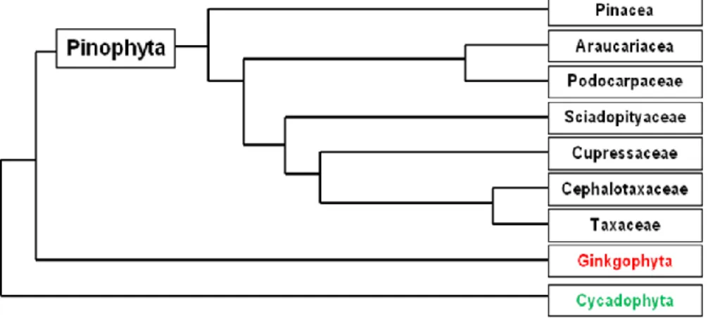
Conifers include six to eight families, with a total of 65-70 genera and 600-630 species (Catalogue of Life: 2007 - Conifer database). The most distinct families are shown in the phylogenetic diagram (Fig. 1). For a classification by linear sequence based on molecular data see the work of Christenhusz et al (2011).
Fig. 1 - A schematic representation of the Pinophyta phylogeny (from Quinn and Price 2003).
The earliest conifer fossils are found in the Upper Carboniferous (Stewart and Rothwell 1993, Farjon 2008) and many of the extant families can be recognized by the late Triassic or early Jurassic (Singh 2006). Major fossil orders of conifers or conifer-like plants include the Cordaitales, Vojnovskyales, Voltziales and in some cases the Czekanowskiales.
Examples of contemporary conifers include: Pines (Pinus spp.), Spruces (Picea spp.), Cowtail Pine (Cephalotaxus spp.), Cypress Pine (Callitris spp.), Firs (Abies spp.), Larches (Larix spp.), Bald Cypresses (Taxodium spp.), Yellowwood (Podocarpus spp.), Yews (Taxus spp.), Arbor vitae (Thuja spp.), Junipers (Juniperus spp.), Cedars (Cedrus spp.), Douglas-firs (Pseudotsuga spp.) and Golden Larch (Pseudolarix spp.), among others.
3
Pinus is the largest extant genus of the Pinaceae family (Fig. 2) naturally occurring
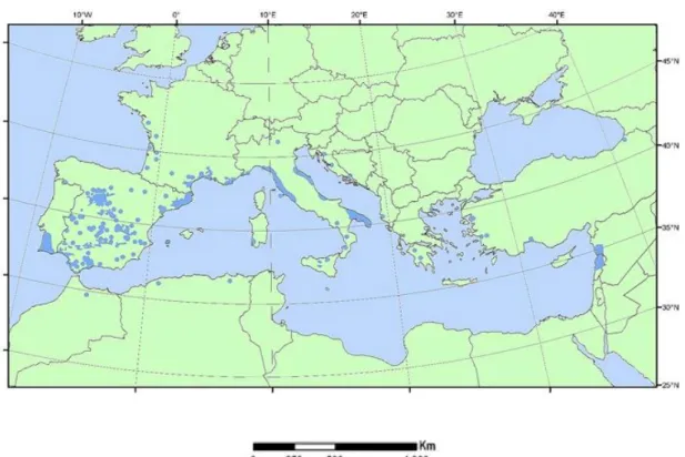
almost exclusively in the Northern Hemisphere, but introduced and widely naturalized in both hemispheres (Procheş et al. 2012) with over 100 widely recognized species (Richardson 1998, Farjon 2001, Alves-Freitas et al. 2011). Stone pine (Pinus pinea L.) is an economically important conifer distributed across the Mediterranean Basin (Fig. 3). In Portugal it is cultivated mainly for the production of nut seeds (pinion) for the food consumer market.
Fig. 2 - Taxonomic hierarchy of Pinus pinea L. (from Integrated Taxonomic Information System (ITIS), 2012).
Fig. 3 - The distribution map of Pinus pinea L. throughout the Mediterranean Basin and adjacent zones (from Euforgen.org).
Taxonomic Hierarchy
Kingdom
Plantae
– Planta, Vegetal, plants, plantes SubkingdomViridaeplantae
– green plantsInfrakingdom
Streptophyta
– land plantsDivision
Tracheophyta
– vascular plants, TracheophytesSubdivision
Spermatophytina
– Spermatophytes, seed plants, Phanérogames InfradivisionGymnospermae
– Gymnosperms, Gymnospermes, Gimnosperma ClassPinopsida
– conifersOrder
Pinales
– pines FamilyPinaceae
– pines GenusPinus
L. – pines4
The total area covered by stone pine is around 380,000 ha (Varol and Tel 2010). The Iberian Peninsula accounts for approximately 75% of Pinus pinea stands, the largest area occurring in Spain with an area of 464,000 ha (BDN 2008), followed by Portugal with an area of 130,300 ha (IFN5 2010).
Stone pine trees can reach a maximum height of 25 meters, and the trunk is often short and slightly sinuous. The canopy has a horizontal spread and ascending branches that gives their adult crown a characteristic umbrella-like shape (Fig. 4). From this feature is derived one of the most common names in English: umbrella pine. Other common names that occur are: pinheiro manso (Portuguese); pino piñonero, pino manso, pino
doncel, (Spanish); pin parasol, pin pignon (French) and pino domestico (Italian).
Fig. 4 - Pinus pinea L. trees in a typical pine stand in Alcácer do Sal district, Portugal.
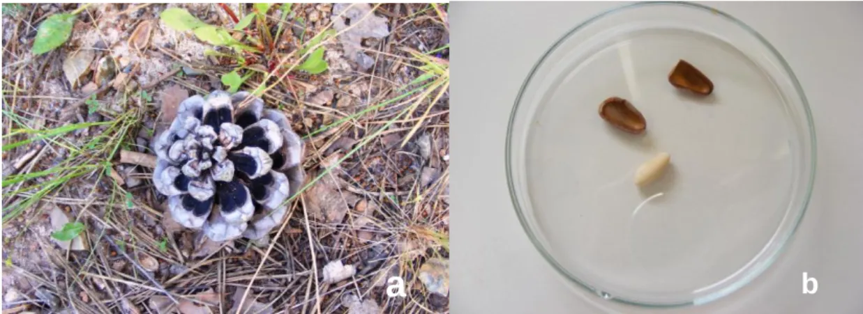
Some P. pinea tree characteristics like glabrous twigs and green needles that occur in fascicles of two can be found in typical pine stands. Like other pine species, P. pinea is monoecious, although fecundation rarely occurs within the same tree since, in most cases, the male gametes and female cones are formed in different branching systems and maturation is sometimes not coincident on the same tree. Pollen grains are mainly transported by wind (anemophilous pollination) and fecundation takes place 2 years after pollination (Singh 2006). Cones reach maturity after 3 years and are 8-14 cm in length (Fig. 5a). Figure 5b shows a typical P. pinea seed without the seed-coat. They are heavy and mostly dispersed by small mammals (Fady et al. 2004). Seed production
5
commences after 12 to 18 years depending whether tree occurrence is in isolation or in stands (Moussouris and Regalo 1999).
Fig. 5 - Mature cone of a P. pinea tree (a) and seed without the seed-coat (b).
P. pinea occurs either in arid inland or coastal areas affected by salinity stress (Correia
et al. 2010), but also grows in harsh environments. This species is highly sensitive to climate variants (De Luis et al. 2009b) and faces regeneration problems in many localities.
Prior to the anthropogenic range expansions of the last few thousand years, stone pine was probably confined to the Iberian Peninsula, since is the only area that can be found away from ancient trade routes (Rikli 1943). There still remains a great deal of controversy regarding the natural expansion and origin of this species. Richardson (1998) stated that it is “impossible to determine its natural range". Agrimi and Ciancio (1994) have located in the western Mediterranean zone the origin of P. pinea, despite the fact that some theories hold that the species is native not only to the western Mediterranean but also to the eastern Mediterranean Basin (Barbéro et al. 1998). P.
pinea was extensively planted around the Mediterranean Sea throughout history by the
Etruscans, the Greeks, the Romans and the Arabs (Fady et al. 2004, Evaristo et al. 2007).
Although it has been cultivated since the Roman period for timber (for construction and ship-building), the most economically important product derived from the tree is its seed, the pine nut. World production of pine nuts is about 20,000 tons/year (Nergiz and Donmez 2004). The main countries where pine nuts are traditionally produced and consumed are Portugal, Spain, Italy, Tunisia and Turkey.
Concerning seed production, in the case of P. pinea, as a nut-producing tree, seed yield is the main clonal selection criterion (Mutke et al. 2005). Due to the rise of traditional agriculture and long-distance trading, this species could have experienced a
6
further reduction in its genetic diversity, which may be explained by the recent expansion of the species or low mutation rates (Vendramin et al. 2007). Among widely distributed sexually-reproducing trees, stone pine may be considered an exception due to its low level of genetic diversity. Nevertheless, it should be emphasized that whereas some species can survive in harsh environments, they do not represent the norm (Godt and Hamrick 1997). An understanding of the current adaptive diversity of P. pinea is a prerequisite for outlining its potential distribution and the consequences that it may face from environmental changes in the future.
The economic exploitation of pine nuts and the existence of great potential for P. pinea improvement are some of the reasons for the development of genetic breeding programs, based on the identification of excellent genotypes by establishing clonal banks with different origins (Alonso et al. 2006). The implementation of in situ conservation networks, as is the case with many other forest trees around the world, is essential. Despite the fact that the rate of survival of P. pinea in forest fires is considerably higher than that of other Mediterranean pines (Rodrigo et al. 2004), wild fires and overgrazing are still the greatest risks faced by stone pine forests. Fire protection and preventive measures to reduce these risks should also be introduced for the conservation of this typical Mediterranean pine (Fady et al. 2004).
According to the US Food and Drug Administration, there is a growing worldwide market for pine nuts, due to the fact that consumption reduces the risk of coronary heart disease, which is attributed to the high linoleic acid content (Nergiz and Donmez 2004). Also, the seeds have a high nutritional content, being particularly rich in proteins and vitamins (Savage 2001), and are thus considered a food supplement. In addition to their nutritional value, stone pine nuts have been considered as an aphrodisiac in ancient times, by Roman poets and Greek physicians (Moussouris and Regalo 1999).
Other products of economic value include resin, bark (for tannin extraction), and pine cone shells (for fuel) (Khaldi et al. 2011). P. pinea is also cultivated for environmental protection: ecological restoration (afforestation of coastal areas and dunes), protection of agricultural crops (Fady et al. 2004), can potentially help mitigate desertification problems (Correia et al. 2010), provide food and shelter for local wildlife (Montero et al. 2004), and is currently considered as a viable alternative for use for abandoned farmland (Cortizo et al. 2009). It is also an aesthetically attractive tree which is planted in parks and gardens throughout the world. It has been successfully introduced in North Africa as well as Argentina, Australia, South Africa and the USA.
7
The great importance of the species derives from its environmental, aesthetic, and soil-conservation uses, its high economic value and its ability to survive intense fire damage. Due to its importance and that of applications, interest in the species from forest managers and researchers has increased considerably.
2. In vitro propagation
In vitro propagation, most frequently termed micropropagation, refers to the application
of plant-cell and tissue-culture technology in order to improve mainly the performance of important agriculture crop species. The growth of commercial micropropagation as an industry occurred during the 1970s and 1980s (Ahamed et al. 2001) and generated a great deal of excitement among researchers, mostly due to cell totipotency through the regeneration of entire plants from single cells (Metivier 2000, Razdan 2003, Iliev et al. 2010).
Plant tissue culture, also known as in vitro culture is the science of growing plant cells, tissue or organs isolated from the mother plant on artificial culture media (George et al. 2008). By manipulating the culture medium composition, the plant cells transferred to the nutrient medium will start to divide and produce cell masses that will eventually produce new plants, often in large numbers (Klimaszewska et al. 2011).
The in vitro growth and development of a plant is determined by a number of complex factors: (a) the genetic structure of the plant (b) nutrients: water, sugars, macro- and micro-elements (c) physical growth factors: light, temperature, pH, O2 and CO2 concentrations, and (d) certain organic substances: plant growth regulators (PGRs) and vitamins.
Several techniques for in vitro plant propagation have been developed, for example, the induction of axillary and adventitious shoots, the culture of isolated meristems and plant regeneration by means of organogenesis and/or somatic embryogenesis. These different plant-tissues culture techniques can present advantages in comparison with traditional methods of agronomic, horticultural and forestry species propagation (Iliev et al. 2010). Some of these benefits include: (1) consistent production of the same genotypes over time, (2) greater genetic gains, (3) flexibility for the rapid deployment of suitable clones given changing breeding goals and/or environmental conditions, and (4) the capability for managing genetic diversity and genetic gain in plantation forestry (Park et al. 1998). Furthermore, the benefits produce plants that require fewer pesticides and which are in some cases pathogen-free (Ahamed et al. 2001).
8
The propagation of superior mature individuals is an effective way of achieving genetic gain by exploiting genetic variance (dominance, additive and epistatic) within a given generation without the need for proceeding through long breeding cycles (Ahuja 1993). Consequently, the regeneration of mature trees, either singly, or as part of conventional breeding programmes, could provide a powerful instrument for improving forestry management (Cortizo et al. 2009), and it is an increasingly essential tool for all propagation programs.
In the case of trees, conventional breeding is not as straightforward since in some cases they have long life cycles, are self-incompatible and very slow to mature (Merkle and Dean 2000, Campbell et al. 2003). Maturation is an important issue since it induces changes in meristem behavior, thus reducing the propagation potential of forest trees; also, the economic value of a tree is better assessed after it reaches maturity (Greenwood 1995, von Aderkas and Bonga 2000, Mitchell et al. 2004).
3. In vitro propagation in conifers
Conifer cultures are generally initiated from immature or mature zygotic embryos. The uses of germinating embryos cotyledons or primordial shoots excised from seedlings or trees are less frequent (Bonga et al. 2009). In previous, most efforts were focused on propagation by first inducing adventitious shoot formation, primarily from cotyledons, and then roots by means of organogenesis. Success on a commercial scale has been limited to a few species, for example radiata pine (Pinus radiata) (Bhowmik and Matsuiz 2001, Bonga et al. 2003). Developed over the last two decades, somatic embryogenesis is considered a more effective method (Bonga et al. 2003), which is different from organogenesis because organs, such as shoots and roots, have one primary pole or growing point, whereas embryos are bipolar structures with both shoot and root meristems (Bhowmik and Matsuiz 2001, Preece 2003).
Distinct steps in organogenesis include: (1) establishment or bud induction, or both; (2) bud and shoot development and multiplication; (3) rooting of developed shoots; and (4) hardening of plantlets (Saborio et al. 1997). In the case of somatic embryogenesis, the method can produce any number of zygotic-like somatic embryos and plants from one seed, thus providing a standard for mass clonal propagation (Klimaszewska et al. 2007). Although somatic embryogenesis technology has worked well with many conifer species using zygotic embryos as starting material, attempts to achieve the same result with adult conifers have failed (Klimaszewska et al. 2011).
9
Since the genetic potential for features (height growth, branching and crown shape) cannot be evaluated until the tree is fully-grown, it is important to develop methods for propagating conifers in the mature stage (von Aderkas and Bonga 2000). In the last 25 years there have been several reports regarding the in vitro culture of conifers (Horgan 1987, Ewald 1998, Chang et al. 2001, Parasharami et al. 2003, Prehn et al. 2003, 2003, Oliveira et al. 2003, Renau-Morata et al. 2005, Alonso et al. 2006, Zavattieri et al. 2009, Salaj et al. 2007, Bonga et al. 2010).
Within the Pinus genus, successful micropropagation of explants is reported for a number of species such as P. taeda (Mott and Amerson 1981), P. pinaster (Dumas and Monteuuis 1995), P. ayacahuite (Saborio et al. 1997), P. nigra (Özkurt et al. 2008), P.
sylvestris (De Diego et al. 2009), P. maximartinezii (Robledo et al. 2009), P. radiata
(Zhang et al. 2009), P. massoniana (Zhu et al. 2010), P. kesiya (Choudhury and Kumaria 2010), and P. peuce (Stojičić et al. 2012).
Micropropagation of P. pinea via organogenesis has been reported during the last few decades. The most common method, it is based on the induction of shoot buds from cotyledonary explants dissected from mature seeds and in most cases, cultured in media supplemented with some varieties of PGRs (see point 9)
4. In vitro rooting
Conifers have a major role to play in reforestation strategies, and despite the studies that have been published, current research on their vegetative propagation has not been satisfactorily investigated. One of the most common problems encountered in the micropropagation of conifers is the reduction in the ability of cuttings to root (Cortizo et al. 2009, Zhang et al. 2010). This problem is mainly related with maturation, which affects reproductive competence, morphology, and growth rate (Greenwood and Hutchison 1993, Libby and Ahuja 1993, Mitchell et al. 2004).
The rate of spontaneous rooting is generally low in micropropagated cuttings in conifers (Burns et al. 1991, Budimir and Vujicic 1992, Normand et al. 1996, Stojičić and Budimir 2004, Ewald 2007a). In most conifer micropropagation protocols, rooting treatment is required in order to increase the rooting rate (Ragonezi et al 2010b).
5. Adventitious root formation
Esau (1953) defined the term "adventitious root” as a root that arises on an already lateralized root axis or at a place on the plant that is not itself a root (e.g. on a shoot).
10
These roots may occur naturally from stem tissue or may be induced by different conditions: a stressful environment, mechanical damage or the tissue-culture regeneration of shoots (Li et al. 2009). Adventitious root formation (ARF) is essential for plant growth regulation and development since it is a key step in the vegetative propagation of woody or horticultural species (Hess 1994, Sorin et al. 2006). The inability to induce ARF in conventional cuttings or tissue culture is a major limiting factor when cloning plants for genetic improvement and/or commercial applications (Metivier 2000).
ARF is a complex developmental process that consists of three successive but interdependent physiological phases: induction, initiation and expression. Each of these phases has different requirements (Li et al. 2009). The chemical and physical factors that affect rooting include: PGR´s, nutrients (mainly the carbohydrate source), temperature and light (Ragonezi et al. 2010b).
An efficient rooting treatment can lead to a high rate of rooting and higher quality of the root system (Ragonezi et al. 2010b). Root number, length, and the absence of callus at the base of the shoot could have an influence on plant behavior in the ex vitro phase (De Klerk et al. 1999, Hartmann et al. 2002).
In the past few years a number of studies have been published describing attempts to improve rates of in vitro rooting in many species: Pinus armandii var. amamiana (Ishii et al. 2007); Larix sp. (Ewald 2007a); Taxus baccata L. (Ewald 2007b); Juniperus
phoenicea (Loureiro et al. 2007); Pinus pinea L. (Zavattieri et al. 2009, Ragonezi et al.
2010a); Pyrus communis L. (Sun et al. 2009); Teucrium fruticans L. (Frabetti et al. 2009); Olea europea L. (Padilla et al. 2009); Quercus rubra L. (Vengadesan et al. 2009a); Citrus sinensis L. Osbeck × Poncirus trifoliata L. Raf. (Montoliu et al. 2010); and Pistacia vera L. (Benmahioul et al. 2012).
Also, some efforts using biotization procedures (for definition of this, see point 7 of this chapter) to overcome in vitro rooting and improve transplantation survival rates can be cited in different works and for different species: Ragonezi et al. (2012) with P. pinea and P. arhizus; Normand et al. (1996) used Hebeloma cylindrosporum in the rooting and acclimatization phases of P. sylvestris; also with this pine species, Niemi et al. (2000) associated with Pisolithus tinctorius; Sarmast et al. (2012) evaluated the application of Agrobacterium rhizogenes with Araucaria excelsa R. Br. var. glauca; in Gosal et al. (2008) Populus deltoides plantlets were biotized during hardening with
11
with micropropagated Terminalia bellerica Roxb plants inoculated with Piriformospora
indica during ex vitro acclimatization; Vettori et al. (2010) analyzed the effect of Azospirillum brasilense Sp245 on the micropropagation of three fruit rootstocks: Prunus cerasifera×P. spinosa, Prunus persica × P. amigdalus, and MM 106 apple (Northen
Spy×M1); Martins (2008) worked on micropropagated Castanea sativa plants and
Pisolithus tinctorius.
Certain combinations of ectomycorrhizal fungi and proper procedures of co-culture could be exploited for the in vitro root development improvement of stone pine or any other target species. Based on taxonomic and ecological extrapolation, an estimated 86% of terrestrial plant species acquire mineral nutrients via mycorrhizal root symbionts (Brundrett 2009). Pinaceae is considered the oldest extant plant family that is associated with ectomycorrhizal fungi (Hibbett and Matheny 2009).
6. Mycorrhizae
Since the term symbiosis was introduced by De Bary in 1879, the importance of associations among the numerous organisms has increased. Mycorrhizae are symbiotic structures formed between plant roots and fungi. Plants provide photosynthetically-fixed carbon and a habitat for the fungi, whereas mycobionts provide dissolved- and organically-bound nutrients (Smith and Read 2008). Fungal activity represents an important element of active biomass in forest ecology, since establishment, survival and decomposition in the forest trees dynamics are largely dependent on these organisms (Rosling 2003).
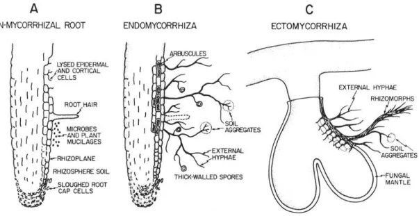
Seven distinct types of mycorrhizae are recognized, but several of them are very similar (Brundrett 2002). Endomycorrhizae and Vesicular-arbuscular mycorrhizae (AM) are the most widespread types. Ectomycorrhizae (ECM) occurs in certain families of Gymnosperms and Dicotyledons and in one Monocotyledon genus (Brundrett 2009). The remaining types of mycorrhizae are restricted to specific plant families. The main differences between a non-mycorrhizal root and a colonized root by either endo- or ectomycorrhizal fungi can be found in Figure 6.
Brundrett (2002) distinguished the various stages in the beginning of the association:
1 Fungi attracted by exudates proliferate on the surface of plants;
2 Fungi develop mechanisms for penetrating living plants without causing harm;
3 The space within living plants becomes an important habitat for these endophytes; providing them with shelter from adverse soil conditions, parasitism and predation;
12
4 Fungi become dependent on the host for energy;
5 Absorptive hyphae within plants increase their surface area and permeability.
Fig. 6 - Morphological changes in roots as they become mycorrhizal and the effects of those changes on the development of a mycorrhizosphere. A - Generalized non-mycorrhizal root with root hairs and indicated sources of organic materials available as substrates for rhizosphere microorganisms. B - Endomycorrhiza with indicated morphological changes such as reduced tissue sloughing, lack of root hairs, presence of external hyphae, thick-walled spores, and associated soil aggregates; no obvious change in surface area. C - Ectomycorrhiza indicating dramatic morphological changes such as development of a fungal mantle plus extensive external hyphae and rhizomorphs and associated soil aggregates, loss of root hairs, and greatly increased branching and surface area (from Linderman 1988).
The main role of this association is the acquisition of nutrients by exploring the soil volume around the host with the aid of the hyphae, which are more responsive and more extensive than the roots themselves (Van der Heijden et al. 2006b, Requena et al. 2007, Turk et al. 2008, Smith and Smith 2011, Cairney 2011, Kiers et al. 2011). Mycorrhizal colonization tends to be reduced when nutrient availability is high (Kiers and van der Heijden 2006) and may become inactive or be lost by attrition. Also, they may experience cycles of dormancy and activity, which is a common feature in the perennial root system of trees (Kottke and Oberwinkler 1986).
6.1 Ectomycorrhizae
Within ectomycorrhizal symbioses, the host (plant roots) and the symbiont (ECM fungi) function collectively as an entity. The development of ECM in plants frequently allows them to establish in habitats that neither symbiont may be able to occupy independently (Nehls et al. 2000). ECM fungi include at least 6000 species (Hibbett et al. 2000) and involve economically important woody plant families and many lineages
13
of fungi that generally belonging to Basidiomycota (agarics, bolets) and Ascomycota (truffles) (Brundrett 2009, Tedersoo et al. 2010a).
ECM symbioses are restricted to <5% of terrestrial plant species (Landeweert et al. 2001) and are ubiquitous in the Pinaceae family (Le Page et al. 1997). This mutualistic relationship among ECM fungi grants conifers an ecological advantage for surviving harsh conditions where climate is strongly seasonal and soils are nutrient-poor (Castro et al. 2010). Other families that are associated with ECM fungi include Fagaceae, Myrtaceae Dipterocarpaceae (Brundrett 2002, Smith and Read 2008, Bonfante and Genre 2010).
For mycorrhizated plants the modifications in both cell organization and physiological and morphological facets are associated with numerous benefits. The fungus improves plant nutrient uptake by means of rhizosphere exploitation and in return it receives carbohydrates that are essential for completion of the fungal life cycle (Bonfante and Anca 2009). The symbiont may also alleviate the environmental stress caused by chemicals, herbivory, pathogens, fire or drought (Smith and Read 2008), and as biofertilizers they may counteract fertilization excess and thus promote sustainable agriculture (Bonfante and Genre 2010).
Ectomycorrhizae are characterized mainly by the presence of a fungal sheath (mantle), which adheres to the root exterior and forms a hyphae structure (Ammarellou and Saremi 2008). The fungus mycelium is connected to the extramatrical hyphae that explore the substrate, and is responsible for nutrition and water uptake (Barker et al. 1998). One of the most striking features of the ECM root is the Hartig net which extends into the root, penetrating between epidermal and cortical cells. The Hartig net forms a crossing point from the inner zone of the mantle through which the symbionts exchange materials (Barker et al. 1998, Bending et al. 2006, Frey-Klett et al. 2007). This is the primary zone for nutrient transfer in the association (Burgess et al. 1994, Dell et al. 1994, Brundrett 2004). In Figure 7 there is a schematic representation of ectomycorrhizal development events.
6.2 Ectomycorrhiza-like structures
As mentioned before, development between the ECM fungi and the host plant involves many changes in root physiology and morphology. Both root elongation and root hair formation are suppressed and short lateral roots go through dichotomous branching that generally culminates in the formation of coralloid structures. In conifers, externally
14
supplied fungal exudates, extracts, or synthetic auxins can partially mimic the effect of mycelium in inducing root proliferation and dichotomous branching of lateral roots (Smith and Read 2008). Normally in the Pinus genus, ECM roots are usually dichotomously branched, as described by Agerer (1987-2002), and in some cases extensive dichotomous and coralloid branching of lateral roots can occur without fungal intervention, and this kind of formation is known as an ectomycorrhiza-like structure (Castro et al. 2010).
Fig. 7 - Schematic representation of ectomycorrhizal development. Morphological events taking place during early (left) and late (right) stages of Ectomycorrhizal formation are indicated (from Baker et al. 1998).
Some research has been carried out on the role of the plant hormones and fungal/root exudates in the production of ECM or ECM-like structures in pine (Slankis 1973, Faye et al. 1980, Rupp and Mudge 1985, Gogala 1991, Castro et al. 2010). Faye et al. (1980), studying P. pinaster, demonstrated that the host plant genome contains all the genetic information required to form an ECM-like organ. In the work of Castro et al. (2010), ARF regenerated by P. pinea microshoots as well as axenic embryo root cultures developed ECM-like structures. It was demonstrated that these structures appeared in all experimental settings tested, with the frequency of dichotomous branching increasing with the reduction of macronutrients in the medium and also in cultures that spent more than one month on the same co-culture medium (which is similar to drought conditions in nature). These findings support those of Kaska et al. (1999), in which nutrient-limiting conditions were required for the formation of both
15
spontaneous and chemically-induced dichotomous and coralloid branching in three different species of pine: P. taeda, P. halepensis and P. muricata. A combined exogenous application of IAA and ethylene induced root responses equivalent to the presence of truffle mycelium in both host plant (Cistus incanus) and the non-host plant (Arabidopsis thaliana) (Splivallo et al. 2009)
PGRs supplied by the ECM fungi have an effect on the thickness, extension and branching of roots, and, when applied experimentally, can induce similar root morphologies as in the absence of fungus (Kaska et al. 1999, Barker and Tagu 2000). By analyzing the anatomical aspects of the ECM-like structures, the similarity between extensive dichotomous branching of lateral roots that grow without fungal presence and those roots derived from the fungal inoculation can be observed. Due to this association, it may be difficult to diagnose ECM roots without histological and anatomical studies. Many theories may be developed on the formation of ECM-like structures and the most evident of them are, nutrient limiting, genetic information and chemically induced roots. Nevertheless, this topic has not been sufficiently investigated and needs to be further examined.
6.3 - Molecular characterization of mycorrhizal fungi
The identification of ECM fungi species is a difficult task. Traditionally, ectomycorrhizae have been identified by using colour, shape and other macroscopic features (Agerer 1987-2002). On the other hand, many fungi species have not been described by morphological methods and are rarely identified using only morphological techniques (Iotti and Zambonelli 2006). Identification remains largely dependent on initial analysis based on morphotyping, and a high level of skill is required by the analyst (e.g. personal experience, ability, rough analysis) (Rosling 2003).
The most reliable approach to the study of ectomycorrhizal community composition is a combination of morphological and molecular identification techniques (Gamper et al. 2009, Walbert et al. 2010, Bahram et al. 2011, Zambonelli et al. 2012).
In the 1990s, a revolution in molecular tools addressed the cultivability issue and substantially enhanced the identification of ECM fungi in situ (Gardes et al. 1991b, Egger 1995, Horton and Bruns 2001, Anderson and Cairney 2004). With the application of these techniques, many common yet unidentified ECM fungi could be taxonomically assigned (Vrålstad et al. 2000, Kõljalg et al. 2002). Since this molecular