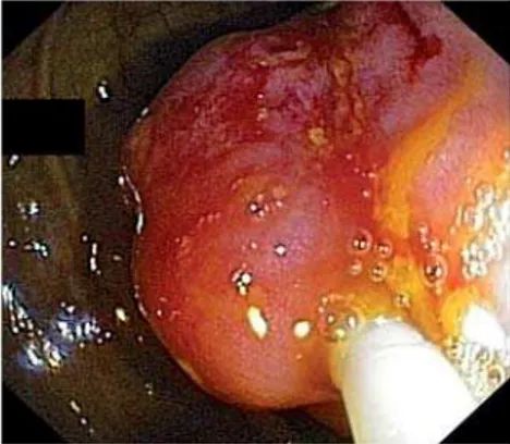
A 52-year-old man with a 1-year history of lower abdominal pain and constipation was referred to our unit for endoscopic re-section of a large lipoma in the ascending colon, which had been detected by ab-dominal computed tomography (CT). Co-lonoscopy revealed a 50-mm, subepithe-lial, broad-based, polypoid mass in the
as-cending colon (
●
" Fig. 1). Using an electro-cautery snare, we transected the upper third of the mass to unroof the lesion (●
" Fig. 2). Fat was observed extruding from the cut surface, consistent with the diagnostic hypothesis (●
" Fig. 3). There were no procedure-related compli-cations. Histopathologic examination of the excised specimen confirmed the di-agnosis of a lipoma. A follow-up endos-copy 1 month later showed a small ulcer at the resection site (●
" Fig. 4), which was completely scarred 3 months later, with no evidence of residual lipoma (●
" Fig. 5).Colonic lipomas are benign, submucosal tumors [1]. Although most colonic lipo-mas remain asymptomatic and need no treatment, large lipomas can cause symp-toms such as abdominal pain, change in bowel habits, bleeding, and intussuscep-tion, and should be removed, preferably endoscopically [1]. Various endoscopic
techniques have been used in the treat-ment of large colonic lipomas, which in-clude snare resection following endo-scopic clipping, looping or injection of the base, endoscopic loop ligation, and en-doscopic submucosal dissection [1–3]. The unroofing technique cuts off only the upper half of the lipoma, while the re-maining adipose tissue is rapidly and completely extruded from the open sur-face [4, 5]. Therefore, this is a simple tech-nique that allows both histological con-firmation and complete treatment with minimal risk of perforation [4, 5]. Never-theless, there are only two case reports of endoscopic resection of lipomas in the duodenum by this technique [4, 5]. To the best of our knowledge, this is the first re-port of the endoscopic resection of a lipo-ma in the colon using this technique. Endoscopy_UCTN_Code_TTT_1AQ_2AD
Competing interests:None
J.-B. Soares, R. Gonçalves, C. Rolanda Gastroenterology Department of Braga Hospital, Braga, Portugal
References
1 Matsushita M, Danbara N, Kawamata S et al. Endoscopic removal of large colonic lipo-mas: difficult submucosal dissection or easy snare unroofing? Endoscopy 2009; 41: 475; author reply 475
2 Geraci G, Pisello F, Arnone E et al.Endoscopic Resection of a Large Colonic Lipoma: Case Report and Review of Literature. Case Rep Gastroenterol 2010; 4: 6–11
3 Koo J, Kaffes A.Endoscopic resection of large colonic lipomas assisted by a prototype sin-gle-use Endoloop device. Endoscopy 2006; 38: 644–647
4 Hizawa K, Kawasaki M, Kouzuki T et al. Un-roofing technique for the endoscopic resec-tion of a large duodenal lipoma. Gastrointest Endosc 1999; 49: 391–392
5 Huang WH, Peng CY, Yu CJ et al. Endoloop-assisted unroofing for the treatment of symptomatic duodenal lipomas. Gastroin-test Endosc 2008; 68: 1234–1236
Bibliography
DOI10.1055/s-0030-1256938 Endoscopy 2011; 43: E407
© Georg Thieme Verlag KG Stuttgart · New York · ISSN 0013-726X
Corresponding author J.-B. Soares, MD
Gastroenterology Department of Braga Hospital Sete Fontes, S. Victor
4701-243 Braga Portugal
Endoscopic resection of a large colonic lipoma by
unroofing technique
Fig. 1 Endoscopic view of the large, subepi-thelial, broad-based, polypoid mass with smooth, yellow surface in the ascending colon.
Fig. 2 The electrocautery snare transecting the upper portion of the mass.
Fig. 3 Fat seen extruding from the mass after the unroofing procedure.
Fig. 4 Follow-up endoscopy 1 month later showing a small ulcer in involution at the re-section site.
Fig. 5 Follow-up endoscopy 3 months later showing scarred mucosa at the resection site.
UCTN
–
Unusual cases and technical notes
E407
Soares J-B et al. Endoscopic resection of a large colonic lipoma by unroofing technique… Endoscopy 2011; 43: E407