Case 8056
Haemorrhagic cellular leiomyoma
M. Rosário Matos , Teresa Margarida Cunha1 2
Department of Radiology, Hospital de Dona Estefânia - Centro Hospitalar de Lisboa Central. 1
Department of Radiology, Instituto Português de Oncologia Francisco Gentil de Lisboa. 2
Genital (Female) Imaging Section:
2010, Apr. 28 Published:
27 year(s), female Patient:
Clinical History
A 27-year-old Caucasian female patient presenting with a large uterine mass on gynecological ultrasound, which showed significant growth in relation to the previous examination.
Imaging Findings
The patient was asymptomatic. She was not taking oral contraceptives and was not pregnant or in postpartum period. Physical examination revealed a non tender pelvic mass. Blood tests were normal.
The annual follow-up gynaecologic ultrasound examination of a previously documented uterine leiomyoma revealed a significantly larger uterine mass, slightly hyper-echoic compared to the myometrium. The patient was then referred for an MR study, which showed an intramural lesion with regular margins, hypo-intense on T1-weighted images (Fig. 2), hyper-intense on T2-weighted images (Fig. 3), enhancing intensively with gadolinium (Fig. 4).
Based on the histological pattern of the tumor, patient age and response to treatment, the final diagnosis was haemorrhagic cellular leiomyoma.
Discussion
Leiomyomas are the most common gynecologic neoplasm, occurring in 20-30% of women in reproductive age. They are predominantly composed of smooth muscle cells and variable amounts of fibrous connective tissue [1, 2].
Haemorrhagic cellular leiomyomas are a histological subtype of leiomyomas. They occur almost exclusively in young women treated with oral contraceptives, during pregnancy or in postpartum period (these features were not present in our case). They are referred as apoplectic leiomyomas" due to the fact that the clinical symptoms are often acute and pathological features are dominated by recent haemorrhages. These lesions are different from extensive or zonal infarcts, commonly termed red degeneration", which coexist with coagulative or ischemic necrosis, with no hypercellularity [3, 4].
Typically, non-degenerated uterine leiomyomas appear on MR imaging as well-circumscribed masses of homogeneously decreased signal intensity compared to that in the outer myometrium on T2-weighted images and of intermediate signal on T1-weighted images. Cellular leiomyomas, which are composed of compact smooth muscle cells with little or no collagen, can have relatively higher intensity signal on T2-weighted images and demonstrate enhancement on contrast enhanced images [1].
MR imaging can help differentiate cellular leiomyomas from degenerated T2-weighted
hyper-intense leiomyomas, as degenerated leiomyomas often show irregular, peripheral, or minimal enhancement compared with that of myometrium, depending on the degeneration within the tumor [5].
The differential diagnosis also includes lesions with extensive oedema affecting their signal intensity, which can be high on T2 weighted images and demonstrate marked enhancement. Microscopy will show fluid in the stroma of the leiomyoma, often in association with collagen [2].
Finally, altough it has been suggested that an irregular margin of a uterine leiomyoma at MR imaging is related to a sarcomatous transformation, the ability of MR imaging to allow
differentiation of cellular (or degenerated) leiomyoma from leiomyosarcoma of the uterus has not been yet assessed. The diagnosis is established histologically [1, 5].
Final Diagnosis
Haemorrhagic cellular leiomyoma
Figures
Figure 1 No title
Ultrasound: sagittal trans-abdominal (a) and trans-vaginal (b), showing a large posterior
uterine mass, slightly hyperechoic compared to the myometrium.
Ultrasound: sagittal trans-abdominal (a) and trans-vaginal (b), showing a large posterior
uterine mass, slightly hyperechoic compared to the myometrium.
Figure 2 No title
Figure 3 No title
MR T2-weighted axial (a) and sagittal (b) images show a well circumscribed, hyperintense
heterogeneous posterior intramural uterine mass.
MR T2-weighted axial (a) and sagittal (b) images show a well circumscribed, hyperintense
heterogeneous posterior intramural uterine mass.
Figure 4 No title
Contrast-enhanced T1-weighted image shows intense enhancement of the mass.
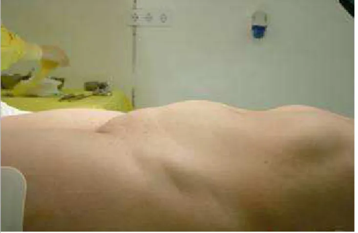
Clinical inspection reveals a visible swelling on lower abdomen.
Figure 6 No title
Intra-operative aspect (a). The gross specimen of myomectomy (b) shows dark red softening
macroscopic appearance.
MeSH
[C13.371.852.762] Uterine Neoplasms
Tumors or cancer of the UTERUS.
References
[1] Murase E, Siegelman ES, Outwaker EK, Perez-Jaffe LA, Tureck RW (1999) Uterine
leiomyomas: histopathologic features, MR imaging findings, differential diagnosis and treatment Radiographics 19: 1179-97
[2] Ueda H, Togashi K, Konishi I, Kataoka ML, Koyama T, Fujiwara T, Kobayashi H, Fujii S, Konishi J (1999) Unusual Appearances of Uterine Leiomyomas: MR Imaging Findings and Their Histopathologic Backgrounds Radiographics 19:131-145
[3] Norris HJ, Hilliard GD, Irey NS (1988) Hemorrhagic cellular leiomyomas ("apoplectic leiomyoma") of the uterus associated with pregnancy and oral contraceptives Int J Gynecol Pathol 7:212-24
[4] Hock YL, Goswami P, Rollason TP (2000) Mitotically active haemorrhagic cellular (apoplectic) leiomyoma Eur J Gynaecol Oncol 21:28-9
[5] Yamashita et al (1993) Hyperintense Uterine Leiomyoma at T2weighted MR Imaging: Differentiation with Dynamic Enhanced MR Imaging and Clinical Implications Radiology 189:721-725
Citation
M. Rosário Matos , Teresa Margarida Cunha1 2
Department of Radiology, Hospital de Dona Estefânia - Centro Hospitalar de Lisboa Central. 1
Department of Radiology, Instituto Português de Oncologia Francisco Gentil de Lisboa. (2010, 2
Apr. 28)