Márcia Figueiredo Gonçalves
Licenciatura em Bioquímica
Understanding how dendritic cell glycans affect antitumor
immune responses
Dissertação para obtenção do Grau de Mestre em Bioquímica para a Saúde
Orientador: Dr. Paula Alexandra Quintela Videira,
Assistant Professor,
NOVA Medical School, NOVA University of Lisbon, Portugal Co-orientador: Dr. Sandra Van Vliet,
Assistant professor,
VU University Medical Center Amsterdam, The Netherlands Co-orientador: Dr. Yvette van Kooyk,
Professor,
VU University Medical Center Amsterdam, The Netherlands
Júri:
Presidente: Prof. Doutor Sebastião Rodrigues Arguente: Prof. Doutora Irís Caramalho Vogal: Prof. Doutora Paula Alexandra Quintela Videira
| I
Bibliographic elements
The work described here has originated:
o Provisional patent application
Videira, PA. (2015) Order for international patent – U.S. Patente N/REF. 150316-PP
“A NOVEL DENDRITIC CELLS POPULATION, METHOD OF PRODUCTION AND
USE THEREOF “
o Manuscript under preparation
“Cell surface Sialic acid removal from dendritic cells improves antigen cross-presentation
and boosts anti-tumor immune responses” Mariana Silva*, Zélia Silva*, Graça Marques,
| III
Acknowledgment
I would like to present my genuine acknowledgements to all those who in one way or another, allowed, facilitated and motivated this thesis.
The Department of Immunology, Glycoimmunology Group - CEDOC at the NOVA Medical School, NOVA University of Lisbon, Portugal, and the Department of Molecular Cell Biology and Immunology at the VU UNIVERSITY MEDICAL CENTER AMSTERDAM, for hosting me in their laboratories.
European mobility – Erasmus program (SMT), fellowship which funded six months of this thesis, obtained through Science and Technology Faculty, NOVA University of Lisbon, Portugal.
Dr. Sandra van Vliet, my co-supervisor, for her tireless monitoring, the invaluable advice and for making me feel at home.
Dr. Paula Videira, my advisor, for being the main initiator for this experience, for her support, availability and motivation.
Professor Yvette van Kooyk, my co-supervisor, for her friendliness and welcoming spirit, receiving me with open arms in her laboratory.
Dr. Zélia Silva, for her help and patience.
| IV
To my second family: Helena, Joana, Catarina and Tiago for the friendship, patience, for always being there even when I am unbearable, and for all the right words spoken at the right time. To Jomi, Maria João and Pedro Calixto by the long English sessions.
| V
Abstract
Dendritic cells (DCs) have a unique capacity to induce immune responses against tumor cells. They can phagocyte tumor antigens, maturate and present them to T cells, triggering antigen-specific immune responses that may lead to the elimination of tumor cells. Since they induce long-lasting immunological memory, DCs become an attractive strategy as cellular targets for vaccines in the treatment and/or prevention of cancer. However, the therapeutic results obtained in clinical trials with DCs are scarce and only few patients effectively respond to the DC vaccines. Our group has shown that sialic acid containing glycans play an important functional role in ex vivo generated DC. Here we
aimed to establish an in vitro model to assess specific antitumor responses. To achieve this,
an enzymatic treatment of monocyte-derived DCs (moDCs) was performed using sialidase to cleave surface sialic acids. The maturation profile of the moDCs was characterized by flow cytometry and cytokine expression. The results show that sialidase treatment can upregulate co-stimulatory molecules on surface of moDCs stimulated with Toll like receptor (TLR) agonists. To understand whether sialidase treatment affected the TLR signaling, we have used HEK cells stably transfected with TLRs 2, 4 and 7/8. The data showed that desialylation of moDCs does not affect the signaling via these receptors. To investigate the functional impact of sialidase treatment in the capacity of moDCs to present antigen and to activate antigen specific T cells, sialidase treated and untreated moDCs were co-cultured with CD8+ T cell clones specific for peptides derived from the gp100 tumor
antigen. Our results show that desialylated HLA02:01+ DCs are superior in
cross-presentation of the peptide to gp100280–288 specific CD8+ T cells. In addition, sialidase
treatment also increased the DC capacity to induce CD4+ T cells proliferation. Together,
these data indicate that moDCs with altered cell surface sialic acids, through a sialidase treatment, have a better immunostimulatory potential which could improve anti-tumor immune responses.
| VII
Resumo
As células dendríticas (DCs) têm a capacidade única de induzir respostas imunitárias contra as células tumorais, fagocitando antigénios tumorais e apresentando-os às células T, provocando respostas imunitárias específicas que conduzem à eliminação de células de tumorais. Por induzirem memória imunológica de longa duração, as DCs são uma estratégia atrativa para o tratamento e/ou prevenção do cancro. No entanto, os resultados terapêuticos obtidos em ensaios clínicos com DCs são escassos e pouco eficientes. O nosso grupo demonstrou que ácidos siálicos que contêm glicanos desempenham um papel funcional importante em DCs geradas ex vivo. Com o objetivo de
estabelecer um modelo in vitro para avaliar a resposta anti-tumoral específica realizou-se
um tratamento enzimático a DCs derivadas de monócitos (moDCs) com sialidase, enzima que cliva ácidos siálicos na superfície celular. O perfil de maturação de moDCs foi caracterizado por citometria de fluxo e expressão de citocinas. Os resultados mostram que a sialidase pode regular positivamente a expressão de moléculas co-estimuladoras na superfície de moDCs estimuladas com agonistas de Toll like receptors (TLRs). Para
percebermos se o tratamento com sialidase afeta a sinalização dos TLRs foram usadas células HEK transfectadas de forma estável com TLRs 2, 4 and 7/8. Os dados mostraram que a desialilação não afeta a sinalização através estes recetores. Para investigar o impacto funcional da sialidase na capacidade de moDCs em apresentar um antigénio e ativar células T, moDCs foram tratadas, ou não, com sialidase e cultivadas com clones de células T CD8+
específicas para os péptidos derivados do antigénio tumoral gp100. Os resultados mostram que DCs HLA*02:01+ desialiladas exibem maior cross-presentation do péptido gp100
280-288
às células T CD8+ específicas. Além disso o tratamento com sialidase também aumenta a
capacidade de DCs de induzir a proliferação de células T CD4+. Em conjunto, os resultados
indicam que moDCs com menos ácidos siálicos na superfície, têm melhor potencial imuno-estimulador, com maior capacidade de induzir respostas imunes anti-tumorais.
| IX
Index
1) Introduction 1
1.1) Cancer 1
1.2) Immune system 2
1.2.1) Innate and adaptive immunity 2
1.2.2) T lymphocytes 4
1.3) Dendritic cells 6
1.3.1) Origin, differentiation and classification 6
1.3.2) Antigen recognition and uptake 7
1.3.3) Antigen processing 10
1.3.4) DC maturation 11
1.3.5) Antigen presentation by DCs to T cells 12
1.4) Anti-tumor immunity 13
1.5) Immunotherapy 15
1.6) Vaccines based on DCs 16
1.6.1) DC vaccine optimization 17
1.7) Glycosylation 18
1.7.1) Glycoproteins 19
1.7.2) Sialic acids 20
1.8) Hypothesis, Aims and Scopes 21
2) Materials & Methods 25
2.1) Human peripheral blood 25
2.2) Isolation of monocytes from buffy coats by Ficol and Percol gradient 25
2.3) Generation of monocyte-derived dendritic cells (moDCs) 27
2.4) Sialidase treatment 27
2.5) Lectin binding assay 27
2.6) MoDC stimulation 28
2.7) Toll-Like Receptor (TLR) test 29
2.8) Enzyme-linked Immunosorbent Assay (ELISA) 30
2.9) Real-Time PCR (qPCR) 31
| X
2.11) Binding of gp100 peptide to T2 cell line 33
2.12) Activation of CD8+ T cell clones 34
2.13) CD4+ T cell Proliferation 35
2.14) Statistical analysis 36
3) Results 37
3.1) Sialidase protocol optimization 37
3.2) Removal of sialic acid from the moDC surface by sialidase treatment increased co-stimulatory molecule expression when stimulated with TLR-ligands 39
3.3) Cytokine secretion is not affected in moDCs treated with sialidase 41
3.4) Sialidase treatment and TLR activation 44
3.5) Desialylation effect in gp100 short peptide binding to HLA-A2 in T2 cell line 45
3.6) Sialidase treatment of moDCs improves their ability to activate tumor antigen
gp100-specific T cells 47
3.7) Sialidase treatment of moDCs improves proliferation of autologous CD4+ T cells 49
4) Discussion and conclusions 51
| XI
Index of figures
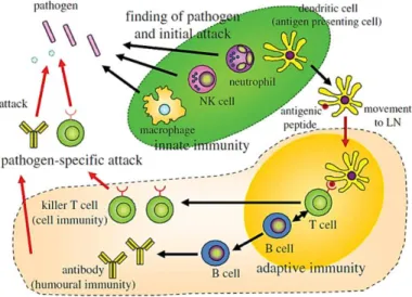
Figure 1 - Immune System: Innate immunity and adaptive immunity. Addapted from Akira
et al., (Akira, 2011). 4
Figure 2 – Sites for therapeutic intervention that allow regulated anti-tumoral immunity.
Increased presentation of tumor antigens by mature dendritic cells leads to more effective adaptive immune response mediated by T cells and help to prevent immunosuppression. Adapted from Mellman et al., (Mellman, Coukos e Dranoff,
2011). 13
Figure 3 - Key points to improve DC vaccination in cancer patients. Adpated from Pham et
al., (Lee et al., 2012). 18
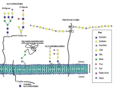
Figure 4 - Glycoconjugates classes expressed in human cells. Adapted from Reis et al.,
(Reis et al., 2010). 19
Figure 5 - Layout of separation by ficoll gradient. 25
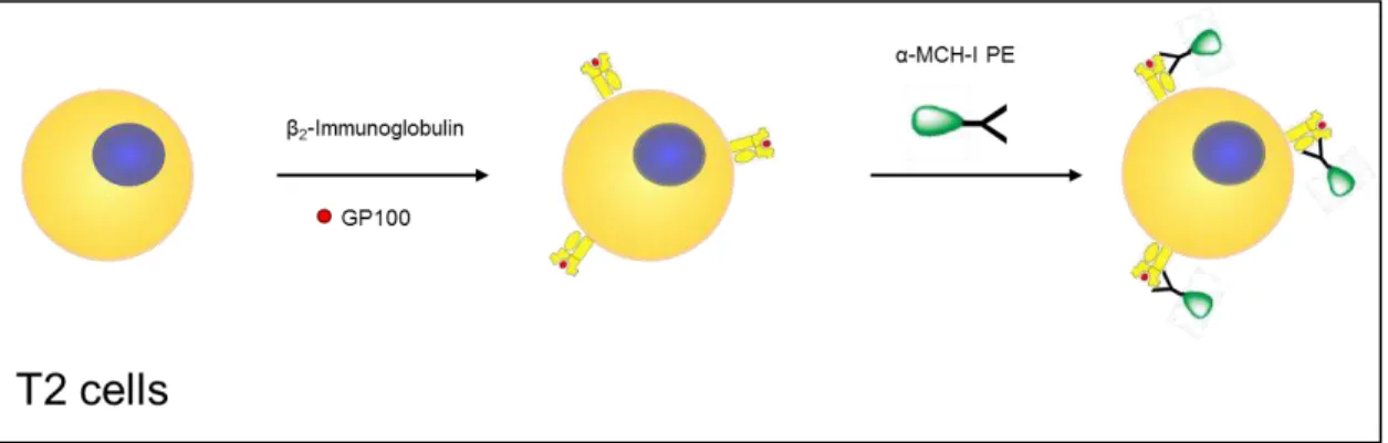
Figure 6 - Summary of the MHC-I binding assay with T2 cells. Prior to the assay T2 cells
were treated with sialidase or left untreated. After a wash step T2 cells were resuspended in serum-free RPMI-1640 medium with β2-microglobulin and plated in the presence of gp100280–288 (short) peptide. After 4 hours of incubation at 37 °C the
cells were stained with PE-labelled anti-HLA-A2+ antibody. 34
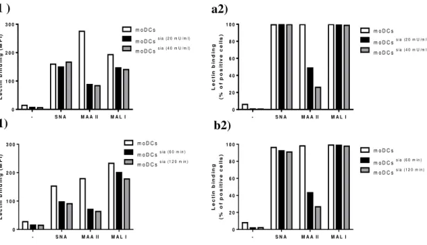
Figure 7 - Lectin binding to cell surface of moDCs. MoDCs were stained with Sambucus
nigra lectin (SNA - recognizing α2,6-sialic-acids) and Maackia amurensis lectin (MAA II - recognizing α2,3-sialic-acids on O-glycans and MAL I - recognizing α 2,3-sialic-acids on N-glycans) following sialidase treatment. a) Immature mo-DCs were treated with sialidase at two different concentrations (20 mU/ml and 40 mU/ml) or left untreated, for 60 min at 37 °C (n=1). Control results are represented in white, 20 mU of sialidase in black and 40 mU in grey: a1) Mean fluorescence intensity (MFI); a2) the percentage of positive cells. b) Immature mo-DCs were treated with 20 mU/ml of sialidase at two different incubation times (60 min and 120 min) or left untreated for 60 min at 37 °C (n=1). Controls results are represented in white, 60 min of incubation in black and 120 min of incubation in grey: b1) Mean fluorescence intensity (MFI) ; b2) the percentage of positive cells. The results were obtained by flow cytometry, the results displayed in a) and b) are derived from different donors. 38
Figure 8 - Expression of maturation markers, CD80, CD83 and CD86, in moDCs after
| XII
cytometry. The results are the average mean fluorescence intensity (MFI) of 15 independent assays. Statistical significance (*p ≤ 0.05; **p ≤ 0.01; ***p ≤ 0.001) refers to the difference between untreated (white bar) and sialidase-treated moDCs
(black-bar). 40
Figure 9 - Plant lectin binding assay. moDCs (day4 after of differentiation) were treated
with sialidase (60 min at 37 °C) or left untreated and then were labelled with the different plant lectins (SNA, MAA II and MAL I). lectin binding was evaluated by flow cytometry. The results are the average mean fluorescence intensity (MFI) (left side) or percentage of positive cells (right side) of 16 independent assays. Statistical
significance (**p ≤ 0.01; ***p ≤ 0.001) refers to the difference between untreated
(white bar) and sialidase-treated moDCs (black bar). 41
Figure 10 - Cytokine secretion. MoDCs were treated with sialidase (black bar) for 60 min
at 37 °C or left untreated (white bar). After over-night incubation with different TLR-ligands the supernatant was harvested and cytokine production was measured by ELISA. The results are the mean ± SEM of 9 independent experiments. 42
Figure 11 - Cytokine production analysed by ELISA (left side) and qPCR (right side).
moDCs were treated with sialidase (60 min at 37 °C) or left untreated. After incubation for 6 h (grey bar and blue bar) or over-night (white bar and black bar) with different TLR-ligands, the supernatant was harvested and the cytokine production was measured by ELISA. cell pellets were lysed and cytokines mRNA levels were measured by qPCR. The results are of 1 independent experiment. 43
Figure 12 –Toll-like receptor test. HEK cells were treated with sialidase for 60 min at 37
°C or left untreated. After incubation over-night with different TLR-ligands the supernatant was harvested and the IL-8 production was measured by ELISA. TLR-2 results are the mean ± SEM of 4 independent experiments, TLR-4 results are of one representation experiment out of 3 and the TLR-8 results are the mean ± SEM of 3 independent experiments. Statistical significance (*p ≤ 0.05) refers to the difference
between untreated (black line) and sialidase-treated (blue line) HEK cells. 45
Figure 13 –gp100 peptide bound MHC-I is more stable on sialidase treated T2 cells. a) T2
binding assays were performed by incubating cells, treated (blue line) or not (black bar) with sialidase, without peptide (ss why not just call this zero?) or with different
concentrations of gp100 short peptide in the presence of β-microglobulin for 4h at 37
| XIII
experiments and statistical significance (*p ≤ 0.05) refers to the difference between
untreated and sialidase-treated T2 cells. 46
Figure 14 - Improved production of IFNy by CD8+ T cell clones. a) gp100-specific CD8+ T
cells were co-cultured overnight with gp100-loaded moDCs, that have previouslybeen treated (black bar)or not (white bar). b) gp100-specific CD8+ T cellswere co-cultured
overnight with gp100-loaded moDCs and LPS, after treatment (black bar) or not (white bar) with sialidase. c) gp100-specific CD8+ T cellswere co-cultured for 3 days
with gp100-loaded moDCs after treatment (black bar)or not (white bar) with sialidase. d) gp100-specific CD8+ T cells were co-cultured for 3 days with gp100-loaded moDCs
and LPS after treatment (black bar) or not (white bar) with sialidase. Different concentrations of long gp100 peptide, which is cross-presented to CD8+ T cells, were
used. The short gp100 peptide was used as a positive control for the functionality and antigen specificity of CD8+ T cell clone. The secretion of the IFN-γ cytokine was
measured by ELISA (n=3). Results were normalized to the IFN-γ levels after incubation with short peptide without sialidase treatment (a) and c)) or with short peptide without sialidase treatment and with LPS (b) and d)). 49
Figure 15 - Sialidase treatment of moDCs improves proliferation of autologous CD4+ T
cells. The proliferation of purified CD4+ T cells was measured by [3H]thymidine
uptake assay, after a challenge with different ratios of moDCs that were treated (black bar) or not treated (white bar) with sialidase, in the absence a) or in the presence b) of LPS for 3 days (n=3). Results were normalized to the condition without moDCs and without sialidase treatment (a) or without moDCs ,without sialidase treatment and with LPS (b). Graphs show the mean ± SEM of at least 3 independent experiments. 50
Index of Tables
Table 1 - Exogenous/synthetic ligands of Toll-like receptors and respective PAMPs.
Adapted from Bhardwaj et al.,28. 9
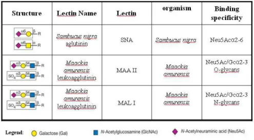
Table 2 – Lectin specificity. Representation of the structure, the lectin name, the organism
origin and its binding specificity. Adapted from Varki, et al.,59. 28
Table 4 - Sample dilutions to cytokine measured by sandiwch-ELISA. 31
Index of Appendixes
| XV
Abbreviations
Ag – Antigen
AMV RT - Avian Myeloblastosis Virus Reverse Transcriptase APCs – Antigen Presentation Cells
BCR – B Cell Receptor
BSA – Bovine Serum Albumin CLR – C-type Lectin Receptor CTLs – Cytotoxic T Lymphocytes DCs – Dendritic Cells
DC-SIGN - DC-specific ICAM-3 grabbing non-integrin dNTP - Deoxyribonucleotides Phosphated
ELISA - Enzyme-Linked Immunosorbent Assay ER – endoplasmic reticulum
FDA - Food and Drug Administration FITC - Fluorescein isothiocianate GalNAc - N-acetylgalactosamine
GAPDH - Glyceraldehyde 3-phosphate dehydrogenase GM-CSF - Granulocyte macrophage colony-stimulating factor GMO – Genetically modified
| XVI
HEK – Human Embryonic kidney HLA – Human Leukocyte Antigen HPV – Human Papillomavirus HSCs – Hematopoietic Stem Cells IFN-γ – Interferon-gamma
IL – Interleukin Io – Iomycin
LCs – Langerhans cells LPS – Lipopolysaccharide
MAA II - Maackia amurensis agglutinin (O-glycan)
MACS - Magnetic-activated cell sorting MAL I - Maackia amurensis lectin (N-glycan)
mDC – Myeloid Dendritic Cell MFI – Mean Fluorescence Intensity
MHC – Major Histocompatibility Complex moDCs – monocyte-derived dendritic cells Neu5A - N-acetylneuraminic acid
NF-ᴋB - Nuclear factor kappa β
NK – Natural Killer cell
NOD – nucleotide-binding oligomerization domain receptors ON – overnight
| XVII
PBA - PBS/0.5% BSA
PBLs – Peripheral blood lymphocytes
PBMCs – Peripheral blood mononuclear cells PBS – Phosphate buffered saline solution pDC – Plasmacytoid Dendritic Cell
PE – Phycoerythrin
PMA – Phorbol Myristate acetate PRRs – Pattern Recognition Receptors
qPCR - quantitative polymerase chain reaction RT – Room Temperature
Siglecs - Sialic acid-binding Ig-like lectins SNA - Sambucus nigra agglutinin
TAP – Transporter for Antigen Presentation TCR – T Cell Receptor
Th – helper T lymphocyte TLR – Toll-like Receptor
TMB - 3,3′,5,5′-tetramethylbenzidine TNF-α – Tumoral Necrosis Factor –α
| 1
1) Introduction
1.1) Cancer
For a good functioning of the human body a balance must exist between proliferation and cell death (Baehrecke, 2002). However, due to external or internal conditions, mutations occur during the generation of new cells which could lead to disruption of this homeostasis since these cell lose the ability to respond to stimulus (Kumar, V., Abbas A. K., Fausto, N. & Mitchell, 2007).
Transformation of normal cells into cancer cells is associated with phenotypic changes that affect the behavior of cells and consequently other biological processes. Hanahan and Weinberg (Hanahan e Weinberg, 2011), described the main aspects that characterize cancer: evasion of apoptosis, inadequate signs to inhibit cell growth, the ability to proliferate indefinitely, acquiring modifications that allow invasion of other tissues and tumor vascularization. All of them allow tumor cell survival and uncontrolled cell proliferation that leads to invasion of other tissues and subsequent metastasis (Bogenrieder e Herlyn, 2003; Goldsby, R.A., Kindt, T. J., Osborne, B., Kuby, 2003; Hanahan e Weinberg, 2011; Kumar, V., Abbas A. K., Fausto, N. & Mitchell, 2007), the main cause of death in people with cancer (EL, 1997).
These characteristics (Hanahan e Weinberg, 2011) distinguish cancer cells from normal cells, potentially allowing the tumors to be recognized as "foreign" by the immune system (Chen, Irving e Hodi, 2012; Gros et al., 2014; Mellman, Coukos e Dranoff, 2011;
Rooij, van et al., 2013). However, tumors are rarely rejected spontaneously, reflecting their
| 2
In 2012, worldwide (World cancer research fund international) 14.1 million new cancer cases were diagnosed of which 8.2 million died. It is estimated that in 2035 the number of persons with cancer will increase dramatically to 24 million.
Conventional therapies such as surgical resection and a combination of chemotherapy and radiotherapy are approaches that are often accompanied by unintended collateral damage and with high toxicity to healthy tissues and have not been sufficiently effective in the fight against cancer. It is of great importance to develop a therapy that uses the patient's own immune system and, in that way, ensures the safety of patients while being effective in eliminating the tumor.
1.2) Immune system
The immune system has the ability to respond to different external aggressions, in particular antigenic natures that are foreign to the body, whether a microorganism or macromolecule, and various internal injuries, or modified tumor cells. Thus, you can define immunity as a set of defense mechanisms that our body has to protect itself from attacks, to maintain the immune homeostasis. The immune system under normal conditions doesn´t present any kind of response to host cells, a situation that it is known as immunological tolerance (Arosa, A. F., Cardoso, E.M., 2011).
1.2.1) Innate and adaptive immunity
The reaction of the immune system can be divided into two types of interconnected pathways: innate immunity and adaptive immunity.
| 3
known as phagocytic cells, which phagocytose the foreign body and destroy it (Akira, 2011).
This is an immediate response with a broad spectrum specificity, recognizing only molecular patterns stored in microorganisms or PAMPs (Pathogen-associated molecular patterns) through specific receptors on phagocytic cells (macrophages and DCs). These receptors are called pattern recognition receptors (PRRs) and include the Toll-like receptors (TLRs) and C-type lectin receptors (CLRs), such as the mannose receptor. TLRs are able to recognize a wide variety of PAMPs. This recognition results in the activation of phagocytic cells that can lead to the internalization of the microorganism, as well as the release of cytokines and inflammatory mediators, triggering an inflammatory process (Arosa, A. F., Cardoso, E.M., 2011). In contrast to adaptive immunity, each time the body is exposed to the pathogen, the immune response is always the same, since there is no memory on previous exposures.
Cytokines released by cells from inflamed tissues, such as interleukin-1 (IL-1) and interleukin-6 (IL-6) are important in activation of immune response and cell interaction. Then, mononuclear cells and lymphocytes, that in the meantime, are attracted to the inflammatory
focus, are activated and start releasing their own cytokines (IL-1, IL-2, IL-4, TNF-α, IFN- γ,
etc.) enhancing and promoting the migration and activation of certain cells more directly involved in the immune response (Akira, 2011; Arosa, A. F., Cardoso, E.M., 2011).
| 4
Figure 1 - Immune System: Innate immunity and adaptive immunity. Addapted from Akira et al., (Akira, 2011).
While the B cell receptor recognizes the antigen in its native form (not processed), the T cell receptor only recognizes the antigen in a fragmented form (for example peptides) presented in the context of MHC (Major Histocompatibility Complex) molecules. This recognition results in activation and differentiation of B lymphocytes, which will produce antibodies specific to the antigen, and the activation and differentiation of T lymphocytes, which lead to the formation of effector cells and regulatory cells of the immune response (Arosa, A. F., Cardoso, E.M., 2011).
1.2.2) T lymphocytes
T cells are a lymphocyte lineage originated from bone marrow hematopoietic precursors which complete their maturation in the thymus. These cells are roughly divided into four groups: helper T lymphocytes (Th), cytotoxic T cells (CTL), regulatory T cells (Treg) and natural killer T lymphocytes (NKT) (Arosa, A. F., Cardoso, E.M., 2011).
| 5
which is restricted by antigen presentation in MHC class II molecules. After activation, the Th lymphocytes begin a differentiation process, and can develop into four types of lymphocytes: Th1, Th2 and Th17 (Goldsby, R.A., Kindt, T. J., Osborne, B., Kuby, J, 2007). The Th1 lymphocytes are characterized by production of IL-2, IFN-γ and TNF-β.
The function of IL-2 is to regulate the growth of Th1 cells as well as CD8+ T lymphocytes.
The Th2 lymphocytes are characterized by production of IL-4, IL-5, IL-9, IL-10 and IL-13 (anti-)inflammatory cytokines responsible for regulating the humoral response against extracellular pathogens and allergens (Goldsby, R.A., Kindt, T. J., Osborne, B., Kuby, J, 2007).
The CTL express CD8, and their function is to eliminate other cells, particularly when they have become a tumor or are infected. The CTL response is restricted by antigen presentation in MHC class I molecules and is characterized by production of the cytokines
IFN-γ and TNF-α and lytic enzymes (perforin and granzymes), responsible for the
elimination of target cells (Arosa, A. F., Cardoso, E.M., 2011).
The Treg derived from a separate “lineage” of lymphocytes (not Th) and function as
controllers of the immune response and play an important role in the maintenance of immunological tolerance to innocuous antigens in the periphery and in the prevention of autoimmunity, allergies and, in general, are responsible for the suppression of immune responses (Lehtimäki e Lahesmaa, 2013).
The NKT cells contain mixed features of T and NK lymphocytes and are determinants of infectious and autoimmune diseases. NKT cells express a particular type of TCR that recognizes glycolipid antigens in the context of non-classical MHC CD1d and once activated are cytotoxic and produce cytokines such as IL-4 and IFN-γ (Wu e Kaer, Van, 2009). Although NKT can directly eliminate tumor cells, it is believed that their effectiveness arises from the effect on NK cells, CD8+ T cells and DCs, leading,
| 6
T cell activation begins with the recognition by the TCR/CD3 of antigenic peptides exposed on the surface of APCs in association with MHC molecules. The signal from the TCR /CD3-MHC-peptide interaction is the primary activation signal ("signal 1"), which confers specificity to the adaptive response. However, this signal in itself is not effective to induce strong activation that causes the naive CD4+ T and naive CD8+ T lymphocytes to
enter the cell cycle and to proliferate, making it necessary to receive accessory signals. These accessories signals are transmitted by the CD28 receptor, considered "Signal 2", which will lead to the formation of CD4+ T and CD8+ T lymphocytes with different
phenotypic and functional characteristics than the initial naive T lymphocyte (Arosa, A. F., Cardoso, E.M., 2011).
1.3) Dendritic cells
1.3.1) Origin, differentiation and classification
DCs were identified for the first time by Paul Langerhans in 1968, who mistakenly thought they were part of the nervous system (Langerhans cells, LCs) (Goldsby, R.A., Kindt, T. J., Osborne, B., Kuby, J, 2007). This concept held until the mid-twentieth century. In 1973 Ralph Steinman and Zanvil Cohn observed a cell population in the spleen with a dendritic shape and demonstrated that this was a new class of cells with immunomodulatory functions in the immune system (Paczesny S., 2003).
Currently, it is known that DCs originate from CD34+ hematopoietic stem cells
(HSCs) from the bone marrow. In physiological stress conditions, monocytes can differentiate into immature DCs in the presence of the stimulating factor, granulocyte macrophage colony (GM-CSF) and from a variety of other cytokines (Mogensen, 2009).
| 7
surface markers, location in the body. Both mDCs and pDCs perform specific functions and specific inflammatory stimulus that induce differentiation (Arosa, A. F., Cardoso, E.M., 2011).
The pDCs can be derived from myeloid precursor and are in the blood and lymphoid organs (Sathe et al., 2013). These cells are characterized by their extraordinary
ability to produce interferon type 1 (IFN-α / β) following viral infection or after interaction
with TLRs 7 and 9 agonists. From a functional standpoint, pDCs have an enormous plasticity and can induce Th responses (Th1 and Th2), or tolerance via induction of Treg
cells. Moreover, they are still capable to perform antigen cross-presentation via MHC class I, due to their special endosomal compartments (Arosa, A. F., Cardoso, E.M., 2011; McKenna, Beignon e Bhardwaj, 2005).
Likewise, mDCs are from the myeloid lineage and can be found in tissues (Langerhans cells and interstitial DCs) and in peripheral blood (inflammatory DCs) (Brussel I.V., Berneman Z.N., 2012; Paczesny S., 2003; Zhang, C. e Engleman, 2006). Inflammatory DC are involved in recognition of bacterial structures and production of pro-inflammatory cytokines, including tumor necrosis factor α (TNF-α), IL-6 and IL-12p70 to activate Th1 / Th17 cells, and thus recruiting CTLs (Arosa, A. F., Cardoso, E.M., 2011; Zhang, C. e Engleman, 2006).
It has been further shown that pDCs can enhance the immune response through cross-talk with mDCs through IFN-γ production and CD40L expression, enabling the
production of IL-12p70 by the mDC ( Boudreau J. E., Bonehill A., Thielemans K., 2011).
1.3.2) Antigen recognition and uptake
| 8
maturation process in which immature DC with large endocytic capacity and low expression of MHC-II molecules, differentiate into mature DC characterized by a low endocytosis capacity and a high expression of MHC-II molecules. The immature DCs, lying in organs and peripheral tissues, have a great ability to endocytose and process endogenous and exogenous antigens while mature DCs are specialized in the activation of T lymphocytes in secondary lymphoid organs.
Generally, endogenous antigens presented by DCs in the context of MHC-I activate CD8+ T lymphocytes, while exogenous antigens are presented by DCs in the context of
MHC II activate CD4+ T lymphocytes. DCs also have the unique ability to present
exogenous antigens in vivo to CD8+ T lymphocytes via MHC-I, in a process called
cross-presentation (Arosa, A. F., Cardoso, E.M., 2011; J. E. Boudreau, A. Bonehill, K. Thielemans, 2011).
1.3.2.1) Receptors in the antigen recognition and uptake
The designated PAMPs, which are highly conserved structures including microbial lipids, polysaccharides, nucleic acids and viral RNA, are recognized by immature DCs through PRRs. These receptors are highly diverse, which includes TLRs, nucleotide-binding oligomerization domain (NOD-like) receptors and CLRs (Kanazawa, 2007; Mogensen, 2009).
After antigen recognition, their internalization is mediated by a large number of endocytic and phagocytic receptors including CLRs and integrins.
The TLRs are transmembrane proteins which are present in DCs, macrophages, fibroblasts and epithelial cells. They are involved in recognition of antigens and subsequent activation of cell signaling pathways. mDCs express TLR 1–8, which when stimulated, upregulate activation markers (CD80, CD86, MHC class I and II), produce pro-inflammatory cytokines (TNF-α, IL-1, IL-6, IL-12), chemokines, adhesion molecules (ICAM-1) and prime antigen-specific CD4+ and CD8+ T cells (Gnjatic, Sawhney and
| 9
maturation and activation, thereby determining the onset of immune responses (Arosa, A. F., Cardoso, E.M., 2011).
Table 1 - Synthetic ligands of Toll-like receptors and respective PAMPs. Adapted from Gnjatic et al.,(Gnjatic, Sawhney and Bhardwaj, 2010).
Receptor Pathogen Associated ligands (PAMPs) Synthetic ligands
TLR ½ Triacylated lipopeptides (Bacteria and Mycobacteria)* Pam3CysK4
TLR 4 Lipopolysaccharide (LPS) (Gram-negative bacteria); LPS
TLR 7 Viral ssRNA (Influenza, VSV, HIV, HCV) Guanosine analogs; imidazoquinolines
(e.g.R848, Resiquimod®); Loxoribine.
TLR 8 ssRNA from RNA virus Imidazoquinolines (e.g.R848,
Resiquimod®); Loxoribine.
NOD-like receptors are located in cytoplasm of the DC and are capable of binding to bacterial peptidoglycans (Kanazawa, 2007).
| 10
1.3.3) Antigen processing
Before antigen detection, DCs are in an immature stage. After recognition, DCs phagocytose the antigen and then enter an activation process, maturation, and migrate to the lymph nodes where a T cell immune response specific for the antigen is initiated. During the process of maturation and migration, DCs process the antigen into smaller fragments that may be presented to T lymphocytes (Hamdy et al., 2011).
DCs process endogenous and exogenous antigens, presenting them to T lymphocytes in the form of antigenic peptides bound to MHC molecules (Hamdy et al.,
2011). This processing is different based on the origin and the molecular nature of antigen, whereby presentation occurs via three mechanisms: i) via MHC class I or cytosolic (endogenous); ii) via MHC class II or endocytic (exogenous); iii) presentation of lipid antigens coupled to CD1 molecules (Goldsby, R.A., Kindt, T. J., Osborne, B., Kuby, J, 2007).
Extracellular antigens are captured by endocytosis, phagocytosis and pinocytosis, which enter the endocytic pathway, forming endosomes that subsequently undergo maturation and fusion with lysosomes. In lysosomes hydrolytic enzymes cleave the antigen into smaller molecules, peptides, which are then coupled to II molecules. The MHC-peptide complex is transported to the cell surface during the process of dendritic cell maturation for antigen presentation to CD4+ T naive lymphocytes (Goldsby, R.A., Kindt, T.
J., Osborne, B., Kuby, J, 2007).
| 11
1.3.4) DC maturation
In peripheral tissues DCs are usually in an immature state and practically devoid of immunostimulatory activity. However, after a stimulus (danger signal - antigen) they acquire different morphological, phenotypic and functional properties. In an immature state, DCs have a low ability to stimulate immune responses and a high power to capture antigens. When the maturation process starts, DCs lose their phagocytic receptors and increase their migration from peripheral tissues to secondary lymphoid organs where they present antigen to naive lymphocytes, culminating in the acquisition of immunostimulatory potential (maturation). antigen processing is regulated in a coordinated manner through maturation of the DC, which causes a decrease in pH of the endosomes, whereby processing is facilitated, enabling the transport of the MHC-peptide complex to the cell surface (Arosa, A. F., Cardoso, E.M., 2011).
Indeed, mature DCs express high levels of costimulatory molecules, as well as MHC molecules and synthesize high levels of IL-12 which enhances the ability to induce innate (NK cells) and adaptive (T and B cells) responses (Brussel I.V., Berneman Z.N., 2012). This process is continuous and highly regulated by signal transduction pathways that can be triggered directly through recognition of pathogens via PRRs or indirectly by exposure to other inflammatory mediators produced by immune cells, which results in increased membrane expression of costimulatory molecules (Brussel I.V., Berneman Z.N., 2012; Neves, 2010). The increased expression of MHC class I and class II and costimulatory molecules CD40, CD83, CD80 and CD86 during maturation is crucial in order to establish the immunological synapse and consequent stimulation of lymphocytes (Arosa, A. F., Cardoso, E.M., 2011).
The stimuli that induce maturation include products of microorganisms that bind to pattern recognition receptors, immune complexes that act on Fc receptors, inflammatory molecules released from the host cells, particularly CD40L, TNF-α, IL-1β, IL-6 e IFN-γ,
| 12
1.3.5) Antigen presentation by DCs to T cells
In the lymph nodes, DCs present antigen to CD4+ and CD8+ T cells via MHC II and
MHC-I, respectively. This interaction results in the activation of CD8+ T lymphocytes and
the differentiation of CD4+ T lymphocytes in their different effector and/or regulatory cells.
For this it is necessary that the antigenic recognition via MHC and interaction between costimulatory molecules on DCs and their respective ligands on T cells occurs (J. E. Boudreau, A. Bonehill, K. Thielemans, 2011).
In addition, DCs also have a unique ability to present exogenous antigens via MHC-I molecules. This function is referred to as cross-presentation of antigens. Exogenous antigens can be derived from apoptotic tumor cells or apoptotic infected cells (viral or bacterial). These antigens are degraded by the proteasome and coupled to MHC-I molecules for presentation to naive CD8+ T lymphocytes. This process ensures that DCs are
able to create a cytotoxic immune response against tumor or infected cells. However, the cross-presentation process can yield, by the CD8+ T cells both effective immunity
(cross-priming) and tolerance (cross-tolerance). Nevertheless, this mechanism has been crucial in the formulation of anti-tumor vaccines (Arosa, A. F., Cardoso, E.M., 2011; Goldsby, R.A., Kindt, T. J., Osborne, B., Kuby, J, 2007; Hamdy et al., 2011).
The lipid antigens present in microorganisms (mycobacteria) or endogenous tissues, are presented to the lymphocytes by DCs through CD1 molecules. These are essential in presenting specific glycolipids to NKT cells. Plasmacytoid DC do not have CD1 molecules (Arosa, A. F., Cardoso, E.M., 2011; Goldsby, R.A., Kindt, T. J., Osborne, B., Kuby, J, 2007).
| 13
In conclusion, DCs are central in the initiation and regulation of adaptive immune responses which makes these cells a promising target in anti-tumor treatments (Benencia et al., 2012).
1.4) Anti-tumor immunity
Activating the immune system for therapeutic benefit in cancer has long been a goal in immunology and oncology.
The generation of an effective anti-tumor immune response is a complex multistep process and the understanding of this matter provides a rationale for immunotherapeutic strategies (Buckanovich et al., 2008; Mellman, Coukos e Dranoff, 2011; Pardoll, 2012).
Advances in the understanding of how tolerance, immunity and immunosuppression regulate anti-tumor immune responses suggest that active immunotherapy represents a means to obtain a durable and long-lasting response in cancer patients.
For anti-tumor immunity to be effective three distinct steps must be achieved, either spontaneously or therapeutically: immunization, T cell response and blocking immunosuppression (fig.2) (Mellman, Coukos e Dranoff, 2011).
| 14
The anti-tumor response must begin with the uptake of tumor-associated antigens, exogenously or captured from dead or dying tumor cells by immature dendritic cells.
The dendritic cells process the captured antigen for presentation or cross-presentation on MHC class II and class I molecules, respectively, and migrate to draining lymph nodes. However, during capture and presentation it is necessary an adequate stimulus (activation signal) to mature dendritic cells is present. This stimulus depends of the concentration of the antigen, and the intensity and duration of the interaction with lymphocytes.
Activation signals could be therapeutically supplied in an exogenous (for example, TLR - ligands) or endogenous manner: dying or necrotic tumor cells release factors (for example, high mobility group proteins or ATP) that are thought to result in the immunogenic maturation of dendritic cells (Mellman, Coukos e Dranoff, 2011). Thus it initiated the antitumor response mediated by anti-cancer effector T cells.
The T cells of the immune system must first be able to recognize cancer cells as foreign, to generate a population of CTLs that can traffic to and infiltrate tumors wherever they reside, and specifically bind to and kill cancer cells (Chen, Irving e Hodi, 2012). However, if there is no accompanying maturation, DCs can induce tolerance leading to the elimination of T cells, T cell anergy or differentiation of Treg cells (Chen, Irving e Hodi,
2012).
In addition to the activation of CD8+ and CD4+ T cells, dendritic cells may also
trigger antibody and NK or NKT cells responses, which may contribute to the tumor immunity (Mellman, Coukos e Dranoff, 2011)
Additionally, tumors may downregulate their expression of MHC class I molecules or their expression of target tumors antigens (Hamanishi et al., 2007; Kooi et al., 1996).
| 15
Therefore, the importance of the maturation state of DCs is essential and key to whether the type of anti-tumor response is adequate and effective.
1.5) Immunotherapy
The conventional treatment for cancer includes routinely surgical resection and a combination of chemotherapy and radiotherapy. These approaches are often accompanied by unintended collateral damage and highly toxic to healthy tissues, which are offset by only marginal improvements in prognosis in patients with advanced cancers. This unfortunate balance has driven the development of new therapies aimed at achieving tumor elimination both safely and efficiently.
Over the last decade, increasing evidence supports a therapeutic utility of the immune system by immunotherapy. The aim of this strategy is to enable, restore, manage and still complement the patient's own immune system to control tumor growth and dissemination (Aris e Barrio, 2015). Sometimes pre-existing anti-tumor T cells can be ineffective in the elimination of the tumor for several reasons: due to their low frequency, the selection of tumor cells to escape recognition by the immune system, or even because these lymphocytes are functionally disabled (Aris e Barrio, 2015).
Among other immunotherapy approaches, vaccines can be prophylactic and therapeutic (Palucka, Banchereau e Mellman, 2010). Prophylactic (or preventative) vaccines have been used with considerable success for the prevention of cancers of viral origin, such as hepatitis B virus and human papillomavirus (HPV), where the etiological agent is known. In contrast, the development of therapeutic vaccines to treat existing disease has proven problematic. The long history of failure has tainted the entire strategy of immunotherapy in the eyes of many oncologists.
The idea of a therapeutic cancer vaccine began with the discovery that patients can harbour CD8+ and CD4+ T cells specific for cancer-testis or differentiation antigens
| 16
amplify the frequency and strength of these pre-existing responses or perhaps induce some
de novo reactions. Therapeutic vaccines can be administered as adjuvant therapy after
excision of tumors, with the aim of overcoming immunosuppression produced by the tumor and its microenvironment, to stimulate specific immune effectors that can destroy tumor cells or to increase the immunogenicity of the tumor to induce long-lasting immunity.
Additionally, clinic-pathological studies have demonstrated a strong association between prolonged patient survival and the presence of intra-tumoral CD8+ cytotoxic T
cells and an IFN-γ gene signature (Galon et al., 2006; Zhang et al., 2003). Thus, if
vaccination could trigger these types of T cell responses, a clinical benefit might be expected.
In general, vaccines require two critical components, the source of Antigen and adjuvant (Batchu et al., 2005). Therapeutic vaccines include the use of different antigenic
sources, such as, antigen peptides, proteins, nucleic acids, tumor cells lysates, recombinant virus or irradiated whole cells (Aris e Barrio, 2015).
1.6) Vaccines based on DCs
Alternatively, since the mid-90s, DCs have been used in clinical trials as cellular mediators for therapeutic vaccination of cancer patients (Anguille et al., 2014). The use of
DCs in an immunotherapeutic strategy is based on its ability to initiate cellular immune responses through the stimulation of naive T cells. Immature DCs are good at antigen uptake and processing, but for a stimulatory T-cell response they must mature to become fully activated DCs, which express high levels of cell surface-related MHC antigen and costimulatory molecules. Because of their ability to stimulate T cells, DCs act as a link between innate immunity and adaptive immunity in anti-tumor immune responses (Banchereau e Steinman, 1998).
| 17
immunotherapy and have been used in the treatment of more than 20 malignancies, most commonly melanoma, renal cell carcinoma, prostate cancer, and colorectal carcinoma (Palucka, Ueno e Banchereau, 2011; Ridgway, 2003). Since tumor antigen-loaded DCs are expected to be able to stimulate tumor-specific CTLs and to overcome T cell tolerance in tumor patients, the development of DC vaccines that can consistently eliminate minimal residual neoplastic disease remains an important goal in the field of tumor immunology (Banchereau e Palucka, 2005).
However, much skepticism has been shown due to the uncertainty of their clinical efficacy, since only some patients have an effective response. However, the clinical benefit of DC-based immunotherapy is small, but real, whereby 8.5% of patients with melanoma achieved an objective response. The DC therapy is as effective as dacarbazine, the standard of care chemotherapy, where 5-15% of patients show an objective response (Anguille et al.,
2014).
In 2010, the FDA approved Sipuleucel-T, the first DC-based vaccine for the treatment of metastatic castrate resistant, hormone refractory prostate cancer (Beer et al.,
2011).
1.6.1) DC vaccine optimization
The key step in this approach lies in producing a DC vaccine capable of eliciting an immune response that is capable of destroying the tumor. The vaccines based on DCs are composed of DCs that are generated from peripheral blood precursors (i.e., monocytes, HSCs) or bone marrow progenitor cells and are educated ex vivo with tumor antigens prior
| 18
Despite this simplistic picture (fig.3), there are several strategies used to activate DCs and to obtain a more effective vaccine. The use of immature DCs or mature DCs, the way to induce DC maturation, the type of tumor antigen, the techniques used to load tumor antigens to DCs, routes of administration, and dosing schedules are all being investigated (Figdor et al., 2004).
It has been demonstrated by several authors (Boog et al., 1989; Cabral et al., 2013;
Crespo et al., 2009; Jenner et al., 2006) that changes in the glycosylation of the dendritic
cell surface, more specifically sialylation, influences the subsequent activation of these cells and their role in induction of immune responses.
1.7) Glycosylation
Glycosylation is a post-translational modification more commune in proteins of eukaryotic cells has a frequency of 50% (Hang e Bertozzi, 2005; Wopereis et al., 2006).
This process involves covalent attachment of one or more glycans consisting of
Figure 3 - Key points to improve DC vaccination in cancer patients. Adpated from Lee et al.,(Lee et al.,
| 19
monosaccharides to a protein, lipid, carbohydrate or any other organic component, forming a glycoconjugate (Reis et al., 2010; Varki et al., 2009).
Glycoconjugates are involved in many physiological and pathological processes, including the processes of differentiation, cell migration and signaling, host-pathogen interactions and tumor invasion and metastasis (Campbell e Yarema, 2005; Reis et al.,
2010).
Glycans classes which are found in eukaryotic cells are defined based on the nature of the glycan binding to the carrier molecule (protein or lipid), forming in this way, various glycoconjugates (fig.4). Thus, glycoconjugates can be classified into proteoglycans, glycosphingolipids, glycosylphosphatidylinositol bound proteins and glycoproteins (Li e Richards, 2010; Varki et al., 2009).
1.7.1) Glycoproteins
Glycoproteins are defined as glycoconjugates in which one or more oligosaccharide chains are covalently linked to a protein (Ohyama, 2008; Reis et al., 2010; Varki et al.,
2009). There are two types of glycans associated with glycoproteins based on the type of
| 20
attachment: N-glycans and O-glycans. Both types of glycosylation can exist simultaneously within the same molecule and in the same cell. An N-glycan is an oligosaccharide covalently bound to the nitrogen atom of asparagine residues within a type amino acid sequence Asn-X-Ser / Thr (X being any amino acid except proline). O-glycan is a oligosaccharide covalently bound to the oxygen atom of a serine or threonine residue in an amino acid sequence (Li e Richards, 2010; Taniguchi, N., et al., 2008; Varki et al., 2009).
There are many ways in which the O-glycans may be elongated and processed with the addition of terminal residues to these structures, namely the addition of sialic acid, fucose and/or sulfate. These terminal residues often determine the biological function as well as recognition properties of modified glycans (Varki et al., 2009).
1.7.2) Sialic acids
Sialic acids (Sias) constitute a large family of terminal monosaccharides, which include N-acetylneuraminic acid (Neu5Ac) and derivatives thereof, which typically are attached to the expressed glycoconjugates on the cell surface of animal tissues and certain microorganisms (Varki e Schauer, 2009). Sias are involved in many cellular functions, both in physiological and pathological processes, including the regulation of the immune system, triggering infection and progression of certain diseases (Varki, A., Angata, T, 2006; Varki, A., et al. 2009) .
Sias are unique sugars that usually occupy the terminal position of the glycan chains and may be modified by external factors, such as pathogens, or upon specific physiological cellular events (sialidases). At cell surface, sialic acid-modified structures form the fundamental key determinants for a number of receptors with known involvement in cellular adhesiveness and cell trafficking, such as the selectins and thesialic acid-binding Ig-like lectins (Siglecs).
| 21
binding specificity for terminal sialic acids (Aarnoudse et al., 2006). Siglecs are expressed
predominantly on cells of the immune system (Crocker, Paulson and Varki, 2007), such as human immature DCs and are in particular highly expressed on tolerogenic DCs.
Siglecs are thought to play a role in both positive and negative regulation of immune responses (Crocker, Paulson e Varki, 2007; McMillan e Crocker, 2008) and cells that express high levels of siglecs, such as DCs, are crucial for initiation and differentiation of immune responses (Bax et al., 2011).
1.7.3) Desialylation
Interestingly, sialic acid-modified structures are involved in all DC functions, such as antigen uptake, DC migration, and capacity to prime T cell responses. Sialic acid content changes along DC differentiation and activation and these changes have important implications in DC function.
Videira et al. (Videira et al., 2008) observed that the removal of sialylated structures
through treatment with a sialidase diminishes the endocytic capacity of moDCs, suggesting a maturation trigger of these cells. Boog et al. (Boog et al., 1989) showed that removal of
sialic acids in non-responder types of APC in mice restores specific failure of T cells to respond to nominal antigen or autoantigen, leading to the idea that sialylation contributes to dampening of immune functions. Crespo et al, (Crespo et al., 2009) indicated that sialidase
treatment increases the expression of MHC and of co-stimulatory molecules and affects some functionality of DCs resulting in onset of maturation
1.8) Hypothesis, Aims and Scopes
| 22
it is still unclear how sialic acid content affects DC activation and their capacity to prime T cells.
In this thesis, the hypothesis is: the capacity of human dendritic cells to activate antigen specific T cells is most effective when cell surface sialic acids are removed from dendritic cells. We also hypothesized that TLR signaling is improved in desialylated DCs.
The aims of this thesis were:
o To characterize moDCs with altered sialic acid content, by an extrinsic enzymatic
treatment with sialidase, evaluating their maturation profile and cytokine expression;
o To analyze the effect of sialic acid shortage in TLR-mediated signaling. To address
this we used HEK-cell lines overexpressing Toll-like receptors.
o To investigate effect of sialidase treatment on gp100 peptide (melanoma antigen)
binding to MHC-I. For this T2 cells (TAP-deficient) were used.
o To evaluate the ability of dendritic cells with altered sialic acid content to modulate
T cell-specific responses.
To study CD8+ T-cells activation, we used primary human moDCs treated or
untreated with sialidase that were co-cultured with CD8+ T-cell clones, with
a specific TCR for the gp100 peptide, in the presence of two types of gp100 peptide: a gp100 short peptide (YLEPGPVTA) and a gp100 long peptide (YLEPGPVTANRQLYPEWTEAQRLDC). The amino acid binding motif for HLA-A*is underlined. HLA-A*0201, an allele of MHC class I, is frequent in the Caucasian population (~50%). Therefore, these epitopes also have the potential to be recognized by gp100 specific CD8+ T cell clones.
| 23
To study CD4+ T cell proliferation, primary human moDCs treated or
untreated with sialidase were co-cultured with different ratios of CD4+
T-cells isolated from PBLs.
| 25
Figure 6 - Layout of separation by ficoll gradient.
2) Materials & Methods
2.1) Human peripheral blood
Human monocytes were isolated from buffy coats of healthy donors (Sanquin, Amsterdam, The Netherlands) through a Ficoll and subsequent Percoll gradient. Informed consent was given by all donors for the use of their blood samples.
2.2) Isolation of monocytes from buffy coats by Ficol and Percol gradient
Peripheral blood mononuclear cells (PBMCs) were isolated by sequential density gradient centrifugation. The first step in this process consists of diluting 50 ml of buffy coat in 130 ml of phosphate buffered saline solution (PBS; BRAUN) containing 1% citrate (PBS-Citrate). This solution was mixed very gently and added to a 50 ml tube containing 10 ml of Ficoll. Afterwards the Ficoll gradient was centrifuged at 700 x g for 30 minutes, without brake.
| 26
400 x g for 10 minutes, and subsequently at300 x g, for 4 minutes. Afterwards cells were resuspended in 50 ml RPMI-1640 medium (Gibco, UK) supplemented with 10% Fetal Calf Serum (BioWhittaker) and counted using trypan blue (1:10), in order to determine the total number of live PBMCs. Cell count was always performed using the Neubauer chamber (104). The total number of live PMBCs is determined as follows:
Total no. live cells = cells counted (without coloration) × DF × 104 × solution final volume (ml)
(1)
Before starting with the Percoll isolation the cell were centrifuged once more at 300 x g, for 4 minutes.
The next process was aimed at separating the monocytes and lymphocytes using a Percoll gradient. For this isolation to be successful it is essential to perform the entire process at room temperature (22 °C) because the Percoll is very temperature-sensitive.
PBMCs were resuspended in RPMI-1640 medium supplemented with 10% Fetal Calf Serum (22 °C) at a concentration of 10 million cells per ml. 50 ml tubes were prepared with 15 ml Percoll (Percoll ,1.5M NaCl, MQ water) each and 15 ml of PBMCs were added slowly and carefully on top of the Percoll layer. The Percoll gradient was subsequently centrifuged at 400 x g for 40 minutes, at 22 °C (acc=4, dec=1, added delay 10 minutes). The ring of cells, containing 70% to 96% monocytes depending on the starting amount in PBMC, was collected and transferred to new 50 ml tubes. Tubes were filled up to 50 ml with PBS-citrate, and centrifuged at 400 x g, for 10 minutes, at 22 °C with brake. The cells were washed at least three more times in 50 ml PBS-Citrate and centrifuged at 300 x g, for 4 minutes. Finally, the cells were resuspended in 50 ml RPMI-1640 medium supplemented with 10% Fetal Calf Serum and counted using a trypan blue (1:10), as describe above in
(1). The percentage of live “monocytes” from PBMCs is determined as follows:
% of live monocytes from PBMCs = 𝒕𝒐𝒕𝒂𝒍 𝒏𝒖𝒎𝒃𝒆𝒓 𝒐𝒇 𝒎𝒐𝒏𝒐𝒄𝒚𝒕𝒆𝒔
| 27
2.3) Generation of monocyte-derived dendritic cells (moDCs)
After Percoll isolation, the monocytes were resuspended in RPMI-1640 medium supplemented with 10% Fetal Calf Serum (FCS), Interleucine-4 (IL-4, 500 U/ml), and granulocyte macrophage colony-stimulating factor (GM-CSF, 800 U/ml) (both from BioSource/Invitrogen, Carlsbad, CA, USA). 15 million monocytes were seeded per T75 flask in 12 ml medium. The cells were kept in culture at 37 ºC, in a humidified atmosphere with 5% CO2, to promote the differentiation into dendritic cells (DCs). After 4-5 days the
monocytes have differentiated into dendritic cells (moDCs).
2.4) Sialidase treatment
MoDCs (0.5 million of cells) were incubated for 60 min at 37 °C with 25 mU/ml of sialidase (Roche, USA) in serum-free RPMI-1640 medium or DMEM medium (Gibco, UK) and subsequently, washed and resuspended in serum-free RPMI-1640 medium or DMEM medium. In parallel, a control lacking sialidase was incubated under the same conditions containing the same amount of cells.
2.5) Lectin binding assay
The binding of sialic acid-specific plant lectins (Table 1), Sambucus nigra
agglutinin (SNA) and Maackia amurensis agglutinin I and II (MAA II and MAL-I, all from
Vector Laboratories, Burlingame, CA) to moDCs with or without sialidase treatment (describe in 2.4 section) was determined by flow cytometry. The cells were incubated with the lectins at a final concentration of 5 µg/ml for 30 minutes at 37°C. Lectins were diluted
in Hank’s buffered saline solution (HBSS, Gibco, UK) containing 0.5% of BSA (Roche,
| 28
with HBSS/BSA, the samples were transferred to FACS tubes and analyzed in a FACSCalibur (BD Biosciences, San Diego, CA, USA). If the samples could not be measured on the same day, the cells were fixed in 0.5 % of paraformaldehyde in HBSS/BSA.
2.6) MoDC stimulation
After sialidase treatment of the moDCs, as describe in 2.4 section, treated and untreated moDCs were plated (1x10⁵ cells/well) and incubated overnight, at 37 °C in the
presence or absence of Toll-like Receptor (TLR) stimulation.
The TLR ligands (Table 1) used were: Lipopolysaccharide (LPS (Escherichia coli) - TLR4-ligand, 10 ng/ml; Sigma-Aldrich), Triacylated lipopeptide (Pam3CysK4 -
TLR2-ligand, 5 μg/ml; Invivogen), or Resiquimod (R848 - TLR7/8-ligand, 5 μg/ml; Invivogen).
TLR ligands were diluted in RPMI-1640 supplemented with 1000 U/ml penicillin/streptomycin (Lonza), 2 mM glutamine (Lonza) and 10% FCS (BioWhittaker) –
RPMI-1640 complete medium.
Table 2 – Lectin specificity. Representation of the structure, the lectin name, the
organism origin and its binding specificity. Adapted from Varki, et al.,(Varki et al.,
| 29
After O/N incubation, moDCs were centrifuged at 300 x g for 2 minutes and 65 μl
of supernatant was harvested and frozen at -80 °C for future evaluation of cytokine
secretion by ELISA. Subsequently, cells were resuspended in 100 μl of PBA (PBS -Fisher
Scientific, USA, with 0.5 % of BSA and Azide) and transferred to a V-bottom plate, whereby each original well was divided into two separate wells in the V-bottom plate. After washing at 300 x g for 3 minutes, cells were incubated for 30 minutes at 4 °C with phycoerythrin-conjugated antibodies to CD markers (BD Biosciences, diluted in PBA) for the analysis of DC maturation. The CD markers that we used were CD83, CD80 and CD86 (all from BD Biosciences). After incubation, cells were washed with 100 µl PBA, centrifuged at 300 x g for 3 min and then resuspended in 100 µl of PBA and transferred to FACS tubes. Cells were analyzed on the FACS Calibur.
2.7) Toll-Like Receptor (TLR) test
The Human Embryonic Kidney (HEK) cells (kindly provided by Douglas Golenbock, University of Massachusetts Medical School, Worcester, USA) is a genetically modified cell line that expresses TLR-2 (HEK-TLR2), TLR-4 (HEK-TLR4), or TLR-8 (HEK-TLR8). They can be used to determine TLR activation upon the stimulation with respective TLR ligand by assessing IL-8 production (only cytokine secreted by these type of cells). These cell lines were grown in DMEM medium supplemented with 1000 U/ml penicillin/streptomycin, 10 % FBS and 0.5 mg/ml geneticin (G418). 2.5 million of cells were treated or left untreated, as described in 2.4 section.. After washing, cells were plated (1x10⁵ cells/well in 100 μl of DMEM medium supplemented with 1000 U/ml
penicillin/streptomycin and 10% FBS) in a 96 wells flat bottom plate and allowed to adhere for 90 min. Afterwards 50 µl of medium was removed and 50 µl of TLR ligand were added in different concentrations: LPS (Escherichia coli, TLR4-ligand, 100-1 ng/ml; Sigma-Aldrich), Pam3CysK4 (TLR2-ligand, 5-0.5 μg/ml; Invitrogen) or R848 (TLR8-ligand,
| 30
atmosphere with 5% CO2. After incubation 50 µl of supernatant was harvested for IL-8
analysis by sandwich ELISA.
2.8) Enzyme-linked Immunosorbent Assay (ELISA)
The cytokine levels in supernatants (IL-10, IL-12p70, IL-8, IL-6, tumor necrosis factor alpha (TNF-α), interferon-γ (INF-γ)) were determined by Sandwich ELISA using an antibody pairs from Life Technologies. The capture antibody was resuspended in coating buffer (50 mM Na2CO3, pH 9.7) (APPENDIX I) and added to the 96 well ELISA plate
(100 µl/well) overnight (ON) at 4 ºC. After two washes with 200 µl/well ELISA washing buffer (PBS 1x containing 0.05% Tween20 (Sigma Aldrich)) wells were blocked with 200 µl/well of blocking buffer (PBS 1x and 1% BSA) for 30 minutes at 37 ºC. After two more washes, 100 µl of samples were added, in duplicate, to each well. Depending on the type of cytokine measured, the sample dilution was adjusted (Table 4). Human cytokine Standards were diluted in blocking buffer (APPENDIX I) to make a standard curve. In addition, 50 µl of the detection antibody diluted in blocking buffer (APPENDIX I) was added to each well. Plates were sealed and incubated for 2 hours at room temperature (RT) under mild shaking. After washing 4 times with ELISA washing buffer, 100 µl/well of streptavidin-HPR (Invitrogen, 1:10.000 dilution in ELISA washing buffer) was added and incubated at RT for 30 minutes. After extensive washing with ELISA washing buffer (6 times) 100 µl/well of
substrate solution (100 µl of 10 mg/ml 3,3′,5,5′-tetramethylbenzidine (TMB), 1 µl 30%
H2O2 in 10 ml of substrate buffer (0.1 M citric acid/0.1 M sodium acetate, pH 4.0)). The
reaction was stopped by the addition of 50 μl of 0.8 M H2SO4. Plate were read at 450 nm
| 31
Table 3 - Sample dilutions to cytokine measured by sandwich-ELISA.
Cytokine Dilution
IL-6 1:100 IL-8 TLR-2 : 1:10 IL-8 TLR-4 : 1:2 IL-8 TLR-8 : 1:2 IL-10 1:20 IL-12p70 1:20
TNF-α 1:200
INF-γ 1:3
2.9) Real-Time PCR (qPCR)
For determining the mRNA expression levels of interleukin (IL)-6, IL-10, IL-12p40 and TNF-α) in moDCs a qPCR was used. MoDCs were treated prior with sialidase or left untreated (shown in 2.4 section) and after over-night incubation with TLR stimulation (shown in 2.6 section) cells were lysed and mRNA was isolated at indicated time-points using an mRNA Capture kit (Roche, Indianapolis, IN, USA). In short, lysates were incubated with biotin-labeled oligo(dT)20 for 5 min at 37°C and subsequently transferred to
streptavidin-coated tubes and incubated for 5 min at 37°C.
After washing twice, cDNA was synthesized using the Reverse Transcription
System kit (Promega, Madison, WI, USA) following manufacturer’s guidelines. In brief, 30
μl of the reverse transcription mix was added (5 mM MgCl2, 1× reverse transcription
buffer, 1 mM of deoxyribonucleotides phosphated (dNTP), 0.4 U recombinant RNasin ribonuclease inhibitor, 0.4 U of avian myeloblastosis virus reverse transcriptase (AMV
RT), 0.5 μg random hexamers in nuclease-free water) and incubated for 10 min at room
temperature, 90 min at 42°C, and 5 min denaturing at 99°C.