Evaluation of the genotoxic and antigenotoxic potential
of
Melissa officinalis
in mice
Natália Cassettari de Carvalho
1, Maria Júlia Frydberg Corrêa-Angeloni
1, Daniela Dimer Leffa
1,
Jeverson Moreira
2, Vanessa Nicolau
2, Patrícia de Aguiar Amaral
2, Ângela Erna Rossatto
2and Vanessa Moraes de Andrade
11
Laboratório de Biologia Celular e Molecular, Programa de Pós-Graduação em Ciências da Saúde,
Unidade Acadêmica de Ciências da Saúde, Universidade do Extremo Sul Catarinense, Criciúma, SC, Brazil.
2Laboratório de Estudos Etnofarmacológicos, Curso de Farmácia, Unidade Acadêmica de Ciências da
Saúde, Universidade do Extremo Sul Catarinense, Criciúma, SC, Brazil.
Abstract
Melissa officinalis (L.) (Lamiaceae), a plant known as the lemon balm, is native to the east Mediterranean region and west Asia. Also found in tropical countries, such as Brazil, where it is popularly known as “erva-cidreira” or “melissa”, it is widely used in aqueous- or alcoholic-extract form in the treatment of various disorders. The aim was to investi-gatein vivo its antigenotoxicity and antimutagenicity, as well as its genotoxic/mutagenic potential through comet and micronucleus assaying. CF-1 male mice were treated with ethanolic (Mo-EE) (250 or 500 mg/kg) or aqueous (Mo-AE) (100 mg/kg) solutions of anM. officinalis extract for 2 weeks, prior to treatment with saline or Methyl methanesulfonate (MMS) doses by intraperitoneal injection. Irrespective of the doses, no genotoxic or mutagenic ef-fects were observed in blood and bone-marrow samples. Although Mo-EE exerted an antigenotoxic effect on the blood cells of mice treated with the alkylating agent (MMS) in all the doses, this was not so with Mo-AE. Micronucleus testing revealed the protector effect of Mo-EE, but only when administered at the highest dose. The implication that an ethanolic extract ofM. officinalis has antigenotoxic/antimutagenic properties is an indication of its medicinal rele-vance.
Key words: Melissa officinalis, comet assay, micronucleus test, genotoxicity, antigenotoxicity. Received: September 11, 2010; Accepted: February 17, 2011.
Introduction
Medicinal plants and their derived forms (extracts, syrups, etc.) have been the basis of medical therapy for cen-turies. Traditionally used in the treatment of several human disorders, their pharmacological and therapeutic properties are attributed to various chemical constituents isolated from their crude extracts (Pereiraet al. 2009). Notwith-standing, their correct use requires the manipulation of plants selected for their efficacy and safety, based either on folk tradition or scientific validation (Tovart, 2009). The use of herbal infusions to cure various disorders is very common in Brazilian folk medicine, often replacing mod-ern forms. Although the diversity of plant species in Brazil is a potential source of biologically active compounds, the effects on human health and genetic material are often un-known. Not all are harmless, some even presenting toxic
and mutagenic substances in their phytochemical composi-tion (Bresolin and Vargas, 1993; Sá-Ferreira and Vargas, 1999; Fernandes and Vargas, 2003). On the other hand, there are indications that the protective action on genetic material can lead, not only to its repair, but also the preser-vation of its integrity (Berhowet al.2000; Fernandes and Vargas, 2003; Souzaet al.2004). Interest in such popular usage has recently gained strength, through recent knowl-edge that chemicals, such as proteases and antioxidants may prevent or reduce the development of cancer by block-ing genetic damage (Berhowet al.2000; Hernández-Ce-rueloset al.2002; Souzaet al.2004).
Originally native to the east Mediterranean region and west Asia,Melissa officinalis(L.) (Lamiaceae) (lemon balm) is also encountered in certain tropical countries, such as Brazil, where it is popularly known as ‘erva-cidreira’ and ‘melissa’ (Souzaet al. 2004). Aqueous and alcoholic ex-tracts from the aerial part ofMelissa officinalisare tradi-tionally used in the treatment of fevers and colds, indiges-tion associated with nervous tension, hyperthyroidism, depression, mild insomnia, epilepsy, headaches,
tooth-www.sbg.org.br
Send correspondence to Vanessa Moraes de Andrade. Laboratório de Biologia Celular e Molecular, Universidade do Extremo Sul Catarinense, Av. Universitária 1105, Bairro Universitário, 88806-000 Criciúma, SC, Brazil. E-mail: [email protected].
aches, and so on (Carnatet al.1998; Herodezet al.2003; Salah and Jäger, 2005; Dastmalchiet al.2008). Further-more, its antioxidant activity has been described by various authors (Mimica-Dukic et al. 2004; Souza et al. 2004; Canadanovic-Brunetet al.2008).
Phytochemical studies carried out withM. officinalis have demonstrated the numerous constituents, viz., poly-phenolic compounds (rosmarinic acid, caffeic acid and protocatechuic acid), essencial oils (citral), monotherpe-noid aldehides, sesquiterpenes, flavomonotherpe-noids (luteolin) and tannins (Carnatet al.1998; Guginskiet al.2009). Pharma-cological investigation concerning its essential oil has re-vealed that, besides this being an efficient antibacterial and antifungal agent (Mimica-Dukicet al.2004), it is also en-dowed with intrinsic anxiolytic properties (Pereiraet al. 2005). The antioxidant and antitumoral properties are im-plied from a literature that is mainly addressed to core com-ponents, other than extracts or infusionsper se(Souzaet al. 2004; Pereiraet al.2009).
It is worth while emphasizing that, in many studies, only the effects of isolated phytochemicals and complete mixtures inin vitrotests are taken into consideration. In most cases the antimutagenic action attributed to certain plants is, in fact, due to intrinsic compounds, mainly flavo-noids (Czeczot and Kusztelak, 1993; Hernández-Ceruelos et al.2002; Gomes-Carneiroet al.2005).
However, data concerning the action of complex mix-tures, such as teas and juices of vegetable and fruit origin, the predominant form of intake, are scarce, whereby the im-portance of their study. Considering the widespread thera-peutic usage ofMelissa officinalis, mainly in south Brazil, the aim was to investigate the mutagenic and genotoxic ac-tivities of its aqueous and ethanolic extracts, by way of micronucleus testing and comet assaying. In their absence, the antimutagenic and antigenotoxic potential were also in-vestigated.
Material and Methods
Plant material
Aerial parts ofM. officinaliswere collected in Grão Pará, SC, Brazil in September 2008. A voucher specimen (number CRI 7380) was deposited at the Herbarium Pe. Dr. Raulino Reitz, Universidade do Extremo Sul Catarinense, Criciúma, SC, Brazil. The parts were allowed to dry under air circulation (40 °C) for 3 days.
Preparation of extracts
The ethanolic extract was obtained according to me-thodology proposed by the Brazilian Pharmacopoeia (Far-macopéia Brasileira, 2001). This was prepared by soaking 200 g of dried pharmacogen (ground in a knife mill) in 1 L of solvent (water-alcohol solution of ethanol 45%) for about 15 days at room temperature, protected from the light, with stirring once a day, and no renewal of the liquid
extractor. The liquid product was then gauze-strained and filtered, to then be completed to a volume of 1000 mL with the same solvent. The extract was first evaporated to dry-ness under reduced pressure, whereupon the dry form was diluted in water, in order to furnish two different doses for testing (250 and 500 mg/kg) (Müzell, 2006). This aqueous extract was obtained by infusion of 0.6 g of dry pharma-cogen in 20 mL of distilled hot water, immediately before use, its dose (100 mg/kg) being based on the calculation of total solids.
Phytochemical screening
Phytochemical analysis of flavonoids, tannins, an-thraquinones, alkaloids, saponins, coumarins and cardiac glycosides, from the aerial parts of the plant was according to methods described by Harborn (1998). Thin-layer chro-matography analysis was according to Wagner and Bladt (2009). The aluminum chloride colorimetric method was used for the quantitative determination of flavonoids (Far-macopéia Brasileira, 2001). Each plant extract in methanol (0.5 mL of 1:10 g) was separately mixed with 1.5 mL of methanol, 0.5 mL of 2% aluminum chloride, 0.1 mL of 1 M potassium acetate and 2.8 mL of distilled water. After standing at room temperature for 30 min, reaction-mixture absorbance was measured at 425 nm with a biospectro Model SP-22 UV/Visible spectrophotometer.
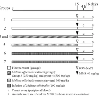
Animals and treatments
CF-1 male mice (weighing 40-50 g) were obtained from the breeding colonies of the University of South Santa Catarina, UNESC, Criciúma, SC, Brazil. The animals were kept in plastic cages in an experimental room under con-trolled conditions of temperature (22 ± 2 °C), humidity
(55±10%), 12-h light/dark cycles andad libitumaccess to food and water. They were randomized at the beginning of the experiment. The study design was approved by the Ani-mal Ethical Committee of UNESC (protocol number 043/2008), and the experiments undertaken in accordance with the ethical principles of the Brazilian College of Ani-mal Experimentation – COBEA.
(40 mg/kg b.w.) on the 15thday. Individuals in groups 3 and
4 received anM. officinalis ethanolic extract (10 mL/kg b.w. per day by gavage), prepared in two different doses, 250 mg/kg (group 3) and 500 mg/kg (group 4), for 2 weeks prior to treatment with MMS. Individuals in group 5 re-ceived only a Melissa officinalis ethanolic extract (500 mg/kg by gavage) prior to a 0.9% NaCl i.p. injection on the 15thday. Individuals in groups 6 and 7 received the
aqueous extract of Melissa officinalis (100 mg/kg), 10 mL/kg b.w. per day by gavage, for 2 weeks prior to a 0.9% NaCl or MMS i.p. injection on the 15thday. The mice
were killed by cervical dislocation, 24 h after treatment, for evaluation of micronucleated polychromatic erythrocytes (MNPCEs) in the bone-marrow. For comet assaying, sam-ples of peripheral blood were collected from mouse tail-tips by a slight incision, 4 h after MMS treatment.
Comet assay
Single-Cell Gel Electrophoresis, or the comet assay, is a highly sensitive method for assessing DNA damage formation and repair, both at clinically relevant and low doses. Alkaline-comet assaying was undertaken as de-scribed by Singhet al.(1988). Peripheral blood samples were collected from the tail of each animal in heparinized microtubes, 4 h after MMS treatment. Briefly, 5 mL of
whole blood was embedded in a layer consisting of 95mL
of 0.75% low-melting-point agarose gel on frosted slides, and then immersed in a lysis buffer (2.5M NaCl, 100 mM EDTA, and 10 mM Tris [pH 10.0-10.5] with freshly added 1% Triton X-100 and 10% dimethyl sulfoxide), for a mini-mum of 1 h and a maximini-mum of one week. Subsequently, the slides were incubated in a freshly prepared alkaline buffer (300 mM NaOH and 1 mM EDTA, pH > 13) for 20 min.
The nuclei were electrophoresed for 20 min at 25 V (0.90 V/cm) and 300 mA, whereupon the alkali was neu-tralized with 0.4 M Tris (pH 7.5). After neutralization, the slides were fixed (15% w/v trichloroacetic acid, 5% w/v zinc sulfate, 5% glycerol), washed in distilled water, and dried overnight. The gels were re-hydrated for 5 min in dis-tilled water, and then stained for 15 min (37 °C) with a solu-tion containing the following sequence: 34 mL of Solusolu-tion B (0.2% w/v ammonium nitrate, 0.2% w/v silver nitrate, 0.5% w/v tungstosilicic acid, 0.15% v/v formaldehyde, 5% w/v sodium carbonate) and 66 mL of Solution A (5% so-dium carbonate). The staining was stopped with 1% acetic acid, whereupon the gels were air-dried (Villela et al. 2006). The visual classification method of Collinset al. (1997) was applied to assess the extent of DNA damage. Cells were scored from 0 (undamaged) to 4 (maximally damaged), according to tail intensity (size and shape), this resulting in a single DNA damage score (damage index) for each sample and, consequently, for each group. Thus, a damage index (DI) of the group could range from 0 (com-pletely undamaged) 100 cells (x 0) to 400 (maximum dam-age) 100 cells (x 4). The percentage of damage frequency (DF) was calculated for each sample on the basis of the number of cells with a tail versus those without.
The micronucleus test
Micronucleus assaying was according to the U.S. En-vironmental Protection Agency Gene-Tox Program (Ma-vourninet al.1990). Slides were prepared from smears of bone-marrow from both femurs, suspended in foetal calf serum. These were air-dried, xed in methanol, stained in 10% Giemsa and then coded for blind analysis. One thou-sand polychromatic erythrocytes were analyzed per mouse. The proportion of PCE and NCE (normochromatic erythro-cytes) in 200 erythrocytes/animal was calculated, to detect possible cytotoxic effects. The slides were scored blindly, using a light microscope with a 100x immersion objective.
Statistical analysis
Variable normality was assessed using the Kolmo-gorov-Smirnov test. Micronucleus testing and comet assay-ing involved multiple pair-wise comparison between experimental groups and positive and negative controls, with the Student t-test at a significance level of 0.05. The statistical package applied was BioEstat 5.0. The percent-age of reduction in the frequency of MMS induced DNA damage was calculated according to Azevedoet al.(2003) and Waterset al.(1990).
Results
The phytochemical analysis of theM. officinalis ex-tract (Table 1) indicated the presence of fenolic com-pounds, tannins, flavonoids, alkaloids and coumarins, but not of other secondary metabolites, such as anthraquinones and saponins.
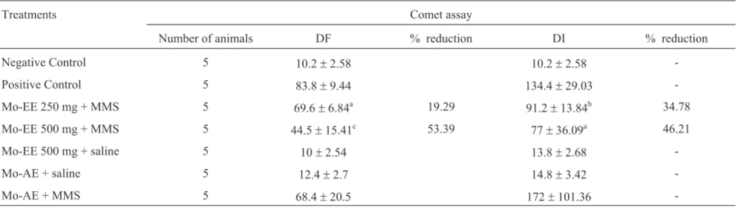
According to comet assay results, DNA damage in both parameters (damage index and frequency) in mouse peripheral blood, following ingestion of 500 mg/kg of an ethanol extract (Mo-EE) and 1.000 mg/kg of an aqueous extract (Mo-AE), both ofM. officinalis, during the 15 days of treatment, presented no statistically significant differ-ence (Table 2). Thus, it can be deduced that in both treat-ments, genotoxic effects on mouse peripheral blood cells
were null. When the antigenotoxicity of both was evalu-ated, a significant decrease in MMS-induced DNA damage (DI and DF) was observed in mice receiving 250 mg/kg and 500 mg/kg of Mo-EE. The reduction in the former case was 19.3% (Student t-test, p < 0.05) for DF, and 34.8% (Student t-test, p < 0.01) for DI, in relation to the positive control (MMS 40 mg/kg), and in the latter 53.4% (Student t-test, p < 0.001) and 46.2% (Student t-test, p < 0.05) for DF and DI, respectively. The analysis of DNA damage after pre-treatment with the Mo-AE extract and Mo-EE infusion, in-dicated no diminution in MMS chemically induced DNA damage.
According to MN testing of mouse bone-marrow cells the low frequencies of micronucleated cells presumes the meager effects of Mo-EE 500 mg/kg and Mo-AE pre-treatments (Table 3), thereby indicating the virtual absence of mutagenic or cytotoxic effects. In other words, no statis-tically significant difference in the frequency of MN PCE or the ratio of PCE to NCE, between the negative control and the groups that ingested extracts could be detected. When evaluating antimutagenicity in Mo-EE and Mo-AE, a significant decrease in the frequency of MMS-induced MNPCE was observed only in mice that had received 500 mg/kg of Mo-EE (p = 0.05 - Student t-test). Likewise, for Mo-AE, there was no statistically significant reduction in the frequency of MN PCE.
Discussion
Over recent years, it has been widely reported that diet is a highly important factor in terms of either cancer in-duction or prevention. As already known, certain dietary micronutrients, such as flavonoids, carotenoids and others, can play an important role in the modulation or prevention of cancer development (World Cancer Research Fund, 2008). The ingestion of plant extracts with medicinal prop-erties represents a plausible alternative in the treatment of various pathological states in economically unprivileged countries. However, in the absence of a scientific basis,
Table 1- Phytochemical screening.
Test Melissa officinalis
Phenolic compounds
W-Ext + KOH 3% +
W-Ext + FeCl31% +
Tannins
W-Ext + gelatine
-W-Ext + FeCl33% +
Flavonoids qualitative
W-Ext + MgO/HCl +
Flavonoids quantitative UV 425 nm
CH3OH-Ext 0.572
Alkaloids
W-Ext + Mayers Reagent
-W-Ext + Bertrand’s Reagent
-W-Ext + Dragendorff’s Reagent
-W-Ext + Bouchard’s Reagent +
Antraquinones
W-Ext + KOH 3%
-Coumarins
CH3CH2OH-Ext + KOH/UV 365 nm + Saponins
W-Ext + HCl (persistent foam)
-W-Ext – water extract./ CH3OH-Ext – methanol extract / CH3CH2OH-Ext – ethanol extract / (-)negative / (+)positive.
Table 2- Detection of lesions in DNA (mean±SD) using the comet assay (DF and DI) in peripheral blood cells of mice exposed to methyl
metha-nesulfonate (MMS, 40 mg/kg) and different doses ofMelissa officinalis.
Treatments Comet assay
Number of animals DF % reduction DI % reduction
Negative Control 5 10.2±2.58 10.2±2.58
-Positive Control 5 83.8±9.44 134.4±29.03
-Mo-EE 250 mg + MMS 5 69.6±6.84a 19.29 91.2±13.84b 34.78
Mo-EE 500 mg + MMS 5 44.5±15.41c 53.39 77±36.09a 46.21
Mo-EE 500 mg + saline 5 10±2.54 13.8±2.68
-Mo-AE + saline 5 12.4±2.7 14.8±3.42
-Mo-AE + MMS 5 68.4±20.5 172±101.36
-SD = standard deviation; DF = damage frequency; DI = damage index; Mo – EE = Eehanol extract ofMelissa officinalis;Mo – AE = aqueous extract of
such usage may give rise to serious adverse effects (Souza et al.2004). An evaluation of the role of plant extracts, in-fusions and the main relevant phytochemical antigenotoxic and antimutagenic compounds involved, is essential, through the evidential participation of the latter in the pre-vention of cancer and other disorders (Berhowet al.2000; Surh and Fergunson, 2003; Patelet al.2007). In this study, the efficacy of two different extracts of M. officinalis against a well-known genotoxic compound, were tested. The aim was to investigate new potential antigenotoxic agents from natural sources, for possible use in prevention.
M. officinalisis invested with compounds with anti-oxidant, antinociceptive and antitumoral properties (Sousa et al.2004; Canadanovic-Brunetet al.2008; Guginskiet al. 2009; Pereiraet al.2009). However, as to date no intrinsic antigenotoxic activity has been demonstrated., an investi-gation was undertaken, not only of this aspect, but also of possible antimutagenic and antigenotoxic effects against DNA damage induced by MMS, an alkylating agent.
Through comet assaying, the first and extremely im-portant observation was the absence of DNA strand breaks, as well as the induced formation of micronuclei in mice that had received two kinds ofM. officinalis extract. Further-more, comet assaying and MN testing indicated the absence of genotoxic or mutagenic properties in ethanolic and aque-ous extracts, respectively. Pereiraet al.(2005) and Furtado et al.(2008, 2010) are alone in evaluating the genotoxic ac-tivity of a certain compound present inM. officinalis, viz., rosmarinic acid (RA). The former showed, through comet assaying, that in brain and peripheral blood samples from rats treated with RA and sacrified 3 h later, there were no significant differences in any of the parameters used to as-sess DNA damage, in comparison with the vehicle-treated control group. The second group investigated the capacity of RA to prevent chemically induced chromosome break-age and DNA dambreak-age, using the same assays as in the pres-ent study, although with V79 cells (Furtadoet al.2010). It was shown that, although exerting no genotoxic effect, RA significantly reduced micronucleus frequency and the ex-tent of DNA damage induced by doxorubicin. With
micro-nucleus assaying, Furtadoet al.(2008) evaluated both the mutagenic and antimutagenic potential of RA on peripheral blood cells in Swiss mice. On the other hand, mutagenic as-saying revealed no increase in micronuclei frequency in an-imals treated with various concentrations of RA, when compared to negative controls, whereas in treatments with these concentrations combined with DXR, there was a sig-nificant reduction in micronucleus frequency compared to animals treated with DXR alone.
On investigating the protective effect ofM. officinalis in peripheral blood cells by comet assaying, and in bone-marrow PCE by MN testing, it was clearly indicated that an M. officinalisethanolic extract was more efficient in revert-ing the antigenotoxic damage (comet assayrevert-ing) than anti-mutagenic damage (MN testing) induced by MMS. However, as revealed by both test forms, the aqueous ex-tract obtained from the infusion ofM. officinalisin hot wa-ter (as a tea) was ineffective.
The already described antioxidant activity of M. officinalis extracts (Mimica-Dukic et al. 2004; Canada-novic-Brunetet al.2008; Pereiraet al.2009), has been at-tributed mainly to intrinsic phenolic compounds. These form one of the largest and most ubiquitous groups of plant metabolites, their antioxidant, anti inflammatory, antimuta-genic and anticarcinoantimuta-genic activities currently being of widespread interest (Atouiet al.2005; Frankeet al.2005; Geethaet al.2005). Pereiraet al.(2009) used encephalic tissue in the thiobarbituric acid reactive substances (TBARS) assay and determined the quantity of phenolic compounds in the plant extracts, in order to verify a possi-ble relationship with antioxidant activity. They found that an aqueous extract ofM. officinalis was the most active against TBARS production induced by all the tested agents, when compared with ethanolic and methanolic extracts therefrom. Interestingly, the inhibition of lipid peroxidation by the three increased with phenol content. However, in the 1’-1’ diphenyl-2’ picrylhydrazyl (DPPH) method, the three different extracts obtained from this plant (aqueous, ethanolic and methanolic) presented similar antioxidant po-tential. Furthermore, Mimica-Dukicet al.(2004), when
ex-Table 3- Effects ofMelissa officinalispre-treatments (gavage) on MNPCE frequencies (mean±SD) in the bone marrow of mice, following
intra-peritoneal injection with saline or methyl methanesulfonate (MMS) 40 mg/kg.
Treatment Number of analyzed cells PCE/NCE ratio MNPCEs p values
Negative Control 1000 230.6±4.51 3.6±0.89
-Positive Control 1000 231.8±7.73 5.2±1.79 0.05a
Mo-EE 250 mg + MMS 1000 233.8±9.42 3.6±1.67 0.09
Mo-EE 500 mg + MMS 1000 239.0±3.46 2.5±2.52 0.05a
Mo-EE 500 mg + saline 1000 236.8±7.89 3.2±2.28
-Mo-AE + saline 1000 227.0±5.39 3.2±1.79
-Mo-AE + MMS 1000 226.4±5.13 6.4±1.67 0.15
amining the oil of this plant, noted that, besides possessing high radical scavenging capacity, it exerted outstanding protective activity in lipid peroxidation processes. This was found to be positively correlated with a mainly mono-terpene ketone and aldehyde content. These results are in accordance with data reported by other authors, thereby demonstrating that the antioxidant activity of certain La-miaceae species, fruits and vegetables correlated positively with phenolic compound content (Ivanova et al. 2005; Katalinicet al.2006; Pérez-Pérezet al.2006).
In the present study, the ethanolic extract of M. officinalispresented both antigenotoxic and antimutagenic activities. As phenolic compounds have been shown to be endowed with these antioxidant properties, their involve-ment can be inferred.
Phenolic compounds are capable of protecting bio-logical systems in various ways (Edenharderet al.2002; Kellyet al.2003). They have a dual effect on phase I and phase II enzymes, repressing some (mainly in phase I) and stimulating others (mainly in phase II) (Szaeferet al.2004). Certain flavonoids, such as hesperetin, can selectively in-hibit human cytochrome P450, thereby reducing the ab-sorption or elimination of toxic compounds (Doostdaret al. 2000). Other phenolic compounds, such as limonoids, are inducers of the detoxifying enzyme gluthatione S-trans-ferase. The stimulation of detoxifying enzymes can facili-tate the elimination of toxic compounds, thereby signifi-cantly affecting the toxic potential of endogenous and exogenous chemicals (Kellyet al.2003). However, since MMS is a directly acting alkylant, thus not requiring meta-bolic activaton, the antimutagenic activity ofM. officinalis is not due to interaction with activating enzymes (Limaet al.2001). Frankeet al.(2005), with the aim of evaluating whether orange juice could reduce DNA damage induced by MMS in mice, showed that, under their experimental conditions, this really occurred. The authors showed that the components of orange juice are biologically effective, including in the role of targets for toxicants and in modulat-ing metabolization/detoxification routes.
It is likely that phenolic compounds can be methyl-ated by alkylating agents, instead of conjugation enzymes, thereby protecting reducing DNA from alkylation. MMS can methylate nucleophilic regions of DNA, as well as amino acid molecules, especially in nitrogen atoms. The methylation of phosphate groups accounts for a minor per-centage of total MMS methylation (< 1%). MMS geno-toxicity is mediated by base modifications, which weaken the N-glycosylic bond, thereby leading to depurination/de-pyrimidination of DNA strands and the appearance of al-kali-labile abasic sites (AP sites). The removal of AP sites by AP endonucleases cleaves the DNA adjacent to these sites, thereby generating DNA strand breaks (Horvathova et al.1998; Boiteux and Guillet, 2004; Frankeet al.2005). To a minor extent, MMS can also act as a weak oxidative stress inducer, as observed by Horvathova et al. (1998),
who tested the effect of a synthetic antioxidant (Stobadine, SBT) in this case.
Two assays were undertaken to elucidate the mecha-nism of antigenotoxic/antimutagenic action in M. officinalis. An antimutagenic effect, as revealed by MN testing, could mainly be due to either the diminished induc-tion of damage or increased repair. Through comet assay-ing, it was possible to measure the amount of DNA damage after treatment with a mutagen, thereby indicating whether an antigenotoxic effect is due to the reduced formation of primary DNA lesions or not. Herein, it was possible to show their obvious induction by MMS, as well as to dem-onstrate that these effects can be reduced by pre-treatment with an ethanolic extract. On the other hand, MN testing re-vealed a significant reduction only at the highest dose of the extract. Consequently, anM. officinalisextract was effec-tive in preventing MMS-induced DNA lesions. In the pre-treatment, phenolic compounds maybe competed as target sites for alkylation, whereas the absence of protection by theM. officinalis, aqueous extract against the frequency of DNA lesions and mutations induced by MMS, could be due to possible interactions among the compounds contained within the extract itself.
Taken together, it can be inferred that an ethanolic ex-tract ofM. officinalisis endowed with antigenotoxic/anti-mutagenic properties, and its use in pre-treatment dimin-ishes the induction of DNA damage by an alkylant agent. Potential differences in antigenotoxic/antimutagenic activ-ity by the various extracts still need to be clarified.
Acknowledgments
This work was supported by UNESC (Universidade do Extremo Sul Catarinense) and CNPq (Conselho Na-cional de Desenvolvimento Científico eTecnológico).
References
Atoui AK, Mansouri A, Boskou G and Kefalas P (2005) Tea and herbal infusions: Their antioxidant activity and phenolic profile. Food Chem 89:27-36.
Azevedo L, Gomes JC, Stringheta PC, Gontijo AMMC, Padovani CR, Ribeiro LR and Salvadori DMF (2003) Black bean (Phaseolus vulgarisL.) as a protective agent against DNA damage in mice. Food Chem Toxicol 41:1671-1676. Azevedo L, Lima PLA, Gomes JC, Stringheta PC, Ribeiro DA
and Salvadori DMF (2007) Differential response related to genotoxicity between eggplant (Solanum melanogena) skin aqueous extract and its main purified anthocyanin (del-phinidin)in vivo. Food Chem Toxicol 45:852-858. Berhow M, Wagner E, Vaughn S and Plewa M (2000)
Character-ization and antimutagenic activity of soybean saponins. Mu-tat Res 448:11-22.
Bresolin S and Vargas VMF (1993) Mutagenic potencies of me-dicinal plants screened in the Ames test. Phytother Res 7:260-262.
Canadanovic-Brunet J, Cetkovic G, Djilas S, Tumbas V, Bogda-novic G, Mandic A, Markov S, Cvetkovic D and Cana-danovic V (2008) Radical scavenging, antibacterial, and antiproliferative activities of Melissa officinalis (L.) ex-tracts. J Med Food 11:133-143.
Carnat AP, Carnat A, Fraisse D and Lamaison JL (1998) The aro-matic and polyphenolic composition of lemon balm (Me-lissa officinalisL. subsp.officinalis) tea. Pharm Acta Helv 72:301-305.
Collins A, Dusinska M, Franklin M, Somorosyska M, Petroyska H, Duthie S, Fillion L, Panyotidis M, Raslová K and Vau-ghan N (1997) Comet assay in human biomonitoring stud-ies: Reliability, validation, and applications. Environ Mol Mutagen 30:139-146.
Czeczot H and Kusztelak J (1993) A study of the genotoxic poten-tial of flavonoids using short-term bacterial assays. Acta Biochim Pol 40:549-554.
Dastmalchi K, Dorman HJD, Oinonen PP, Darwis Y, Laakso I and Hiltunen R (2008) Chemical composition andin vitro antio-xidative activity of a lemon balm (Melissa officinalisL.) ex-tract. LWT – Food Sci Technol 41:391-400.
Doostdar H, Burke D and Mayer RT (2000) Bioflavonoids: Selec-tive substrates and inhibitors for cytochrome P450 CYP1A and CYP1B1. Toxicology 144:31-38.
Edenharder R, Sager JW, Glatt H, Muckel E and Platt KL (2002) Protection by beverages, fruits, vegetables, herbs, and flavo-noids against genotoxicity of 2 acetylaminofluoreno and 2-amino-1-methyl-6-phenylimidazol [4,5-6] pyridine (PhIP) in metabolically competent V79 cells. Mutat Res 521:57-72.
Farmacopéia Brasileira (2001) Farmacopéia Brasileira. 4th edi-tion. Atheneu, São Paulo, 294 pp.
Fernandes JBF and Vargas VMF (2003) Mutagenic and anti-mutagenic potential of the medicinal plantsM. laevigataand C. xanthocarpa. Phytother Res 17:269-273.
Franke SIR, Pra D, Erdtmann B, Henriques JAP and da Silva J (2005) Influence of orange juice over the genotoxicity in-duced by alkylating agents: Anin vivoanalysis. Mutagene-sis 20:279-283.
Furtado MA, Almeida LCF, Furtado RA, Cunha WR and Tavares DC (2008) Antimutagenicity of rosmarinic acid in Swiss mice evaluated by the micronucleus assay. Mutat Res 657:150-154.
Furtado RA, Araújo FRR, Resende FA, Cunha WR and Tavares DC (2010) Protective effect of rosmarinic acido on V79 cells evaluated by the micronucleus and comet assays. J Appl Toxicol 30:254-259.
Geetha T, Malhotra V, Chopra K and Kaur IP (2005) Anti-mutagenic and antioxidant/prooxidant activity of Quercetin. Ind J Exp Biol 43:61-67.
Gomes-Carneiro MR, Dias DMM, De Oliveira ACAX and Paum-gartten FJR (2005) Evaluation of mutagenic and antimuta-genic activities ofa-bisabolol in the Salmonella/microsome assay. Mutat Res 585:105-112.
Guginski G, Luiz AP, Silva MS, Massaro M, Martins DF, Chaves J, Mattos RW, Silveira D, Ferreira VMM, Calixto JB,et al. (2009) Mechanisms involved in the antinociception caused by ethanolic extract obtained from the leaves of Melissa
officinalis (lemon balm) in mice. Pharmacol Biochem Behav 93:10-16.
Harborn JB (1998) Phenolic compounds. In: Harborne JB (ed) Phytochemical Methods: A Guide to Modern Techniques of Plant Analysis. Chapman & Hall, London, pp 40-106. Hernández-Ceruelos A, Madrigal-Bujaidadar E and De La Cruz C
(2002) Inhibitory effect of chamomile essential oil on the sister chromatid exchanges by daunorubicin and methyl methanesulfonate in mouse bone marrow. Toxicol Lett 135:103-110.
Herodez SS, Hadolin M, Skerget M and Zeljko K (2003) Solvent extraction study of antioxidants from balm (Melissa officinalisL.) leaves. Food Chem 80:275-282.
Horvathova E, Slamenova D, Hlincikova L, Mandal TK, Ga-belova A and Collins AR (1998) The nature and origin of DNA single-strand breaks determined with the comet assay. Mutat Res 409:163-171.
Ivanova D, Gerova D, Chervenkov T and Yankova T (2005) Polyphenols and antioxidant capacity of Bulgarian medici-nal plants. J Ethnopharmacol 96:145-150.
Katalinic V, Milos M, Kulisic T and Jukic M (2006) Screening of 70 medicinal plant extracts for antioxidant capacity and total phenols. Food Chem 94:550-557.
Kelly C, Jewell C and O’Brien NM (2003) The effect of dietary supplementation with the citrus limonoids, limonin and no-milin on xenobiotic-metabolizing enzymes in the liver and small intestine of the rat. Nutr Res 23:681-690.
Lima PLA, Delmanto RD, Sugui MM, Eira AF, Salvadori DMF, Speit G and Ribeiro LR (2001) Letinula edodes (Berk.) Pegler (Shiitake) modulates genotoxic and mutagenic ef-fects induced by alkylating agents in vivo. Mutat Res 496:23-32.
Mavournin KH, Blakey DH, Cimino MC, Salamone MF and Heddle J (1990) Thein vivomicronucleus assay in mamma-lian bone marrow and peripheral blood. A report of the U.S. Environmental Protection Agency Gene-Tox Program. Mutat Res 232:29-80.
Mimica-Dukic N, Bozin B, Socovik M and Simin N (2004) Antimicrobial and antioxidant activities of Melissa officinalisL. (Lamiacea) essential oil. J Agric Food Chem 52:2485-2489.
Müzell DP (2006) Propriedades biológicas de extratos deMelissa officinalis L. (lamiaceae) em ratos wistar. Dissertação de Mestrado em Biologia Celular e Molecular, Faculdade de Biociências da Pontifícia Universidade Católica do Rio Grande do Sul, Porto Alegre, 70 pp.
Patel D, Shukla S and Gupta S (2007) Apigenin and cancer chemoprevention: Progress, potential and promise. Int J Oncol 30:233-245.
Pereira P, Tysca D, Oliveira P, Da Silva-Brum LF, Picada JN and Ardenghi P (2005) Neurobehavioral and genotoxic aspects of rosmarinic acid. Pharmacol Res 52:199-203.
Pereira RP, Fachineto R, Prestes AS, Puntel RL, Boschetti TK, Athayde ML, Büerguer ME, Morel AF, Morsch VM and Rocha JBT (2009) Antioxidant effects of different extracts from Melissa officinalis, Matricaria recutita and Cymbopogon citratus. Neurochem Res 34:973-983. Pérez-Pérez EM, Rodrígez-Malaver AJ, Padilla N,
Sá-Ferreira ICF and Vargas VMF (1999) Mutagenicity of medici-nal plant extracts in Salmonella/microsome assay. Phytother Res 13:397-400.
Salah SM and Jäger AK (2005) Screening of traditionally used Lebanese herbs for neurological activities. J Ethnophar-macol 97:145-149.
Singh NP, McCoy MT, Tice RR and Schneider EL (1988) A sim-ple technique for quantification of low levels of DNA dam-age in individual cells. Exp Cell Res 175:184-191. Souza AC, Alviano DS, Alves PB, Alviano CS and Gattass CR
(2004)Melissa officinalisL. essential oil: Antitumoral and antioxidant activities. J Pharm Pharmacol 56:677-681. Surh Y and Fergunson LR (2003) Dietary and medicinal
anti-mutagens and anticarcinogens: Molecular mechanisms and chemopreventive potential – Highlights of a symposium. Mutat Res 485:1-8.
Szaefer H, Cichocki M, Brauze D and Baer-Dubowska W (2004) Alteration in phase I and II enzyme activities and polycyclic aromatic hydrocarbons-DNA adduct formation by plant phenolics in mouse epidermis. Nutr Cancer 48:70-77.
Tovart RT (2009) Clinical approach to clinical herbal toxicity. Sem Diagn Pathol 26:28-37.
Villela IV, Oliveira IMde, Silva J da and Henriques JAP (2006) DNA damage and repair in haemolymph cells of golden mussel (Limnoperna fortunei) exposed to environmental contaminants. Mutat Res 605:78-86.
Wagner H and Bladt S (2009) Plant Drug Analysis: A Thin Layer Chromatography Atlas. 2nd edition. Springer, Heidelberg, 384 pp.
Waters MD, Brady AL, Stack HF and Brockman HE (1990) Antimutagenicity profiles for some model compounds. Mu-tat Res 238:57-85.
World Cancer Research Fund (2008) Food, nutrition, and the pre-vention of cancer: A global perspective. Nutrition 24:393-398.
Associate Editor: Catarina S. Takahashi