online | memorias.ioc.fiocruz.br
Molecular characterisation of
Sporothrix schenckii
isolates from
humans and cats involved in the sporotrichosis epidemic
in Rio de Janeiro, Brazil
Rosani Santos Reis/+, Rodrigo Almeida-Paes, Mauro de Medeiros Muniz,
Patrícia Morais e Silva Tavares, Paulo Cezar Fialho Monteiro, Tânia Maria Pacheco Schubach, Maria Clara Gutierrez-Galhardo, Rosely Maria Zancopé-Oliveira
Laboratório de Micologia, Instituto de Pesquisa Clínica Evandro Chagas-Fiocruz, Av. Brasil 4365, 21045-900 Rio de Janeiro, RJ, Brasil
An epidemic of sporotrichosis, a subcutaneous mycosis caused by the fungus Sporothrix schenckii, is ongoing in Rio de Janeiro, Brazil, in which cases of human infection are related to exposure to cats. In an attempt to demon-strate the zoonotic character of this epidemic using molecular methodology, we characterised by DNA-based typing methods 19 human and 25 animal S. schenckii isolates from the epidemic, as well as two control strains. To analyse the isolates, the random amplified polymorphic DNA (RAPD) technique was performed using three different prim-ers, together with DNA fingerprinting using the minisatellite derived from the wild-type phage M13 core-sequence. The analyses generated amplicons with considerable polymorphism. Although isolates exhibited high levels of ge-netic relatedness, they could be clustered into 5-10 genotypes. The RAPD profiles of epidemic S. schenckii isolates could be distinguished from that of the United States isolate, displaying 20% similarity to each primer and 60% when amplified with the M13 primer. DNA fingerprinting of S. schenckii isolated from the nails (42.8%) and the oral cavities (66%) of cats were identical to related human samples, suggesting that there is a common infection source for animals and humans in this epidemic. It is clear that cats act as a vehicle for dissemination of S. schenckii.
Key words: Sporotrichosis - zoonotic transmission - RAPD - DNA fingerprinting
+ Corresponding author:rosani.reis@ipec.fiocruz.br Received: 10 June 2008
Accepted: 30 June 2009
Sporotrichosis is a subcutaneous mycosis with a global distribution and an especial prevalence in Latin America and Africa (Rippon 1988, Diaz 1989, Kovarik et al. 2008, Schubach et al. 2008). Sporotrichosis is caused by the dimorphic fungus Sporothrix schenckii, which is associated with plant material and soil. This disease most often affects people with occupations related to soil handling (Diaz 1989) and it results from traumatic im-plantation of the agent into the skin (Rippon 1988).
The sporotrichosis outbreaks reported in the literature have been associated with contaminated plant material. The largest reported outbreak of sporotrichosis occurred in South Africa from 1941-1944, where around 3,000 miners were infected by wood props in the gold mines (Vismer & Eicker 1994). Subsequently, an outbreak in the United States occurred in 1988 in which 84 individuals from 15 states were affected (Dixon et al. 1991). Hajjeh et al. (1997) reported an outbreak of sporotrichosis among tree nursery workers handling sphagnum moss. An out-break in Australia involved individuals in contact with hay prior to infection (O’Reilly & Altman 2006).
The random amplified polymorphic DNA (RAPD) method has been a useful tool in epidemiological inves-tigations, especially for fungi, since prior knowledge
of the genome is not required. DNA techniques were used to examine clinical and environmental isolates of
Sporothrix spp. from the 1988 sporotrichosis epidemic in the United States (Cooper et al. 1992). Recent mo-lecular studies have assessed the genetic diversity of
S. schenckii, demonstrating that different clusters of S. schenckii strains are related to different geographical origins (Marimon et al. 2006, O’Reilly & Altman 2006, Gutierrez-Galhardo et al. 2008). Moreover, Marimon et al. (2007) proposed the existence of three new Sporothrix
species through physiological and molecular studies. Since 1998, there has been a sporotrichosis epidemic affecting humans, cats and in smaller proportions dogs in the metropolitan region of Rio de Janeiro, Brazil, mainly in areas with underprivileged socio-economic conditions and precarious health services. The most frequently af-fected individuals are homemakers taking care of cats with sporotrichosis (Barros et al. 2004). Epidemic iso-lates of S. schenckii from patients and cats have not been compared by molecular typing methods. In this study, the genetic polymorphisms of S. schenckii isolates from humans and associated cats from the sporotrichosis epi-demic in state of Rio de Janeiro (RJ) were analysed us-ing RAPD assays and PCR fus-ingerprintus-ing in an attempt to demonstrate the zoonotic character of the epidemic.
PATienTS, MATeRiALS AnD MeTHODS
with them. Eighteen humans and 16 cats were included. All humans related a history of contact with a sick cat before the onset of symptoms. Ten of the patients (55%) also reported trauma by scratching (6 cases; 33%) or bit-ing (4 cases; 22%) by sick cats on the sites of infection.
Fungi origin - We studied 44 S. schenckii strains, 19 from humans and 25 from cats (Table I). Two additional isolates from humans, one (17845) isolated in RJ, 1996 (before the occurrence of outbreaks), and one (24605) from the Dermatology Department of Columbia Univer-sity, New York, were included in the analyses.
Fungal collection and identification - The isolates were obtained from clinicalmaterials, as pus or biopsies of lesions from humans and felines and nails and swabs of oral cavities from felines. These samples were grown in Sabouraud dextrose agar and Mycobiotic slants (Bec-ton, Dickinson and Company). Fungal identification was performed by conventional methods, which included morphology on PDA slants and microscopic observa-tions of fungal fragments obtained after 15 days of in-cubation at RT. All isolates were tested for conversion to the yeast phase on BHI (brain heart infusion) agar at 37ºC for seven days.
DNA isolation - A single colony of yeast-phase S. schenckii was used to inoculate BHI broth, which was then grown at 37°C on a gyratory shaker at 120 rpm for seven days to achieve sufficient cell mass for DNA ex-traction. Yeast cells were harvested and washed three times in sterile water. Genomic DNA was extracted and purified using the phenol-chloroform-isoamyl al-cohol method (Woods et al. 1993). DNA quantification was done with a Gene Quant pro RNA/DNA Calculator (Amersham Pharmacia Biotech, Cambridge, UK).
PCR-RAPD - For RAPD analysis, each strain was tested with three primers using Kit Ready-To-Go RAPD Analysis Beads (GE Healthcare). DNA amplification
re-actions were carried out in a 25 μL volume containing
20 ng of S. schenckii DNA and 25 pMol/L of primer. The other components were present on the beads. For amplification with primers 1 (5’-GGTGCGGGAA-3’), 4 (5’-AAGAGCCCGT-3’) and 6 (5’-CCCGTCAGCA-3’), the thermal cycler (Peltier PTC200-MJ Research) was programmed for one denaturation cycle at 95°C for 5 min, followed by 45 cycles of denaturation at 95°C for 1 min, annealing at 36°C for 1 min and extension at 72°C for 2 min, followed by a final extension period of 2 min at 72°C. The randomly amplified DNA fragments were separated by electrophoresis on a 1% agarose gel in TBE buffer (0.89 M Tris, 0.89 M boric acid, 0.02 M disodium
EDTA, pH 8.4) and stained with 0.5 μg/mL ethidium
bromide (Roche Molecular Biochemicals). All visual-ised bands were counted and data were scored for the presence or absence of amplification products. Repro-ducibility was confirmed when identical profiles were observed in PCR assays repeated at least three times un-der the same conditions.
PCR fingerprinting -The minisatellite-specific core sequence of the wild-type phage M13
(5-’GAGGGTG-GCGGTTCT-3’) was used as a single primer in the PCR. The amplification reactions were performed as described by Gutierrez-Galhardo et al. (2008).
Computer-assisted data analysis -Similarity coeffi-cients and Dice indices were determined for each isolate for the PCR fingerprinting and RAPD analyses using the NTSYSpc program (version 2.02h, Applied Biostatistic Inc). For clustering, the unweighted pair-group method with arithmetic means (UPGMA) was used.
ReSULTS
DNA conditions and PCR standardisation - Extracted DNA analysed by agarose gel electrophoresis presented a single band without RNA or protein contamination. Six primers (Ready to Go Kit - RAPD analysis beads - GE Healthcare - primers 1, 2, 3, 4, 5 and 6) were initially analysed to determine their discriminatory power. The primers 1, 4 and 6 were selected as they presented dis-criminatory indices of 0.79, 0.87 and 0.64, respectively (data not shown).
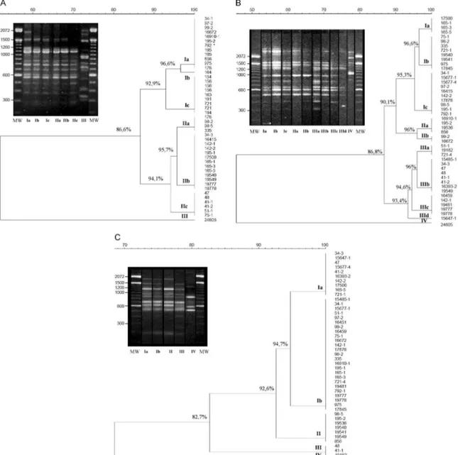
RAPD analysis -Representative RAPD profiles ob-tained with all primers from the 46 S. schenckii isolates indicated considerable polymorphism. Amplification re-actions generated profiles composed of 8-15 bands rang-ing from 314-2357 bp in size. The genetic relationships obtained using the UPGMA method are represented as dendrograms (Fig. 1). The phylogenetic analysis identi-fied 2-3 clonal groups, depending on the primer, which are highly related and contain contain 5-10 genotypes each. The combined data obtained using the three prim-ers (primprim-ers 1, 4 and 6) clustered the 46 S. schenckii
isolates into three clonal groups exhibiting 90% related-ness, except for the US strain. RAPD patterns for the US isolate were completely different from the patterns observed among the other S. schenckii strains analysed by this methodology. The percentage of similarity was about 20% (data not shown).
be-TABLE I
Sporothrix schenckii strains isolated in the epidemic of sporotrichosis in state of Rio de Janeiro, Brazil, used in the study
Case number Strain number Clinical specimen Origin Month/year
1 15485-1 pus human 10/1998
34-1 biopsy cat 10/1998
34-3 nail cat 10/1998
2 15647-1 pus human 12/1998
47 nail cat 03/1999
48 biopsy cat 03/1999
3 15677-1 pus human 01/1999
15677-4 pus human 01/1999
41-1 nail cat a 02/1999
41-2 biopsy cat a 02/1999
51-1 biopsy cat b 05/1999
4 16393-2 pus human 07/1999
97-2 pus cat 08/1999
5 16415 biopsy human 07/1999
99-2 biopsy cat 08/1999
6 16459 biopsy human 07/1999
75-1 biopsy cat 08/1999
7 16672 pus human 09/1999
142-1 nail cat 09/1999
142-2 biopsy cat 09/1999
8 17878 pus human 08/1999
98-2 biopsy cat a 08/1999
98-5 nail cat a 08/1999
335 oral swab cat ba 05/2000
9 16910-1 pus human 11/1999
195-1 biopsy cat 11/1999
195-2 nail cat 11/1999
10 17500 biopsy human 04/2000
165-1 oral swab cat 10/1999
165-3 biopsy cat 10/1999
165-5 nail cat 10/1999
11 19182 pus human 05/2001
721-1 oral swab cat 05/2001
721-4 pus cat 05/2001
12 19481 biopsy human 07/2001
792-1 pus cat 06/2001
13 19536 pus human 07/2001
19540 biopsy human 07/2001
19541 biopsy human 07/2001
19549 pus human 07/2001
856 blood culture cat 07/2001
14 19777 pus human 09/2001
19778 pus human 09/2001
975 pus cat 09/2001
Ref.b 17845 pus human 1996
Ref.b 24605 unknown human unknown
tween the US strain and the strains from RJ was about 64%. It is interesting to note that all samples from cases 6, 8, 9, 10, 11 and 13 (Table I) presented the same DNA fingerprinting when analysed with primer M13.
Correlation between cat and human strains - Total identities between S. schenckii isolates from lesions, nails and oral cavities of cats are listed in Table II with their respective primers. It is notable that when the primers 1 and 6 for the RAPD technique and M13 for DNA finger-printing were used, the same percentage (42.8%) of simi-larity was shown among the S. schenckii isolated from the nails of the cats and the isolates from their owners. With RAPD primer 1, the three strains obtained from the oral cavities of cats were identical to the related patients’ isolates. However, with primers 4 and 6, just one of them
(165-1 and 335, respectively) presented an identical geno-type to the patient’s isolate. Two of these isolates (165-1 and 721-1) were identical based on PCR fingerprinting.
DiSCUSSiOn
nique and DNA fingerprinting using the M13 primer were chosen for this study since they are easy and rapid genotyping methods and they have become quite popu-lar for fungal analysis.
Recently, we demonstrated that 59 human epidemic strains were genetically related through molecular typing with DNA fingerprinting and ITS sequence analysis. These results suggest that the strains isolated from the sporotricho-sis epidemic originated from a common source (Gutierrez-Galhardo et al. 2008). Our data corroborate this hypoth-esis and confirm that cats play a major role in the spread of sporotrichosis in RJ since the S. schenckii isolates from cats are closely related to those isolated from humans.
The DNA profiles of S. schenckii exhibited consid-erable similarity and did not cluster together according to their geographical area. No variation in the genetic patterns of S. schenckii isolates obtained between 1998-2001 in this RJ epidemic was observed. Furthermore, the genetic profile of the reference strain (17845) collect-ed in 1996, before the beginning of the epidemic, was similar to the others. The high similarity among these isolates suggests the possibility that only one virulent genetic population is present in the epidemic environ-ments and that one or more populations of S. schenckii
are maintained in nature for long periods of time.
Our group previously reported the isolation of S. schenckii from the nails of cats with sporotrichosis that infected their owners by scratching. We suggested that the vehicle of transmission could be the cat’s nails (Schu-bach et al. 2001). In this study, we verified this hypothesis by molecular methods, showing between 42.8-57.1% total identity between samples from cat’s nails and humans.
Three S. schenckii isolates from the oral cavity of cats were included in this study. One of them (335 strain) was isolated from an asymptomatic cat that had contact with other cats with sporothrichosis in the same house. In this case (case 8), strain 98-2 was isolated from a symp-tomatic cat of the same household that died nine months before the isolation of strain 335. Both strains (98-2 and 335) presented the same genotypic profile. Strain 335 presented 95.3-100% similarity with the patient strain (strain 17878), suggesting that just one genotype is in-volved in this case and indicating that S. schenckii can persist inside the domiciliary area for prolonged periods of time. Two other isolates from oral cavities were ob-tained from cats with restricted cutaneous lesions. In case 10 we observed 94.7-100% similarity between the strain from the oral cavity of the cat (165-1) and the lesion of the human patient (17500), indicating this pa-tient acquired S. schenckii from its cat. Our data are the first to demonstrate this zoonotic potential through molecular analysis.
In 23-61.5% of our cases, isolates from cats’ skin le-sions and from the associated humans were identical, de-pending on the primer used in the PCR reactions. Reed et al. (1993) have reported one case of zoonotic trans-mission of sporotrichosis as determined by genotypic analysis of the isolates from both the human and the cat involved. The RFLP profiles of the strains from this single outbreak were identical. Our data corroborate the above considerations about the high zoonotic potential of domestic cats in sporotrichosis.
Some patients (45%) included in this work did not re-port any kind of trauma, but they developed sporotricho-sis when they were taking care of their sick animals. The strains isolated from humans and the sick cats showed high genetic relatedness. S. schenckii strains from cases 6 and 10 were identical based on DNA fingerprinting, emphasising again the zoonotic potential of the disease. The infected cats present a high S. schenckii burden in their lesions (Reed et al.1993, Schubach et al. 2004) and can thus act as a source of infection to the human if there is a contact between a pre-existent wound in the patient and the exudates from cat’s lesions.
At present, no teleomorph is known for S. schenckii
(de Beer et al. 2003);however, we found, as with previ-ous studies (Mesa-Arango et al. 2002, O’Reilly & Alt-man 2006, Gutierrez-Galhardo et al. 2008), some genet-ic variability in S. schenckii, especially when analysing strains from different countries. The band pattern of the US strain was totally different from those of Brazil, which is in accordance with results from other studies of
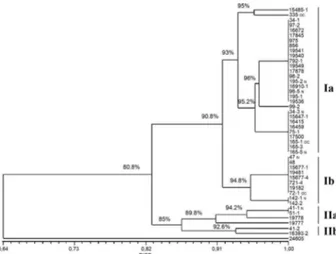
S. schenckii, as well as with other pathogenic fungi, such as Paracoccidioides brasiliensis and Histoplasma cap-sulatum (Muniz et al. 2001, Mesa-Arango et al. 2002, Hahn et al. 2003). However, the genetic variability or Fig. 2: DNA fingerprinting of two controls and 44 epidemic
Sporo-thrix schenckii strains using minisatellite derived from the wild-type
phage M13 core-sequence.
TABLE II
Percentage of identical genotypic profile between Sporothrix schenckii isolates from cats and those from their owners
Primer 1 %
Primer 4 %
Primer 6 %
M13 %
Skin lesion (n = 13) 23 15.3 61.5 38.5
Nail (n = 7) 42.8 57.1 42.8 42.8
polymorphic patterns found in different geographical areas can also be due to different ecological processes or genetic factors that promote these polymorphisms (Me-sa-Arango et al. 2002). Moreover, we cannot discard the possibility that new Sporothrix species are associated with the RJ epidemics, as suggested by Marimon et al. (2007). In addition, the US isolate included in this study most probably represents the real S. schenckii in the new species complex. Studies are in progress in our labora-tory to corroborate these previously reported data.
In conclusion, the RAPD technique is an efficient tool for the investigation of the relationship between S. schenckii isolated from human subjects and felines and can be used to distinguish among 5-10 strain profiles in-cluded in this study. A similar result was obtained when the DNA samples were amplified using primer M13. Our results show a strong correlation between the isolates from humans and their respective cats in RJ and support the idea that contact with cats infected by S. schenckii
constitutes a route of sporotrichosis infection.
ACKnOWLeDGeMenTS
To Andreia Pussenti Derossi and Celuta Sales Alviano, for providing the control strains used in this study.
ReFeRenCeS
Barros MBL, Schubach AO, Valle ACF, Gutierrez-Galhardo MC, Conceição-Silva F, Schubach TMP, Reis RS, Wanke B, Marzochi KBF, Conceição MJ 2004. Cat-transmitted sporotrichosis epi-demic in Rio de Janeiro, Brazil: description of a series of cases.
Clin Infect Dis 38: 529-535.
Cooper CR, Breslin BJ, Dixon DM, Salkin IF 1992. DNA typing of isolates associated with the 1988 sporotrichosis epidemic. J Clin
Microbiol 30: 1631-1635.
de Beer ZW, Harrington TC, Vismer HF, Wingfield BD, Wingfield MJ 2003. Phylogeny of the Ophiostoma stenoceras - Sporothrix
schenckii complex. Mycologia 95: 434-441.
Diaz IAC 1989. Epidemiology of sporotrichosis in Latin America.
Mycopathologia 103: 113-116.
Dixon DM, Salkin IF, Hurd NJ, Haines JH, Kemna ME, Coles FB 1991. Isolation and characterization of Sporothrix schenckii from clinical and environmental sources associated with the largest US epidemic of sporotrichosis. J Clin Microbiol 29: 1106-1113.
Gutierrez-Galhardo MC, Zancopé-Oliveira RM, Valle AC, Almeida-Paes R, Tavares PMS, Monzon A, Mellado E, Rodriguez-Tudela JL, Cuenca-Estrella M 2008. Molecular epidemiology and an-tifungal susceptibility patterns of Sporothrix schenckii isolates from a cat-transmitted epidemic of sporotrichosis in Rio de Ja-neiro, Brazil. Med Mycol46: 141-151.
Hahn RC, Macedo AM, Fontes CJF, Batista RD, Santos NL, Ham-dan JS 2003. Random amplified polymorphic DNA as a valuable
tool for epidemiological studies of Paracoccidioidesbrasiliensis.
J Clin Microbiol 41: 2849-2854.
Hajjeh R, McDonnell S, Reef S, Licitra C, Hankins M, Toth B, Padhye A, Kaufman L, Passarell L, Cooper C, Hutwagner L, Hopkins R, McNiel M 1997. Outbreak of sporotrichosis among tree nursery workers. J Infect Dis176: 499-504.
Kovarik CL, Neyra E, Bustamante B 2008. Evaluation of cats as the source of endemic sporotrichosis in Peru. Med Mycol 46: 53-56.
Marimon R, Cano J, Gene J, Sutton DA, Kawasaki M, Guarro J 2007.
Sporothrix brasiliensis, S. globosa and S. mexicana, three new
Sporothrix species of clinical interest. J Clin Microbiol 45:
3198-3206.
Marimon R, Gene J, Cano J, Trilles L, Dos Santos Lazera M, Guarro J 2006. Molecular phylogeny of Sporothrix schenckii. J Clin
Mi-crobiol 44: 3251-3256.
Mesa-Arango AC, Reyes-Montes MR, Pérez-Mejía A, Navarro-Bar-ranco H, Souza V, Zúñiga G, Toriello C 2002. Phenotyping and genotyping of Sporothrix schenckii isolates according to geo-graphic origin and clinical form of sporotrichosis. J Clin Micro-biol40: 3004-3011.
Muniz MM, Pizzini CV, Peralta JM, Reiss E, Zancopé-Oliveira RM 2001. Genetic diversity of Histoplasma capsulatum strains iso-lated from soil, animals and clinical specimens in Rio de Janeiro State, Brazil, by PCR-based random amplified polymorphic DNA assay. J Clin Microbiol 39: 4487-4497.
O’Reilly LC, Altman SA 2006. Macrorestriction analysis of clinical and environmental isolates of Sporothrix schenckii. J Clin
Micro-biol 44: 2547-2552.
Reed KD, Moore FM, Geiger GE 1993. Zoonotic transmission of sporotrichosis: case report and review. Clin Infect Dis16: 384-387.
Rippon JW 1988. Sporotrichosis. In JW Rippon, Medical mycology, WB Saunders, Philadelphia, p. 325-352.
Schubach A, Barros MBL, Wanke B 2008. Epidemic sporotrichosis.
Curr Opin Infect Dis21: 129-133.
Schubach TMP, Schubach AO, Okamoto T, Barros MBL, Figueiredo FB, Cuzzi T, Monteiro PCF, Reis RS, Perez MA, Wanke B 2004. Evaluation of an epidemic of sporotrichosis in cats: 347 cases (1988-2001). J Am Vet Med Assoc 224: 1623-1629.
Schubach TMP, Valle ACF, Gutierrez-Galhardo MC, Monteiro PCF, Reis RS, Zancopé-Oliveira RM, Marzochi KBF, Schubach A 2001.Isolation of Sporothrix schenckii from the nails of domestic cats (Felis catus). Med Mycol 39: 147-149.
Vismer HF, Eicker A 1994. Growth of human pathogenic isolates of
Sporothrix schenckii on indigenous and exotic wood species in
South Africa. Mycol Res 98: 121-124.