706
Rev Soc Bras Med Trop 50(5):706-708, September-October, 2017 doi: 10.1590/0037-8682-0042-2017
Case Report
Corresponding author: Dr. Eloy Ordaya Espinoza.
e-mail: [email protected]
Received 10 February 2017
Accepted 5 May 2017
Infective endocarditis due to
Bartonella bacilliformis
associated with systemic vasculitis: a case report
Joshua Peñaiel-Sam
[1],
Samuel Alarcón-Guevara
[1],
Sergio Chang-Cabanillas
[1],
Wilkerson Perez-Medina
[2],
Fernando Mendo-Urbina
[1],[3]and Eloy Ordaya-Espinoza
[1],[4][1]. Escuela de Medicina, Universidad Peruana de Ciencias Aplicadas, Lima, Perú. [2]. Servicio de Reumatología,
Hospital Nacional Edgardo Rebagliati Martins, Lima, Perú. [3]. Servicio de Infectología, Hospital Nacional Edgardo Rebagliati Martins, Lima, Perú. [4]. Department of Medicine, University of Minnesota, Minneapolis, MN, USA.
Abstract
Infective endocarditis due to Bartonella bacilliformis is rare. A 64-year-old woman, without previous heart disease, presented with 6 weeks of fever, myalgias, and arthralgias. A systolic murmur was heard on the tricuspid area upon examination, and an echocardiogram showed endocardial lesions in the right atrium. Bartonella bacilliformis was isolated in blood cultures, deining the diagnosis of infective endocarditis using Duke’s criteria. Subsequently, the patient developed clinical and laboratory features compatible with antineutrophil cytoplasmic antibody-associated vasculitis. This case presents an uncommon complication of B. bacilliformis infection associated with the development of systemic vasculitis.
Keywords: Infective endocarditis. Bartonella bacilliformis. Systemic vasculitis.
INTRODUCTION
Infective endocarditis (IE) is an infection of the endocardial surface that usually occurs in patients with previous anatomical abnormalities, such as rheumatic heart disease, congenital malformations, prosthetic valves, and intracardiac devices. Staphylococcus and Streptococcus spp. are the most commonly identified microorganisms in blood cultures. However, in some cases, conventional cultures are negative due to previous antibiotic use or because some fastidious microorganisms require isolation in special media, such as infections by the HACEK group (Haemophilus spp., Aggregatibacter spp., Cardiobacterium hominis, Eikenella corrodens, and Kingella spp.) and Brucella spp. Additionally, a few Bartonella spp. have been identiied as causes of endocarditis, with Bartonella quintana and Bartonella henselae being the most common1. Bartonella bacilliformis, the etiologic agent of Carrion’s disease (CD), has also been reported as a cause of endocarditis in a pediatric patient with a history of right ventricular-coronary artery istula2. Infections can trigger vasculitis through various direct (e.g., endothelium infection) or indirect (e.g., molecular mimicry) pathways. Antineutrophil cytoplasmic antibody (ANCA)-associated vasculitis (AAV) is a group of vasculitides that predominantly affect small vessels and cause systemic manifestations. AAV has three major variants: microscopic
polyangiitis (MPA), granulomatosis with polyangiitis [(GPA), previously Wegener’s granulomatosis], and eosinophilic granulomatosis with polyangiitis [(EGPA), previously Churg-Strauss syndrome]. Several microorganisms that trigger systemic vasculitis have been described, including Staphylococcus aureus nasal carriage, which has been related to GPA and B. henselae with Henoch-Schönlein purpura3,4. Here, we present the case of a woman diagnosed with B. bacilliformis endocarditis associated with systemic vasculitis.
CASE REPORT
707
Peñafi el-Sam J et al. - Endocarditis due to B. bacilliformis
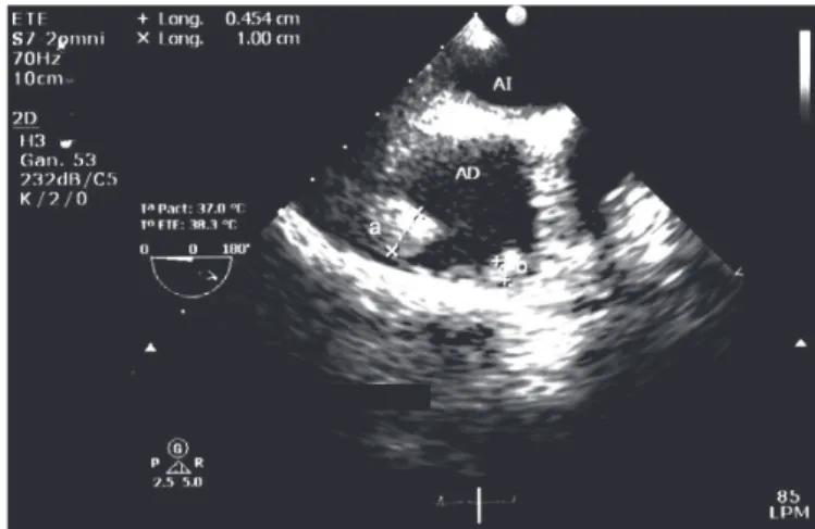
FIGURE 1 - Transesophageal echocardiogram: masses on the lateral wall of
the right atrium, measuring (a) 10 × 18mm and (b) 5 × 10mm on admission.
AI: left atrium; AD: right atrium.
FIGURE 2 - Transesophageal echocardiogram: longitudinal mass located on the lateral wall of the right atrium, measuring 32 × 12mm at 3 weeks of the initial antibiotic regimen administered to the patient. AD: right atrium;
MASA: atrial mass.
Transesophageal echocardiography (TEE) revealed two masses, 10 × 18mm and 5 × 10mm, on the lateral wall of the right atrium (Figure 1). The patient was transferred to the Infectious Diseases ward, where tests were performed, including repeated aerobic and anaerobic blood cultures, peripheral smear for malaria, serology testing for Brucella and Salmonella, bone marrow culture, and enzyme-linked immunosorbent assay (ELISA) test for human immunodei ciency virus, all of which resulting negative. The erythrocyte sedimentation rate, rheumatoid factor, and C-reactive protein were elevated (94mm/h, 24.2IU/ml, and 8.31mg/dl, respectively), and the complement values were normal (C3, 157mg/dl and C4, 18mg/dl). Due to persistent fever of 39°C, worsening anemia (8.9g/dl), and a history of a recent travel to Tarapoto, a jungle region in San Martín, Peru, where CD cases were recently reported, blood samples were sent to the Institute de Medicina Tropical Daniel Alcides Carrión in Lima to rule out CD. The samples revealed negative blood smears (Giemsa stain) but positive blood cultures for B. bacilliformis in Columbia agar (growth at 15 days of incubation). The dei nitive diagnosis of IE due to B. bacilliformis was made using modii ed Duke’s criteria. Treatment with intravenous (IV) ciprol oxacin (400mg/12h) was initiated, but changed to IV ceftriaxone (2g/24h) shortly after, due to lack of clinical improvement.
Despite this therapeutic modification, the patient had persistent anemia and intermittent fever and presented progressive weight loss, paresthesia in extremities, muscular weakness, livedo reticularis, and macular exanthema in the abdomen, dorsum, and lower limbs. Moreover, acute kidney injury (AKI) with creatinine of 1.69mg/dl, proteinuria (0.5g/24h), and the absence of hematuria were noticed. For this reason, Rheumatology evaluated the case and recommended electromyography, which was conclusive for mononeuritis multiplex; immunological markers were positive for p-ANCA (by ELISA). Renal biopsy revealed tubulointerstitial inflammation to predominance of mononuclear cells and moderate-severe thickening of juxtaglomerular and interlobular vessels. Due to the persistence of symptoms, two Bartonella blood cultures were repeated and, again, resulted positive (growth at 20 days and 17 days of incubation). A few coccobacilli were also reported in the blood smear. Furthermore, a follow-up TEE showed growth of the intracardiac masses (Figure 2), prompting a change from ceftriaxone monotherapy to a combination therapy of IV ciprol oxacin (200mg/12h), gentamicin (4mg/kg per 24h), and oral (PO) rifampicin (10mg/kg per 24h). The aminoglycoside was changed later to PO azithromycin (500mg/24h) due to worsening renal function. Additionally, surgical treatment was considered, but the patient did not consent to the procedure. The fever, anemia, and lymphopenia progressively resolved, but the weakness and paresthesias persisted. A multidisciplinary team decided to initiate PO prednisone (20mg/24h) for AAV, based on the symptomatology and immunological and electromyographic i ndings. The patient was discharged afebrile, with progressive clinical improvement and negative blood cultures for B. bacilliformis after 10 weeks of antibiotic treatment. On outpatient evaluation, a follow-up TEE showed absence of vegetations, prednisone dose was reduced to 5mg/day, and PO azathioprine (50mg/24h) was added, resulting in resolution of myalgias and paresthesias.
DISCUSSION
708
Rev Soc Bras Med Trop 50(5):706-708, September-October, 2017
patient had a recent and short epidemiological exposure to a new endemic area in the Peruvian jungle, which could explain her different outcome7. The patient initially received a regimen based on our current guidelines for treating acute B. bacilliformis infection; however, due to the lack of clinical and microbiological responses, we designed a combined therapy based on published antibiotic susceptibility testing of this microorganism8,9. A recent local study showed that 26% of evaluated strains of B. bacilliformis in patients with CD were resistant to ciproloxacin, which could explain the initial treatment failure in our patient10. This study and our case may indicate the need to revise current Peruvian guidelines, which recommend the use of ciproloxacin as irst-line therapy for Oroya fever.
Additionally, the patient showed clinical features compatible with AAV. We considered the case as MPA due to clinical indings (livedo reticularis, low weight, myalgias, arthralgias, paresthesias), AKI, presence of positive p-ANCA, mononeuritis multiplex, absence of granulomas, asthma, and peripheral eosinophilia. Several infections, including IE, cause positive ANCA tests by indirect immunoluorescence but negative ELISA testing. Our patient presented with positive p-ANCA (by ELISA); thus, this inding was most probable because of an AAV1,11. Glomerulonephritis is the most frequent histological inding in this type of vasculitis; however, some cases may consist of interstitial nephritis without glomerular involvement, as shown in this patient12. Endocarditis can cause renal injury through several pathways, such as cortical necrosis and renal infarction (e.g., septic embolism), and due to antibiotic use (e.g., interstitial nephritis), but the most frequent inding is immune complex-mediated glomerulonephritis, characterized by AKI, hypocomplementemia, hematuria, and prominent endocapillary proliferation, among others. The absence of most of these characteristics in our patient made this diagnosis less probable1.
In conclusion, this case report considers B. bacilliformis as a potential etiologic agent of IE in patients with conventional negative blood cultures and previous epidemiological exposure to an endemic area, and as a possible trigger for developing systemic vasculitis.
Acknowledgments
We would like to thank Dr. Jessica Ricaldi (CDC) for helping us in the preparation of this manuscript.
Financial support
This project was self-inanced.
Conlict of interest
The authors declare that there is no conlict of interest.
REFERENCES
1. Holland TL, Baddour LM, Bayer AS, Hoen B, Miro JM, Fowler Jr VG. Infective endocarditis. Nat Rev Dis Primers. 2016;2:16059.
doi: 10.1038/nrdp.2016.59.
2. Breña Chàvez JP, Maguiña Vargas CP, Hernández Diaz HR, Castillo Díaz ME, Pozo Tovar WE. Bartonelosis aguda en niños: Estudio de 32 casos en el Instituto Especializado de Salud del Niño y el Hospital Nacional Cayetano Heredia (Período 1993-2003). Rev Méd Hered. 2006;17(3):122-31.
3. Sakkas LI, Bogdanos DP. Infections as a cause of autoimmune rheumatic diseases. Auto Immun Highlights. 2016;7(1):13.
4. Ayoub EM, McBride J, Schmiederer M, Anderson B. Role of
Bartonella henselae in the etiology of Henoch-Schönlein purpura.
Pediatr Infect Dis J. 2002;21(1):28-31.
5. Maguiña Vargas C, Ugarte-Gil C, Breña Chávez P, Ordaya Espinoza E, Ventosilla López P, Huarcaya Castilla E, et al. Actualización de la enfermedad de Carrión. Update of Carrion’s disease. Rev Med
Hered. 2008;19(1):36-41.
6. Minnick MF, Anderson BE, Lima A, Battisti JM, Lawyer PG, Birtles RJ. Oroya fever and verruga Peruana: bartonelloses unique
to South America. PLoS Negl Trop Dis. 2014;8(7):e-2919.
7. Ministerio de Salud Dirección General de Epidemiología. Dirección Ejecutiva de Vigilancia Epidemiológica. Sala de Situación de
Salud, Perú: Semana Epidemiológica Nº 52; 2013 (Del 22 al 28 de
Diciembre 2013) Lima: Ministerio de Salud; 2013. [Internet]. [citado el 9 de diciembre de 2016]. Disponible en: http://www.dge.gob.pe/ portal/index.php?option=com_content&view=article&id=424
8. Tarazona FA, Maguiña VC, López de Guimaraes D, Montoya LM, Pachas CP. Terapia antibiótica para el manejo de bartonelosis o enfermedad de Carrión en el Perú. Rev Peru Med Exp Salud
Publica. 2006;23(3):188-200.
9. Rolain JM, Brouqui P, Koehler JE, Maguina C, Dolan MJ, Raoult D. Recommendations for treatment of human infections caused by Bartonella species. Antimicrob Agents Chemother.
2004;48(6):1921-33.
10. Mendoza-Mujica G, Flores-León D. Resistencia antimicrobiana de cepas de Bartonella bacilliformis procedentes de regiones endémicas de la Enfermedad de Carrión en el Perú. Rev Peru Med Exp Salud Publica. 2015;32(4):659-66.
11. Veerappan I, Prabitha EN, Abraham A, Theodore S, Abraham G. Double ANCA-positive vasculitis in a patient with infective endocarditis. Indian J Nephrol. 2012;22(6):469-72.