257
Communication/Comunicação
Revista da Sociedade Brasileira de Medicina Tropical 45(2):257-259, mar-abr, 2012
1. Laboratório de Biologia Molecular de Microorganismos, Universidade Federal de Alfenas, Alfenas, MG. 2. Instituto de Biologia, Universidade Estadual de Campinas, Campinas, SP. 3. Departamento de Ciências Médicas, Universidade Federal de Ouro Preto, Ouro Preto, MG. 4. Instituto de Ciências Biológicas, Universidade Federal de Minas Gerais, Belo Horizonte, MG.
Address to: Dr. João Guilherme Lino da Silva. Laboratório Biologia Molecular
de Microorganismos/UNIFAL. Rua Gabriel Monteiro da Silva 714/sala Q107, 37130-000 Alfenas, MG, Brasil.
Phone: 55 35 3299-1475 e-mail: [email protected] Received in 22/11/2010 Accepted in 07/02/2011
Comparison among three polymerase chain reaction assays on detection
of DNA from
Leishmania
in biological samples from patients with
American cutaneous leishmaniasis
Comparação entre três ensaios de reação em cadeia da polimerase na detecção de DNA de
Leishmania
, em amostras biológicas de pacientes com leishmaniose tegumentar americana
João Guilherme Lino da Silva
1, hiago Miranda da Silva
2, Eduardo de Figueiredo Peloso
2, George Luiz
Lins Machado-Coelho
3, Wilson Mayrink
4, Marília Caixeta Franco Ariosa
1, Paulo Márcio de Faria e Silva
1and Marcos José Marques
1ABSTACT
Introduction: he study analyzed positivity of polymerase chain reaction (PCR) on detection of DNA from Leishmania in patients’ samples. Methods:
Extracted DNA was submited to L150/L152, 13Y/13Z, and seminested PCR (snPCR). Results: Results were evidenced by bands of approximately 120, 720, and 670 bp for L150/L152, 13Y/13Z, and snPCR, respectively. L150/L152, 13Y/13Z, and snPCR positivity indexes were 76.9, 56.4, and 69.2 (p>0.05), respectively, for suspected and 93.7, 68.7, and 84.4 (p<0.05), respectively, for conirmed. Conclusions: Preliminary results showed that these assays, mainly L150/L152 and snPCR, can detect Leishmania DNA and carry potential on laboratory diagnosis of leishmaniasis.
Keywords: Leishmania. PCR. Diagnosis.
RESUMO
Introdução: Analisou-se a positividade da reação em cadeia da polimerase (PCR) na detecção de DNA de Leishmania em pacientes. Métodos: DNA extraído foi submetido a L150/L152, 13Y/13Z e PCR seminested (snPCR).
Resultados: Resultados foram evidenciados por bandas de aproximadamente 120; 720 e 670pb para L150/L152, 13Y/13Z e snPCR, respectivamente. Positividades para L150/L152, 13Y/13Z e snPCR foram 76,9; 56,4 e 69,2 (p > 0,05), para suspeitos; e 93,7; 68,7 e 84,4 (p < 0,05) para conirmados, respectivamente. Conclusões: Resultados preliminares mostraram que os ensaios, principalmente L150/L152 e snPCR, podem detectar DNA de
Leishmania e têm potencial para diagnóstico laboratorial das leishmanioses.
Palavras-chaves: Leishmania. PCR. Diagnóstico.
Leishmaniases are caused by an intracellular protozoan of the genus Leishmania, whose promastigote forms can be transmited to humans by the bite of Lutzomyia female insect, also known as sandly1.
Leishmania infection occurs in most Brazilian states, and some authors believe that its dispersal is associated to anthroponotic action. The traditional clinical manifestations for American cutaneous
leishmaniasis (ACL) are single or multiple cutaneous lesions, but there are asymptomatic cases as well1.
The laboratory techniques of diagnosis involve methods of detection of the parasite, such as direct search, isolation in culture, animal inoculation, and histopathology. However, these methods present hurdles related to sensitivity1. On the other hand, immunodiagnostic
techniques, such as the Montenegro skin test (MST), can increase the speed of the diagnosis, but they are unable to distinguish among active, inactive, or past infection2. Historically, a positive MST is
an indicator of previous contact with the parasite through natural inoculation. MST is the most widely used complementary test for the presumptive diagnosis of Leishmania infection. Positivity indexes of 84% and 100% have been estimated in cutaneous and mucocutaneous forms, respectively. Moreover, MST is negative in cutaneous difuse form and in immunocompromised patients2.
In this context, the use of PCR-based techniques has been shown as a new option of diagnosis, mainly due to their speed and high sensitivity1, 3. Moreover, these techniques have been developed
to amplify mini-exon genes4, DNA coding regions for subunits of
the ribosomal RNA5, and kinetoplast DNA-kDNA6 besides other
sequences of nuclear DNA7 in clinical samples for the diagnosis
of leishmaniasis. However, a major concern in the development and implementation of PCR for Leishmania diagnosis is the lack of standardization; many reports have been published, but very few studies have compared the diferent protocols5.
One of the main targets of these protocols is the kDNA that contains minicircles, which usually have the size of 1kb and occur up to 10,000 copies per parasite8. The minicircle kDNA has a
denominated region conserved, with approximately 100–150 bp, and a variable one, with approximately 700-1,000bp. Both regions are targets for primers designed to the PCR8, 9.
In this work, primers directed to the conserved region — L150/ L1529 — and to the variable region — 13Y/13Z8 — were used in the
PCR. In seminested PCR (snPCR), primers LINR4/LIN17/LIN19 were used to target the variable region, although their alignment was with the conserved region6. he positivity of those methods
was assessed based on the analysis of the materials obtained from patients suspected for ACL.
258
Silva JGL et al - PCR on detection of DNA from Leishmania
TABLE 1 -Indices of positivity obtained for the PCR-based techniques applied on biological samples from ACL suspected and conirmed patients.
Positivity index PCR (%)*
ACL suspected (n=39) PAR and/or MST+ (n=32)
PCR technique n % n %
13Y/13Z 22 56.4a 22 68.7a
L150/L152 30 76.9a 30 93.7b
snPCR 27 69.2a 27 84.4a, b
PCR: polymerase chain reaction, snPCR: seminested PCR, ACL: American cutaneous leishmaniasis, PAR: parasitological, MST+: Montenegro skin test, *Chi-square test (signiicant at p<0.05); a, b: equal leters mean positivity indexes that are statistically similar.
he patients were from the regions of Southern, Southwestern (SSMG), and Rio Doce Valley (RDV), State of Minas Gerais. Among those patients, 32 (82%) out of 39 were conirmed positive for ACL by parasitological diagnosis and/or MST. Parasitological diagnosis was done under microscopic analysis of imprints obtained from outer edges of the lesions.
All procedures involving human samples were approved under register number 141/2006 by the Ethics on Human Research Commitee from Universidade Federal de Alfenas.
DNA extraction was carried out using proteinase K digestion of skin biopsies from lesions9. In this methodology, DNA extraction
from samples was performed with 100µL bufer solution (10 mmol/L Tris-HCl and 1 mmol/L ethylenediaminetetraacetic acid, pH 8.0) and 100µg/mL proteinase K (inal concentration), incubated at 56ºC for 3h, and homogenized from time to time. Digestion was stopped by proteinase K inactivation, by boiling it for 15 min. he samples were centrifuged, and the supernatant was used as the Leishmania template DNA source for the PCR reaction. For slides, the methodology used was extraction based on heating10. In this methodology, an
area covering two imprints, previously analyzed on microscope, was scraped from each slide with a toothpick, resuspended in 20µL of double-distilled water, and transferred to 0.5mL microtubes. he samples were heated at 70ºC for 10 min and, then, centrifuged at 12,000 g for 5 min at room temperature; ater which, the supernatant was used as a DNA template.
DNA obtained from the digestion of biopsies as well as the imprints was submited to the following three PCR techniques. Ampliication was carried out in a Perkin-Elmer GeneAmp® PCR
System 9,700 thermocycler under diferent conditions for L150/ L1529, 13Y/13Z8, and snPCR6.
For each reaction, a negative control tube containing no template DNA was included. The positive control was also performed consisting of 10 pg of DNA extracted from axenic cultures of
Leishmania (Viannia) braziliensis MHOM/BR/1975/M2903 and
Leishmania (Leishmania) amazonensis MHOM/BR/1972/M2269. Ampliied products were analyzed in a 1.5% agarose gel, which was stained with ethidium bromide and under 3 V/cm.
Comparative analysis among the positivity indexes obtained by the three PCR techniques was carried out using the Chi-square test, with the level of signiicance set at p<0.05. he analysis was carried out using the Epi Info™ 6.04d (CDC) sotware9.
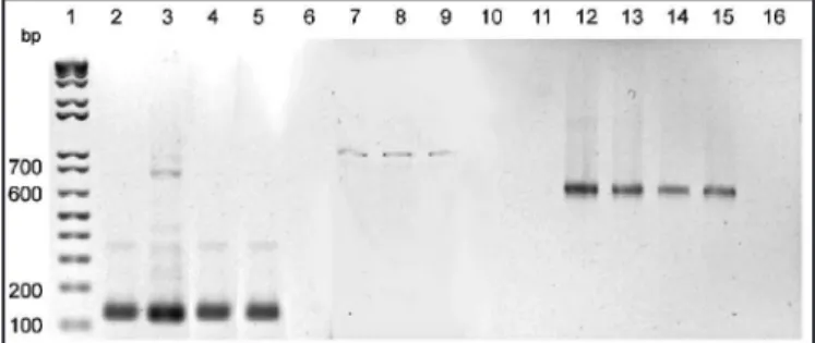
Results were evidenced by a band of approximately 120, 720, and 670 bp for L150/L152, 13Y/13Z, and snPCR, respectively (Figure 1). he positivity indexes obtained by the three techniques can be seen on
Table 1. For ACL suspected cases, a larger percentage of positive results was detected in the L150/L152 PCR (76.9%) in relation to others assays, although without signiicant diference (p>0.05), which was probably due the small number of cases. However, for conirmed cases, in parasitological tests and/or MST, these results were signiicantly diferent (p<0.05).
he performance of L150/L152 in the present work conirmed the results obtained from previous studies9. However, it was noted that
the snPCR technique has the possibility of detecting and amplifying larger sizes of DNA fragments with similar positivity obtained by L150/L152. Larger DNA targets present lower eiciency of ampliication11; however, the opposite was observed in the positivity
index of snPCR from ACL suspected and confirmed patients.
It was proposed that snPCR test is very sensitive on detection of
Leishmania, partly because there can be about 10,000 minicircles per kinetoplast and because its sequences are known for most Leishmania
species3. Results obtained by Nested PCR using other targets as
ITS-1 showed a lower positivity when compared with snPCR using the kDNA5.
Trypanosoma cruzi DNA was tested by L150/L152, 13Y/13Z, and snPCR to verify possible cross-reaction, but speciic fragments were not observed (data not shown). In addition, results of PCR obtained for
Leishmania using 13Y/13Z primers8 as well as snPCR3, 6 did not show
cross-reactivity with other trypanosomatids. Cross-reaction and low speciicity for L150/L15212 were observed. Furthermore, to minimize
risks of contamination, improvements were done in snPCR in relation to the conventional Nested technique13.
Moreover, it was proposed that the control of leishmaniasis in areas of endemicity requires a thorough knowledge of Leishmania
ecology and epidemiology and a sensible method for the detection of the parasite, which can be capable of processing a large number of samples synchronously6. In this context, results obtained in three
PCR assays show that these techniques can be useful for this purpose. Additionally, these methodologies have been shown capable of detecting kDNA from Leishmania in imprints on slides, where the shortage of material is not a limiting factor10.
In conclusion, the preliminary results showed that three PCR assays, mainly L150/L152 and snPCR, can detect Leishmania
DNA, including those in biological samples from ACL patients, and
FIGURE 1 -Representative 1.5% agarose gel showing ampliication products of 120 (lanes 2-6, conserved region with L150/L152 primers), approximately 720 (lanes 7-11, variable region with 13Y/13Z primers), and approximately 670 (lanes 12-16, variable region with PCR Seminested) bp from kDNA minicircles of Leishmania in promastigote cultures and skin biopsies from american cutaneous leishmaniasis patients. Lane 1: Molecular size marker of 100 bp ladder; Lanes 2, 7, and 12: DNA from
259
Rev Soc Bras Med Trop 45(2):257-259, mar-abr, 2012ACKNOWLEDGMENTS
he authors declare that there is no conlict of interest.
CONFLICT OF INTEREST
FINANCIAL SUPPORT
REFERENCES
that they also carry potential application on laboratory diagnosis of leishmaniasis. However, it is important to evaluate a larger amount of samples to verify the trend of L150/L152 and snPCR on producing results with greater positivity and signiicance, in relation to 13Y/13Z. Other studies are in progress to test the ability of the association of restriction fragment length polymorphism (RFLP) and snPCR on distinction of the diferent Leishmania species and to identify Leishmania DNA in other populations like dogs with visceral leishmaniasis.
We thank Mr. Jair Cecílio de Paula for the technical assistance in the Dr. Paulo Araujo Magalhães Leishmaniasis ambulatory.
Conselho Nacional de Desenvolvimento Cientíico e Tecnológico (CNPq) - 19/2004 - 47688.
1. Fagundes A, Schubach A, Paula CC, Bogio A, Antonio LF, Schiavoni PB, et al. Evaluation of polymerase chain reaction in the routine diagnosis for tegumentary leishmaniasis in a referral centre. Mem Inst Oswaldo Cruz 2010;105: 109-112. 2. Weigle A, Labrada LA, Lozano C, Santrich C, Barker DC. PCR-Based diagnosis
of acute and chronic cutaneous leishmaniasis caused by Leishmania (Viannia). J Clin Microbiol 2002; 40:601-606
3. Parvizi P, Mauricio I, Aransay AM, Miles MA, Ready PD. First detection of
Leishmania major in peridomestic Phlebotomus papatasi from Isfahan province, Iran: comparison of nested PCR of nuclear ITS ribosomal DNA and seminested PCR of minicircle kinetoplast DNA. Acta Tropica 2005; 93:75-83.
4. Paiva BR, Secundino NFC, Nascimento JC, Pimenta PFP, Galati EAB, Andrade Junior HF, et al. Detection and identiication of Leishmania species in Field-captured phlebotomine sandflies based on mini-exon gene PCR. Acta Tropica 2006; 99:252-259.
5. Pilati MM, Ferreira AS, Melo MN, Andrade ASR. Comparison of PCR methods for diagnosis of canine visceral leishmaniasis in conjunctival swab samples. Res Vet Sci 2009; 87:255-257.
6. Aransay AM, Scoulica E, Tselentis Y. Detection and identiication of Leishmania
DNA in naturally infected sand flies by Seminested PCR on minicircle kinetoplastic DNA. Appl Environ Microbiol 2000; 66:1933-1938.
7. Noyes H, Reyburn H, Bailey JW, Smith D. A nested-PCR-based schizodeme method for identifying Leishmania kinetoplast minicircle classes directly from clinical samples and its application to the study of the epidemiology of
Leishmania tropica in Pakistan. J Clin Microbiol 1998; 36:2877-2881. 8. Rodgers MR, Popper SJ, Wirth DF. Ampliication of kinetoplast DNA as a tool
in the detection and diagnosis of Leishmania. Exp Parasitol 1990; 71:267-275. 9. Marques MJ, Volpini AC, Coelho GLLM, Pinto JM, Costa CA, Mayrink W, et al.
Comparison of PCR with other laboratorial methods for the diagnosis of American Cutaneous Leishmaniasis. Diagnostic Microbiol Infect Dis 2006; 54:37-43.
10. Volpini AC, Marques MJ, Lopes dos Santos S, Machado-Coelho GL, Mayrink W, Romanha AJ. Leishmania identiication by PCR of Giemsa-stained lesion imprint slides stored for up to 36 years. Clin Microbiol Infect Dis 2006; 12:793-821. 11. Cheng S, Fockler C, Barnes WM, Higuchi R. Efective ampliication of long
targets from cloned inserts and human genomic DNA. Proceed Nat Acad Sci USA 1994; 91: 5695-5699.
12. Rocha LS, Santos CB, Falqueto A, Grimaldi Junior G, Cupolillo E. Molecular biological identiication of monoxenous trypanosomatids and Leishmania from antropophilic sand lies (Diptera; Psychodidae) in Southeast Braz Parasitol Res 2010; 107:465-468.