Conservative treatment of Angle Class III
malocclusion with anterior crossbite
João Hélder Ferreira de Aguiar1
How to cite this article: Aguiar JHF. Conservative treatment of Angle Class III malocclusion with anterior crossbite. Dental Press J Orthod. 2015 July--Aug;20(4):91-8. DOI: http://dx.doi.org/10.1590/2176-9451.20.4.091-098.bbo
Submitted: May 20, 2015 - Revised and accepted: June 16, 2015
Contact address: João Hélder Ferreira de Aguiar
Doutor Rocha, 837, sala 14, Centro – Pedro Leopoldo/MG - Brazil CEP: 33600-000 – E-mail: [email protected]
» The author reports no commercial, proprietary or financial interest in the prod-ucts or companies described in this article.
» Patients displayed in this article previously approved the use of their facial and in-traoral photographs.
1 Specialist in Orthodontics, Associação Brasileira de Ortodontia (ABO/EAP),
Alfenas, Minas Gerais, Brazil. INTRODUCTION
A 15 year and seven-month-old, Caucasian, male pa-tient presented for clinical examination with the chief complaint of not being happy with the results of a four-year treatment with removable appliances and that he felt that his chin was pronouncedly placed forward. His medi-cal and dental history was not signiicant,2 and he reported no parafunctional habits.
DIAGNOSIS
Extraoral clinical examination revealed symmet-ric facial pattern and lip competence, as well as lower lip protrusion in relation to the upper lip. The lateral
view showed a slightly concave profile (Fig 1). Den-tal evaluation revealed anterior crossbite which nega-tively affected facial esthetics. The patient had Angle Class III malocclusion, leveled curve of Spee, nor-mal overbite and, due to anterior crossbite, negative overjet (Figs 1, 2).
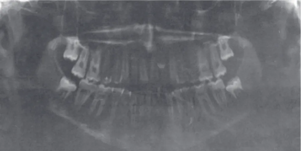
Initial panoramic radiograph showed that the pa-tient had all permanent teeth, including third molars, and that the outlines of tooth roots and alveolar bone were normal (Fig 3). Cephalometrically, (Fig 4 and Table 1) examinations revealed a Class III skeletal pat-tern (ANB = -2o), with a slightly retruded maxilla in relation to the skull base, and a protruded mandible DOI: http://dx.doi.org/10.1590/2176-9451.20.4.091-098.bbo
Angle Class III malocclusion is characterized by anteroposterior dental discrepancy which might be associated or not with skeletal changes. Class III molar relationship is associated with vertical or lingually tipped mandibular incisors and a usually concave profile. These characteristics seriously affect facial esthetics and most frequently are the reason why pa-tients seek orthodontic treatment. This case was presented to the committee of the Brazilian Board of Orthodontics and Facial Orthopedics (BBO) as part of the requisites to become a BBO Diplomate.
Keywords:Angle Class III malocclusion. Crossbite. Corrective orthodontics.
A má oclusão de Classe III de Angle é caracterizada por uma discrepância dentária anteroposterior, que pode ou não estar acompanhada por alterações esqueléticas. Observa-se uma relação molar de Classe III associada ao posicionamento vertical ou retroinclinado dos incisivos inferiores e, geralmente, perfil facial côncavo. Esse aspecto gera grande compro-metimento estético na face, sendo justamente esse o fator que, na maioria das vezes, motiva o paciente a procurar pelo tratamento ortodôntico. O presente caso clínico foi apresentado à Diretoria do Board Brasileiro de Ortodontia e Orto-pedia Facial (BBO) como parte dos requisitos para a obtenção do título de Diplomado pelo BBO.
Figure 1 - Initial facial and intraoral photographs.
Figure 3 - Initial panoramic radiograph.
Figure 4 - Initial cephalometric profile radiograph (A) and cephalometric tracing (B). A
in relation to both (SNA = 84o and SNB = 86o). His growth pattern was balanced, with slight tendency towards predominantly vertical growth pattern (SN-GoGn = 34o, FMA = 29o and Y-axis = 63o). More-over, the position of maxillary incisors was satisfactory (1-NA = 23o and 5 mm), and his mandibular incisors were inclined lingually (1-NB = 16o and 4 mm). Func-tional analysis of tongue position did not reveal any changes during mastication, deglutition or speech.
TREATMENT PLAN
the use of a modiied removable Hawley retainer for the maxillary arch, indicated to be used for 14 to 18 hours a day for one year, followed by overnight use for two more years. A ixed intercanine bar made of 0.016-in untampered stainless steel was placed in the mandibular arch. In that phase, special attention should be given to the development of third molars.
TREATMENT PROGRESSION
Orthodontic treatment was carried out with Edgewise appliance (Rickets, 0.018 x 0.030-in slot) for the max-illary and mandibular arches.4 Initially, double-tube bands were placed on teeth #16 and #26, followed by teeth #17 and #27, and bonding of the other teeth in the maxilla. A Blue Elgiloy 0.016 x 0.016-in multiloop archwire was used for alignment and leveling; all lateral segments were aligned by multiloops segmented arch-wires and nickel-titanium overlay (0.012, 0.014 and 0.016-in). Treatment was completed with the use of a Blue Elgiloy 0.016 x 0.022-in wire of ideal shape and torque. During the active phase of treatment, Class II intermaxillary elastics were used.
In the mandible, the ixed appliance was also ini-tially placed in posterior teeth. Double-tube bands were placed on teeth #36 and #46, followed by teeth #37 and #47, ater which bonding of the other teeth was carried out. A 0.016 x 0.016-in mandibular multiloop archwire was used for alignment and leveling, whereas 0.016 x 0.016-in segmented multiloop archwires were used for alignment of lateral segments, and a 0.016 x 0.022-in
tion in the maxillary arch for one year, with an indication of use of 14 to 18 hours a day, followed by overnight use for the next two years. Ater that, the appliance was used every other night for four months and once a week for another four months, ater which its use was discontinued. In the mandibular arch, an untampered stainless steel 0.016-in intercanine bar was placed ater inal dental examinations were carried out.
RESULTS
After one year and seven months, the achievement of all objectives was confirmed during final exami-nations (Figs 5 to 9). Facial esthetics was satisfactory because of the harmonious forward and downward growth of the face, both in the middle third and in the lower third of the face. Moreover, there was sub-stantial change in exposure of maxillary incisors at smiling, as well as in smile esthetics in the composi-tion of facial beauty.
Normal occlusion of molars and canines was achieved, as well as adequate lateral and protrusive guided occlusion. The anteroposterior relationship between maxilla and mandible remained unchanged, as ANB remained at -2o. Therefore, the projection of maxillary incisors resulted in adequate anterior crossbite and overjet, as planned. Table 1 shows that 1-NA went from 23o and 5 mm to 46o and 10 mm.
Figure 5 - Final facial and intraoral photographs.
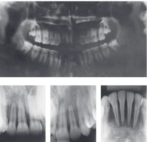
Figure 7 - Final panoramic and periapical radio-graphs of maxillary and mandibular incisors.
Figure 8 - Final cephalometric profile radiograph (A) and cephalometric tracing (B).
Table 1 - Initial (A) and final (B) cephalometric values.
Measurements Normal A B Dif. A/B
Skeletal pattern
SNA (Steiner) 82° 84o 86o 2
SNB (Steiner) 80° 86o 88o 2
ANB (Steiner) 2° -2o -2o 0
Angle of convexity (Downs) 0° -8o -3o 5
Y-axis (Downs) 59° 63o 61o 2
Facial angle (Downs) 87° 88o 89o 1
SN-GoGn (Steiner) 32° 34o 33o 1
FMA (Tweed) 25° 29o 24o 5
Dental pattern
IMPA (Tweed) 90° 77o 81o 4
1.NA (degrees) (Steiner) 22° 23o 46o 23
1-NA (mm) (Steiner) 4 mm 5 mm 10 mm 7
1.NB (degrees) (Steiner) 25° 16o 21o 5
1-NB (mm) (Steiner) 4 mm 4 mm 5 mm 1
1
1- Interincisal angle (Downs) 130° 142o 123o 19
1-APo (Ricketts) 1 mm 4 mm 5 mm 1
Profile Upper lip — S-line (Steiner) 0 mm -2 mm -1 mm 1
Lower lip — S-line (Steiner) 0 mm 0 mm 1 mm 1
Figure 9 - Total (A) and partial (B) superimposition of initial (black) and final (red) cephalometric tracings. B
1. Angle EH. Classiication of malocclusion. Dent Cosmos 1899;41(3):248-64. 2. Langlade M. Diagnóstico ortodôntico. 1ª ed. São Paulo: Ed. Santos; 1993.
3. Langlade M. Otimização das escolhas ortodônticas. 1ª ed. São Paulo:
Ed. Santos; 1995.
4. Ricketts RM. Orthodontic diagnosis and planning: their roles in preventive and rehabilitative dentistry. Michigan: Rocky Mountain Orthodontics; 1982. REFERENCES