RBCCV 44205-1501 DOI: 10.5935/1678-9741.20130079
Reversible pulmonary trunk banding. IX. G6PD
activity of adult goat myocardium submitted to
ventricular retraining
Bandagem ajustável do tronco pulmonar. IX: atividade da G6PD do miocárdio de cabras adultas
submetido ao treinamento ventricular
Renato Samy Assad
1, MD, PhD; Leonardo Augusto Miana
1,2, MD, PhD; Miriam Helena
Fonseca-Alaniz
1, BPh, PhD; Maria Cristina Donadio Abduch
1, VMD, PhD; Gustavo José Justo da Silva
1,3,
PE, PhD; Fernanda Santos de Oliveira
1MD; Luiz Felipe Pinho Moreira
1, MD, PhD; José Eduardo
Krieger
1, MD, PhD
1 Heart Institute (InCor), University of São Paulo Medical School, São Pau-lo, SP, Brazil
2 Medical School, Federal University of Juiz de Fora, Juiz de Fora, MG, Brazil
3 University of Maastricht. Mastricht, Netherlands.
Work carried out at Heart Institute (InCor), University of São Paulo Medical School, São Paulo, SP, Brazil.
Work awarded at the 39th Congress of the Brazilian Society of Cardiovas-cular Surgery, Maceió, April 2012, and at the 67th Brazilian Congress of Cardiology, Recife, September 2012. Presented at the 46th Annual Meeting of the Association for European Paediatric and Congenital Cardiology, Is-tanbul, May 2012.
Funding: SILIMED, Rio de Janeiro, RJ (donation of adjustable banding de-vices and molecular biology kits), FAPESP (research grant nº 2006/50831) Correspondence address:
Renato Samy Assad
Heart Institute (InCor), University of São Paulo Medical School,
Laboratory of Research in Cardiovascular Surgery (LIM-11) / Laboratory of Genetics and Molecular Cardiology
Av. Dr. Enéas de Carvalho Aguiar, 44 – Cerqueira César - São Paulo, SP Brazil – Zip Code: 05403-000
E-mail: rsassad@cardiol.br
Article received on April 14th, 2013
Article approved on August 19th, 2013
Abstract
Objective: Increased glucose 6-phosphate dehydrogenase ac-tivity has been demonstrated in heart failure. This study sought to assess myocardial glucose 6-phosphate dehydrogenase activ-ity in retraining of the subpulmonary ventricle of adult goats.
Methods: Eighteen adult goats were divided into three
groups: traditional (ixed banding), sham, and intermittent (adjustable banding, daily 12-hour systolic overload). Systolic overload (70% of systemic pressure) was maintained during a 4-week period. Right ventricle, pulmonary artery and aortic
pressures were measured throughout the study. All animals were submitted to echocardiographic and hemodynamic
eval-uations throughout the protocol. After the study period, the
animals were killed for morphological and glucose 6-phosphate dehydrogenase activity assessment.
Results: A 55.7% and 36.7% increase occurred in the
in-termittent and traditional right ventricle masses, respectively,
when compared with the sham group (P<0.05), despite less ex
-posure of intermittent group to systolic overload. No signiicant
changes were observed in myocardial water content in the 3 groups (P=0.27). A 37.2% increase was found in right ventricle
wall thickness of intermittent group, compared to sham and tra -ditional groups (P<0.05). Right ventricle glucose 6-phosphate
dehydrogenase activity was elevated in the traditional group,
when compared to sham and intermittent groups (P=0.05). Conclusion: Both study groups have developed similar right
ventricle hypertrophy, regardless less systolic overload expo -sure of intermittent group. Traditional systolic overload for adult subpulmonary ventricle retraining causes upregulation of myocardial glucose 6-phosphate dehydrogenase activity. It may suggest that the undesirable “pathologic systolic overload”
is inluenced by activation of penthose pathway and cytosolic
mature myocardium induced by pulmonary trunk banding (PTB) in order to establish retraining protocols for a more “desirable” physiological hypertrophy.
In physiological conditions, fatty acids, especially the long chain ones, act as the main myocardial energy substrate [5]. Fatty acid oxidation in mitochondria represents approximately 70% of all ATP production in a normal heart [6,7]. On the other hand, several studies have shown that there is greater preference for glucose oxidation by cardiac substrates in heart failure and in a hypertrophic heart [8,9]. However, the change in energy substrate varies according to the etiology and severity of the ventricular dysfunction, where the degree of metabolic modulation plays an important role in determining adaptive or maladaptive function in terms INTRODUCTION
Both adult and adolescent patients diagnosed with congenially corrected transposition of the great arteries (CCTGA) as well as those with transposition of the great arteries (TGA) who underwent either Senning or Mustard surgery can develop right ventricle dysfunction (systemic) [1,2]. Subpulmonary ventricular retraining for anatomical correction, in those patients, still presents disappointing results. Hypertrophy induced by acute pressure overload can lead to foci of cellular necrosis in the hypertrophied myocardium and, consequently, to late ventricular dysfunction [3,4]. At present, it is imperative to understand the molecular mechanisms involved in hypertrophy of the
Abbreviations, acronyms & symbols
ANOVA Analysis of variance Ao Aorta
CCTGA Congenially corrected transposition of the great arteries COBEA Brazilian College of Animal Experimentation DW Dry weight
ECG Electrocardiogram
G6PD Glucose 6-phosphate dehydrogenase IVS Interventricular septum
IW Initially weighed LV Left ventricle
NADPH Nicotinamide adenine dinucleotide phosphate PT pulmonary trunk
PTB Pulmonary trunk banding RV Right ventricle
TGA Transposition of the great arteries
radicals by Nicotinamide adenine dinucleotide phosphate oxi
-dase, an important mechanism in the pathophysiology of heart
failure.
Descriptors: Energy metabolism. Hypertrophy, right ven -tricular. Transposition of great vessels. Cardiac surgical proce-dures. Goats.
Resumo
Objetivo: O aumento da atividade miocárdica da Glicose
6-Fosfato Desidrogenase tem sido demonstrado na insuiciência
cardíaca. Este estudo avalia a atividade miocárdica da Glicose 6-Fosfato Desidrogenase no treinamento do ventrículo subpul-monar de cabras adultas.
Métodos: Foram utilizadas 18 cabras adultas, divididas em
três grupos: convencional (bandagem ixa), sham e intermitente (bandagem ajustável; 12 horas diárias de sobrecarga). A sobre
-carga sistólica (70% da pressão sistêmica) foi mantida durante quatro semanas. As avaliações hemodinâmica e ecocardiográica
foram realizadas durante todo o estudo. Depois de cumprido o
protocolo, os animais foram mortos para avaliação morfológica e
da atividade da Glicose 6-Fosfato Desidrogenase dos ventrículos. Resultados: Apesar de haver sobrecarga sistólica propor-cionalmente menor no ventrículo subpulmonar do grupo in-termitente (P=0,001), ambos os grupos de estudo apresentaram aumento da massa muscular de magnitude similar. Os grupos intermitente e convencional apresentaram aumento da massa
de 55,7% e 36,7% (P<0,05), respectivamente, em comparação ao grupo sham. O conteúdo de água do miocárdio não variou entre os grupos estudados (P=0,27). O ecocardiograma de
-monstrou maior aumento (37,2%) na espessura do ventrículo subpulmonar do grupo intermitente, em relação aos grupos
sham e convencional (P<0,05). Foi observada maior atividade
da Glicose 6-Fosfato Desidrogenase na hipertroia miocárdica do ventrículo subpulmonar do grupo convencional, comparada
aos grupos sham e intermitente (P=0,05).
Conclusão: Ambos os grupos de treinamento ventricular
de-senvolveram hipertroia ventricular, a despeito do menor tempo
de sobrecarga sistólica no grupo intermitente. A maior ativi-dade de Glicose 6-Fosfato Desidrogenase observada no grupo
convencional pode reletir um desequilíbrio redox, com maior
produção de fosfato de dinucleotídeo de nicotinamida e adenina
e glutationa reduzida, um mecanismo importante da isiopato
-logia da insuiciência cardíaca.
of states leading to pathological hypertrophy.
G6PD is the irst and rate-limiting enzyme in the pentose phosphate pathway, which is an alternate and independent pathway from glycolysis, generating nicotinamide adenine dinucleotide phosphate (NADPH) and pentose (ribose-5-phosphate). It catalyzes the conversion of Glucose 6-Phosphate into 6-Phosphogluconolactone. The process is divided into two phases: generation of NADPH and non-oxidative synthesis of pentose. It maintains the level of the NADPH coenzyme, which in turn maintains the level of reduced glutathione, protecting the cells, in normal conditions, from oxidative lesions. Therefore, the more NADPH is needed, the greater the activity level of the G6PD enzyme.
There is evidence that greater G6PD activity leads to increased production of superoxide radicals as well as oxidative stress in diabetes, heart failure, and hypertension. Previous studies of the energy metabolism of acute myocardial hypertrophy in young animals have shown an increase in G6PD activity associated with ventricular dysfunction in continuous systolic overload, compared to intermittent overload [10].
In the subset of the subpulmonary ventricle retraining of adult patients for the double switch operation , it would be also interesting to analyze changes in energy metabolism. This study sets out to evaluate qualitative differences in the process of myocardial hypertrophy induced by continuous and intermittent pressure overload, through biological markers such as G6PD, which show occasional phenotypic changes to the energy metabolism.
METHODS
This study was approved by the Ethics Committee for the Analysis of Research Projects at – InCor University of Sao Paulo Medical School (process: SDC 2660/05/080) and carried out in accordance with the regulations on the use of animals in teaching and research of the Brazilian College of Animal Experimentation (COBEA). Eighteen adult goats with comparable weight (P=0.63) were used, divided into three groups: (1) Traditional group (n=6, weight=26.33 kg ± 2.32 kg, PTB with continuous systolic overload of the RV); (2) Sham or Positive Control group (n=6, weight=26.42 kg±2.63 kg, loose banding, no overload of the RV); and Intermittent group (n=6, weight=25.17 kg ± 2.48 kg, PTB with adjustable device and 12 hours/day of intermittent systolic overload of the RV).
Anesthesia
The animals were fasted for 24 hours before the surgery and received Xylazine 2%, 0.1 mg/kg IM, as preanesthetic medication. Anesthesia was induced with propofol 1% (7 mg/kg), intravenously (IV) for orotracheal intubation. The animals were maintained under mechanical ventilation
(Takaoka Fuji Maximus, São Paulo, SP), with inspired oxygen fraction between 60 and 100% and Isolurane inhalation (1 to 2%). The goats were placed in the right lateral decubitus and monitored with continuous electrocardiogram (ECG) and invasive arterial pressure (auricular artery). Postoperative pain relief was obtained in the irst three days by administering Tramadol chlorhydrate (2 mg/kg, intramuscular).
Surgical Procedure
The goats were prepared for the sterile procedure, as described in previous studies [11,12]. For animals in the Traditional group, pulmonary trunk (PT) banding was performed with cardiac tape (Polysuture, São Sebastião do Paraíso, MG), positioned about 10 mm above the valve. Animals in the Sham and Intermittent groups had an adjustable banding device implanted immediately above the pulmonary valve and ixed to the PT adventitia. The insuflation button was implanted and ixed subcutaneously in the thorax. Hemodynamic monitoring catheters were exteriorized and maintained illed with heparin. The following antibiotics were administered prophylactically to all animals: cefazolin (30 mg/Kg), gentamicin (4 mg/Kg), and Benzathine Penicillin (500,000 IU), intramuscular. The Benzathine Penicillin dose was repeated after two weeks. In addition, Heparin Sodium 5,000 IU was administered daily by subcutaneous injection until the end of the protocol.
Description of the adjustable banding device
The adult model device was developed in collaboration with SILIMED (Indústria de silicone e instrumental médico-cirúrgico e hospitalar Ltda., Rio de Janeiro, RJ), as previously published [11,12]. It has an outer diameter of 24 mm and is 6 mm wide. The inner surface has a distensible silicon layer, 0.6 to 0.8 mm thick, whose expansion compresses the lumen of the PT according to the amount of liquid injected in the insuflation button. This button is implanted subcutaneously so that ine adjustments to the diameter of the banding ring can be made percutaneously.
RV systolic overload protocol and pressure readings Traditional Group
RV training started during the banding implant surgery. The animals remained under continuous systolic overload of the RV for a period of four weeks, with conventional ixed banding adjusted on the day of the surgery in order to reach an RV systolic pressure of about 70% of systemic systolic pressure, provided that there was no more than a 10% drop in systemic systolic pressure. Pressures of RV, PT, and aorta (Ao) were recorded twice a week with the animal conscious and immobilized on a special stretcher.
Sham Group
were taken twice a week during the four weeks of the study and the banding device was kept delated throughout the protocol.
Intermittent Group
The RV training started after approximately 60 hours of convalescence. As in the Traditional group, baseline pressures of RV, PT, and Ao were recorded with the animal conscious and the device completely delated. Subsequently, the adjustable banding device was insuflated with 0.9% saline in order to reach the same RV systolic overload as the Traditional group. The pressures were then measured once again. The RV systolic overload was maintained for a period of 12 hours, after which arterial pressures were read one more time. After that, the device was delated and pressures were measured again. The process of insuflating and delating the device was performed daily for four weeks, with pressures being measured three times a week. On alternate days, the injected volume was the same as the volume calculated on the last day of hemodynamic measurements.
Echocardiographic study
All animals underwent echocardiographic evaluation by the same observer prior to the protocol and weekly after surgery. During the exams, the goats were sedated with Ketamine (10 mg/kg, intramuscular) and kept in the left lateral decubitus. The ACUSON Cypress (Siemens Healthcare, Mountain View, USA) echocardiography machine was used as well as the multi-frequency sector transducer (1.8-3.6 MHz) to capture images and analyze low. The thicknesses of the interventricular septum (IVS) and left ventricle (LV) posterior wall were measured by two-dimensional echocardiogram at the end of diastole through longitudinal parasternal section at the mitral valve cusps [13]. The thickness of the RV free wall was obtained through transverse parasternal section (at the great vessels and at the papillary muscles level) and longitudinal four-chamber section in the region where its boundaries were more easily seen. Finally, the arithmetic mean of those values was obtained.
Weighing cardiac masses
At the end of the protocol, the animals were euthanized for resection of the heart. Anesthesia was deepened and heparin (5 mg/kg) was administered endovenously. Then, potassium chloride was administered until cardiac arrest. At that time, samples of the RV, LV, and interventricular septum were collected, weighed, and immediately placed in tagged plastic containers. The samples were stored in liquid nitrogen (-80ºC) until they could be transferred to the Genetics and Molecular Cardiology Laboratory, where they were kept in a special freezer (Forma Scientiic -80º C). The heart was then removed from the thorax. The great vessels, atria, as well as the cardiac valves and epicardial fat were carefully resected. Ventricular
and septal walls were separated so that cardiac masses could be weighed individually in a METTLER AE-200 (Mettler-Toledo AG, Greifensee, Switzerland) digital scale.
Tissue water content
After weighing the cardiac masses, samples were collected from each one for water content assessment. Every sample was initially weighed (IW), then wrapped in aluminum foil and properly identiied before being placed in an incubator (temperature: 55-60º C). After about 70 hours of dehydration, every sample was weighed once again to determine dry weight (DW). The percentage of water content was then determined by the following formula, assuming the water distribution was homogenous in the septum and ventricles:
WC (%) = 100 – (DW x IW-1 x 100)
Analysis of glucose 6-phosphate dehydrogenase (G6PD) maximum activity
G6PD maximum activity was determined in the septum and ventricle samples obtained. Samples were stored in liquid nitrogen and homogenized in extraction buffer (proportion 1:5 weight/volume). The material kept in ice was homogenized using Polytron (PT 3100, Kinematica AG, Littau-Lucerne, Switzerland), at maximum speed for 30 seconds. Cell remnants were separated by centrifugation (15,000 g, 15 minutes, 4ºC) using a Centrifuge 5417 C/R- Eppendorf (Hamburg, Germany). Enzymatic activity was analyzed using the supernatant of the last centrifugation.
Proteins were quantiied using the BCATM protein assay kit (PIERCE Biotechnology, Rockford, IL, USA). Results were expressed as nmol.min-1.mg-1 of protein present in the extract. The extraction buffer for G6PD contained Tris-HCl (50 mM) and EDTA (1 mM), with a pH of 8.0. The assay buffer (270 mL/sample) was comprised of Tris-HCl (8.6 mM), MgCl2 (6.9 mM), NADP+ (0.4 mM), and Triton X-100 0.05% (volume/volume), with a pH of 7.6. The reaction was initiated by adding 15 mL of Glucose-6-phosphate (1.2 mM) to the enzymatic extract (15 mL sample) and it was monitored for 10 minutes at 25º C. Absorbance was monitored at 340 nm, the extinction coeficient being 6.22 for that particular wavelength.
The biochemical reaction is based on glucose phosphorylation and subsequent oxidation of glucose-6-phosphate to 6-phosphogluconate catalyzed by G6PD. G6PD activity levels were determined indirectly as the total NADPH produced in the pentose phosphate pathway.
Statistical Analysis
by one-way ANOVA. Both analyses were followed by the Bonferroni post hoc test. Systolic overload imposed on the RV, in the Traditional and Intermittent groups, was assessed by calculating the areas under the curve (trapezoidal method). The comparison between those areas was made through unpaired Student’s t-test. Values were expressed as mean ± standard deviation (SD), unless otherwise indicated. The signiicance level was 5% for all cases. Statistical analyses were done using GraphPad Prism v.4 (San Diego, CA, USA) and SigmaStat v.3.11.0 (Systat Software, Inc., San Jose, CA, USA).
RESULTS
Hemodynamics
RV/PT pressure gradient
The Traditional group showed a decrease in gradient after the second postoperative day (from 45.00 mmHg ± 4.90 mmHg to 39.25 mmHg ± 13.05 mmHg), but remained stable until the end of the protocol (limits: 36.33 mmHg ± 4.04 mmHg to 40.00 mmHg ± 7.72 mmHg). During euthanasia, there was a signiicant decrease in the RV/PT gradient in the Traditional group (P<0.05), compared to the values of the fourth week. In the Sham group, as opposed to the others, pressure gradient (limits: 4.67 mmHg ± 2.08 mmHg to 9.40 mmHg ± 4.51 mmHg) was maintained throughout the protocol (P<0.05).
Peak pressure gradients were signiicantly higher in the Intermittent group (limits: 46.67 mmHg ± 6.80 mmHg to 59.00 mmHg ± 8.29 mmHg) (P<0.05), alternating with periods of “rest” of the RV. The RV/PT gradient remained elevated in both study groups compared to the Sham group (P<0.001). The Traditional group was subjected to a signiicantly larger area of systolic overload (23,764 mmHg.h ± 2,192 mmHg.h) than the Intermittent group (17,414 mmHg.h ± 1,144 mmHg.h; P<0.0001). Both Traditional and Intermittent groups had larger areas of systolic overload than the Sham group (3,841 mmHg.h ± 1,298 mmHg.h; P<0.05).
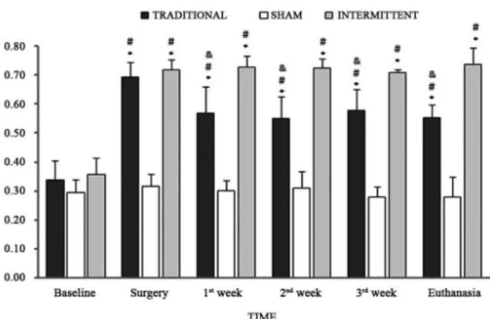
RV/Ao pressure ratio
Baseline RV/Ao pressure ratio of approximately 0.25 was similar in all groups. During surgery, RV/Ao ratio of 0.70 was reached in the stimulated groups. However, after the irst week, the ratio decreased signiicantly in the Traditional group (P<0.05) and remained stable for the remainder of the study. In the Sham group, the baseline RV/Ao ratio remained stable throughout the protocol (P<0.05). In the Intermittent group, the maximum RV/Ao ratio was kept at around 0.7 throughout the study, as opposed to the Traditional group (Figure 1; P<0.05).
Echocardiographic indings
Thickness of the cardiac walls. There was signiicant percentage variation in the thickness of the RV free wall in
the Intermittent group, starting in the irst week of the study when compared to the Sham group (P<0.001), and in the second and fourth weeks of the study when compared to the Traditional group (P<0.01; Figure 2). There was no variation among the groups in the thicknesses of the interventricular septum and the LV posterior wall.
Fig. 1 – Temporal comparison of the RV/Ao maximum ratio in the Traditional, Sham, and Intermittent groups
*P<0.05 compared to Baseline instant in its respective group; # P<0.05 difference between the Traditional and Intermittent groups compared to the Sham group; & P<0.05 difference between the Traditional and Intermittent groups.
Morphological indings
Measurement of cardiac mass
Table 1 shows the weight of the ventricular chamber masses. The Intermittent and Traditional groups showed an increase in RV mass of 57.0% and 36%, respectively, compared to the Sham group (P<0.05). There was no signiicant variation in the weight of the IVS (P=0.09) and LV (P=0.30) masses among the groups.
Water content
There was no signiicant variation in water content of the RV myocardium in the Traditional (79.67% ± 1.25%), Sham (79.16 ± 1.28), and Intermittent (80.61 ± 1.87) groups (P=0.27).
Glucose 6-phosphate dehydrogenase (G6PD) maximum activity
Table 2 shows the mean of absolute values for G6PD maximum activity in the Traditional, Sham, and Intermittent
groups. In the Traditional group, maximum activity levels of this enzyme in the RV were 55.2% higher than in the Sham group and 40.7% higher than in the Intermittent group (P=0.05). These data are graphically represented in Figure 3. No signiicant differences were found between the groups in G6PD maximum activity in the LV (P=0.39) and Septum (P=0.31).
DISCUSSION
This experimental study set out to compare right ventricular hypertrophy in adult goats subjected to intermittent versus traditional systolic overload, highlighting the energy metabolism in the ventricular retraining of mature myocardium. From a morphological standpoint, differences in hypertrophy in favor of the Traditional group were expected due to the higher exposure of the myocardium to hypertrophic stimuli, quantiied by the larger area of systolic overload of the right ventricle.
Fig. 3. – Maximum activity of the Glucose-6-phosphate Dehydrogenase (G6PD) enzyme in the myocardium of the Traditional, Sham, and Intermittent groups. Measures: nmol/min/mg of protein ± standard error. n= 6
Table 2. Maximum activity of the glucose-6-phosphate dehydrogenase (G6PD) enzyme in the Traditional, Sham, and Intermittent groups.
RV LV Septum
Traditional
2.11 ± 0.88 1.85 ± 0.22 0.96 ± 0.28*
Sham
1.36 ± 0.14 1.80 ± 0.17 0.86 ± 0.25
Intermittent
1.50 ± 0.24* 1.71 ± 0.16 1.13 ± 0.32*
P Value
0.05 0.39 0.31
Values = average ± standard deviation; Measures: nmol/min/mg of protein; * n = 5
Table 1. Weight of cardiac masses of the right ventricle (RV), interventricular septum (IVS), and left ventricle (LV), normalized by the weight of the animals in the Traditional, Sham, and Intermittent groups.
RV IVS LV
Traditional N = 6 1.08 ± 0.17 *
0.96 ± 0.19 1.52 ± 0.21
Sham N = 6 0.79 ± 0.15 0.84 ± 0.20 1.35 ± 0.22
Intermittent N = 6 1.24 ± 0.16 #
1.09 ± 0.13 1.47 ± 0.10
P Value
<0.05 0.09 0.30
Even though both ventricular retraining groups were able to promote ventricular hypertrophy of similar magnitude compared to the Sham group, the echocardiographic indings of the Intermittent group showed increased RV free wall thickness. However, the length of time of the hypertrophic process was greater than that found in in young animals submitted to just 96-hour intermittent systolic overload protocol.
Likewise, morphological and/or echocardiographic analyses revealed no changes in septal thickness, which also diverges from previous studies in young animals that showed signiicant increase in the septal mass [10]. Perhaps this deviation can be explained by a more eficient protein production in young as opposed to mature myocardium. All three groups showed no variation in myocardium water content, a sign of cellular edema, suggesting the gain in mass was mainly due to enhanced protein synthesis.
Previous studies from our laboratory have demonstrated impaired functional and morphological RV performance of adult goats submitted to Traditional systolic overload under the same protocol. [11,12]. Likewise, there was a concordance of impaired RV function and increased RV G6PD activity of Traditional group after 4-week study period, corroborating the indings of the 96-hour systolic overload in young animals [10]. Increased G6PD activity indicates an exacerbation of the pentose phosphate pathway and it can mean loss of redox balance, with higher production of NADPH and reduced glutathione, as well as development of oxidative stress derived from superoxide anions associated with NADPH oxidase.
Under pathological conditions, NADPH is produced by activation of G6PD after stimuli from a range of factors, such as angiotensin II, thrombin, and alpha tumor necrosis factor [14,15]. Cardiomyopathy related to protein aggregation and myocardial lesion would be the inal consequence. There is growing evidence that increased G6PD activity is associated with oxidative and reductive stress, with new drugs being developed in order to inhibit its activity [16]. For instance, patients with diabetes mellitus show increased G6PD activity and NADPH levels. This metabolic disorder is associated with endothelial dysfunction due to the inhibition of nitric oxide synthesis [17].
Although the mechanisms through which most of the free radicals are produced in the heart are not completely known, it has been suggested that higher glucose oxidation increases the potential of the mitochondrial membrane thereby augmenting NADPH oxidase activity in the vascular system and, consequently, increasing the production of superoxide anions [18,19]. The latter would act as mediators of diabetic vasculopathy and precursors of myocardial dysfunction related to the disease [20,21]. There is a 10-fold increase in G6PD activity in pacing-induced heart failure compared to normal hearts [22].
This study did not directly assess the production of free radicals associated with NADPH oxidase; however, it can be speculated that the overexpression of G6PD observed in the RV of the Traditional group indicates that the Pentose pathway increases the availability of NADPH, providing the release of free radicals via NADPH oxidase and nitric oxide synthase. Thus, this could lead to myocardial lesion caused by accumulation of superoxide anions and protein aggregation and, subsequently, to ventricular dysfunction should the overload continue. This is corroborated by the fact that functional recovery of hearts with compensated hypertrophy is signiicantly higher than that of non-hypertrophic hearts when myocardial perfusion is intermittently reestablished, a maneuver which prevents or minimizes the accumulation of glycolytic products and H+ ions. Indeed, reestablishment of this subendocardial coronary low during the resting periods of the Intermittent group would avoid the accumulation of the products of glycolysis and H+ ions.
Besides, fatty acid oxidation is directly stimulated during the reestablishment of subendocardial reperfusion in the resting periods from systolic overload as a result of changes in the enzymes and metabolites responsible for the regulation of fatty acid oxidation. Predominance of fatty acid oxidation during periods of rest of the RV could lead to decreased myocardial glucose uptake. On the other hand, increased eficiency of intermittent systolic overload could be related to the release of hypertrophic stimuli and the cascade of protein synthesis, just as in the Traditional group, yet with decreased myocardial energy expenditure. The mechanisms of the hypertrophic process triggered by molecular cascade are likely to happen under good conditions during the 12-hour resting period and ideal oxygen delivery. Therefore, the type of retraining, in terms of level and duration of the systolic overload as well as its impact on the myocardium, must be considered. Even though the increase in G6PD is an unspeciied pathway for production of free radicals, this study has found an agreement between previously shown right ventricular dysfunction in the Traditional group and the increase in G6PD activity, a situation where there is inadequate oxygen delivery due to continuous systolic overload, likely high consumption of ATP, and, hence, larger production of free radicals.
Limitations of the study
There are limitations to implementing the results of this study in human beings. Firstly, metabolism and morphological aspects can differ among species. Secondly, the great arteries were normally related in the animals; hence, the ventricle studied (anatomically right) is not the same as the target population in humans (anatomically left).
end, it would be ideal to also analyze the production of free radicals and/or markers of myocardial lesion since there are several inluences at play from other metabolic pathways. This rational suggests that future studies of the production of oxidized glutathione, free radicals, and apoptosis may provide important information to this research line.
CONCLUSION
This study has shown that ventricular retraining in both groups of adult goats led to right ventricular hypertrophy without myocardial edema. Continuous systolic overload promoted increased G6PD activity in the RV myocardium. This enzymatic hyperactivity may be related to increased production of free radicals caused by greater demand for constant myocardial overload stimulus. Conversely, intermittent systolic overload enabled a more eficient RV hypertrophy, considering the smaller area of systolic overload of the RV and decreased G6PD activity.
ACKNOWLEDGEMENTS
The authors are grateful to SILIMED, Rio de Janeiro, RJ, for the donation of the adjustable banding devices and to FAPESP, for the research grant nº 2006/50831.
Authors’ roles & responsibilities
RSA Design, supervision, and execution of the project; statistical analysis; writing and review of the draft
LAM Execution of the project; surgical procedures; data collection; and writing of the draft
MHFA Processing of samples and energy metabolism analysis MCDA Echocardiographic exams; writing and review of the draft GJJS Statistical analysis and writing of the draft
FSO Assistance in surgical procedures
LFPM Supervision; statistical analysis; and review of the draft JEK Design; energy metabolism analysis; supervision; and review of
the draft
REFERENCES
1. Cochrane AD, Karl TR, Mee RB. Staged conversion to arterial switch for late failure of the systemic right ventricle. Ann Thorac Surg. 1993;56(4):854-61.
2. Mee RB. Severe right ventricular failure after Mustard or Senning operation. Two stage repair: pulmonary artery banding and switch. J Thorac Cardiovasc Surg. 1986;92(3 Pt 1):385-90.
3. Siehl DL, Gordon EE, Kira Y, Chua BHL, Morgan HE. Protein
degradation in the hypertrophic heart. In: Glaumann H, Ballard FJ, eds. Lysosomes: their role in protein breakdown. London: Academic; 1987.
4. Takahashi Y, Nakano S, Shimazaki Y, Kadoba K, Taniguchi K,
Sano T, et al. Echocardiographic comparison of postoperative left
ventricular contractile state between one- and two-stage arterial
switch operation for simple transposition of the great arteries. Circulation. 1991;84(5 Suppl):III180-6.
5. Van der Vusse GJ, Glatz JF, Stam HC, Reneman RS. Fatty acid homeostasis in the normoxic and ischemic heart. Physiol Rev. 1992;72(4):881-940.
6. Taegtmeyer H. Energy metabolism of the heart: from basic concepts to clinical applications. Curr Probl Cardiol. 1994;19(2):59-113.
7. Grynberg A, Demaison L. Fatty acid oxidation in the heart. J Cardiovasc Pharmacol. 1996;28(Suppl 1):S11-7.
8. Huss JM, Kelly DP. Mitochondrial energy metabolism in heart failure: a question of balance. J Clin Invest. 2005;115(3):547-55.
9. Sambandam N, Lopaschuk GD. Brownsey RW, Allard MF. Energy metabolism in the hypertrophied heart. Heart Fail Rev. 2002;7(2):161-73.
10. Assad RA, Atik FA, Oliveira FS, Fonseca-Alaniz MH, Abduch MC, Silva GJ, et al. Reversible pulmonary trunk banding. VI: Glucose-6-phosphate dehydrogenase activity in rapid ventricular hypertrophy in young goats. J Thorac Cardiovasc Surg. 2011;142(5):1108-13.
11. Miana LA, Assad RS, Abduch MC, Gomes GS, Nogueira AR, Oliveira FS, et al. Sobrecarga sistólica intermitente promove melhor desempenho miocárdico em animais adultos. Arq Bras Cardiol. 2010;95(3):364-72.
12. Miana LA, Assad RS, Abduch MC, Silva GJ, Nogueira AR, Aiello VD, et al. Reversible pulmonary trunk banding VIII: Intermittent overload causes harmless hypertrophy in adult goat. Ann Thorac Surg. 2013;95(4):1422-8.
13. Lang RM, Bierig M, Devereux RB, Flachskampf FA, Foster E, Pellikka PA, et al; Chamber Quantification Writing Group; American Society of Echocardiography’s Guidelines and Standards Committee; European Association of Echocardiography. Recommendations for chamber quantification: a report from the American Society of Echocardiography’s Guidelines and Standards Committee
and the Chamber Quantiication Writing Group, developed in
14. Matsui R, Xu S, Maitland KA, Hayes A, Leopold JA, Handy DE, et al. Glucose-6 phosphate dehydrogenase deficiency decreases the vascular response to angiotensin II. Circulation. 2005;112(2):257-63.
15. Li JM, Mullen AM, Yun S, Wientjes F, Brouns GY, Thrasher AJ, et al. Essential role of the NADPH oxidase subunit p47(phox) in endothelial cell superoxide production in response to phorbol ester and tumor necrosis factor-alpha. Circ Res. 2002;90(2):143-50.
16. Gupte SA. Glucose-6-phosphate dehydrogenase: a novel therapeutic target in cardiovascular diseases. Curr Opin Investig Drugs. 2008;9(9):993-1000.
17. Guzik TJ, Mussa S, Gastaldi D, Sadowski J, Ratnatunga C, Pillai R, et al. Mechanisms of increased vascular superoxide production in human diabetes mellitus: role of NAD(P)H oxidase and endothelial nitric oxidase synthase. Circulation. 2002; 105(14):1656-62.
18. An D, Rodrigues B. Role of changes in cardiac metabolism in development of diabetic cardiomyopathy. Am J Physiol Heart Circ Physiol. 2006;291(4):H1489-506.
19. Hink U, Li H, Mollnau H, Oelze M, Matheis E, Hartmann M, et al. Mechanisms underlying endothelial dysfunction in diabetes mellitus. Circ Res. 2001;88(2):E14-22.
20. Park J, Rho HK, Kim KH, Choe SS, Lee YS, Kim JB. Overexpression of glucose-6-phosphate dehydrogenase is associated with lipid dysregulation and insulin resistance in obesity. Moll Cell Biol. 2005;25(12):5146-57.
21. Serpillon S, Floyd BC, Gupte RS, George S, Kozicky M, Neito V, et al. Superoxide production by NAD(P)H oxidase and mitochondria is increased in genetically obese and hyperglycemic rat heart and aorta before the development of cardiac dysfunction. The role of glucose-6-phosphate dehydrogenase-derived NADPH. Am J Physiol Heart Circ Physiol. 2009;297(1):H153-62.