281 Rev Bras Cir Cardiovasc 2010; 25(2): 281-284
RBCCV 44205-1189
Letters to the Editor
Radius of the vessel, resistance and coronary flow
Dear Dr. Braile,
I would like to make some comments regarding the recent article published in BJCVS: Concepts of basic physics that every cardiovascular surgeon should know. Part I - Mechanics of fluids. Rev Bras Cir Cardiovasc. 2010, 25 (1):1-10. [1].
The article comments on flow inside tubular conductors that stenosis with 50% of the diameter of a coronary artery will be reduced by 94% of its flow. This assertion was based on the Hagen-Poiseuille equation, which shows that the flow is inversely proportional to the length of the tube and the viscosity and directly proportional to the fourth power of the radius and the pressure difference between the ends. Throughout the text, there are comments on the vascular resistance of the entire coronary circulation system, but it is not commented on the relative importance of each segment.
The statement of reduced coronary flow of 94% with a stenosis of 50% is mistaken because this equation must take into account all the resistance of a series of tubes. To demonstrate this misconception, it is first necessary to introduce some concepts that unfortunately were not mentioned in that aforementioned article.
The first concept is that of coronary flow reserve. “The coronary flow can increase 4-5 times in relation to basal flow during exercise or use of vasodilator drugs. This increased flow is called coronary vascular reserve. The peak flow at any level of perfusion pressure is essentially function of the sectional area of resistance vessels. The lower the number, or the caliber of the resistive vessels, the lower coronary reserve” [2].
The second concept is of the regulation of coronary circulation. “The coronary flow depends directly on the pressure difference between the arterial side - aorta - and venous side - right atrium. However, the flow varies inversely with the resistance offered by coronary arteries and the adjacent structures. Under normal conditions, the total coronary resistance depends mainly on the small vessels, especially arterioles, while the large epicardial
coronary arteries are responsible for only 2% to 5% of the total resistance. The regulation of coronary flow occurs through extrinsic factors to the arterial bed and other factors that influence intrinsically the tone of the coronary arteries” [3].
Finally, with respect to the concept of coronary reserve and fixed obstructive lesion, I would like to make some comments. In developing a coronary lesion, “the strength of the large vessel, usually very low, rises and becomes a significant fraction of the total resistance of the series. In these conditions, the adjustment of self-regulation acts and influences arteriolar resistance to maintain the proper ratio flow/myocardial oxygen demand. In other words, as the epicardial artery resistance increases as function of the higher degree of stenosis, the arteriolar resistance decreases to maintain the total resistance (and hence flow) at normal levels. But this process of vasodilation has limits, so that there comes a point where further increases in stenosis leads to decreased flow”. “An obstruction of the arterial lumen (coronary) of 50% to 60% is compatible with appropriate irrigation at baseline, due to arteriolar vasodilation. But in this situation a part of the vasodilator reserve is being used to maintain the basal flow. As a result, for an exercise that determines an increase in coronary blood flow three to four times, the flow ceases to be sufficient. Therefore, in the presence of more intense exercise, even with a less severe stenosis, we are faced with an inadequate response to flow coronary, with clear signs and symptoms of myocardial ischemia [4].
282
Letters to the Editor Rev Bras Cir Cardiovasc 2010; 25(2): 281-284
is essentially determined by the sectional area of resistance vessels, that is, the more capillary the bed, the greater the flow through the graft. Nordgaard et al. [8] performed a study on the flow of isolated saphenous vein grafts, double sequential saphenous vein and triple sequential saphenous vein and showed that the average flow was respectively 50 ml/ min, 69 ml/min and 81 ml/min, with significant difference. Therefore, the flow of the graft is increased in proportion to the number of arteries approached sequentially.
A final aspect of sequential arterial grafts, especially the LITA is in relation to the degree of stenosis of the native bed. If the diagonal or the AD has only a moderate injury, there is an important flow competition with the native bed, increasing the chance of partial or total failure of the graft. However, in the presence of lesions of at least 70% in both native beds, the patency of the sequential LITA is similar to isolated LITA to AD [9].
The concepts of basic physics are fundamental, but one should be careful about their use in complex models such as the cardiovascular system.
Grateful,
Roberto Rocha e Silva, São Paulo/SP
REFERENCES
1. Oliveira MAB, Alves FT, Silva MVP, Croti UA, Godoy MF, Braile DM. Conceitos de física básica que todo cirurgião cardiovascular deve saber. Parte I - Mecânica dos fluídos. Rev Bras Cir Cardiovasc. 2010;25(1):1-10.
2. Silva MR. Fisiopatologia da circulação. São Paulo:Editora Atheneu;2000. p.79.
3. Silva MR. Fisiopatologia da circulação. São Paulo:Editora Atheneu;2000. p.80.
4. Silva MR. Fisiopatologia da Circulação. São Paulo:Editora Atheneu;2000. p.85.
5. Zeff RH, Kongtahworn C, Iannone LA, Gordon DF, Brown TM, Phillips SJ, et al. Internal mammary artery versus saphenous vein graft to the left anterior descending coronary artery: prospective randomized study with 10-year follow-up. Ann Thorac Surg. 1988;45(5):533-6.
6. Rocha-E-Silva R, de Pádua Mansur A, Fabri Junior J, Ramos RB, Cunha Filho CE, Dallan LA, et al. Coronary revascularization with the left internal thoracic artery and radial artery: comparison of short-term clinical evolution between elective and emergency surgery. Clinics (Sao Paulo). 2005;60(3):227-32.
7. Rocha-e-Silva R, Santos TS, Rochite CE, Rocha-Filho JA, Mansur AP, Fabri J Jr, et al. Elective vs non-elective radial artery grafts: comparing midterm results through 64-Slice computed tomography. Clinics (Sao Paulo). 2007;62(6):725-30.
8. Nordgaard H, Vitale N, Haaverstad R.Transit-time blood flow measurements in sequential saphenous coronary artery bypass grafts. Ann Thorac Surg. 2009;87(5):1409-15.
9. Silva RR, Truffa MA, Birolli JR, Silva TF, De Mola R, Oliveira JB. CABG late angiographic grafting patency analysis in patients with recurrent symptoms. Rev Bras Cir Cardiovasc. 2009;24(2):138-42.
Answer
I thank for reading and critical evaluation of Full Professor Roberto Rocha e Silva and especially for the opportunity to broaden the focus with regard to fluid dynamics applied to the physiology of the coronary system, which was not in the study’s objective. It is a topic of great importance and deserves to be discussed.
The Hagen-Poiseuille formula is useful when analyzing both an arterial segment as well as it all, from the aorta to the right atrium, or coronary ostium to the coronary sinus, since the formula variables are allocated properly. It becomes easier when we replace the equation IX [1] in VIII [1], which results in:
F =
(PA - PB)
Rh
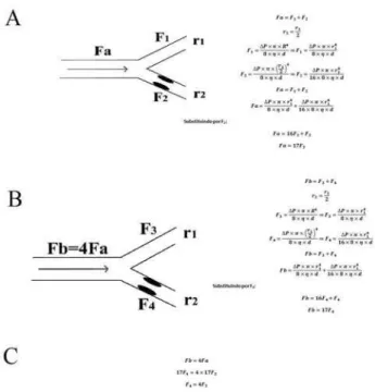
Supposing there is an obstructive lesion in the epicardial coronary artery, with the opening of the coronary arteries of resistance, resultin in a fourfold increase of the coronary blood flow. Figure 1 mathematicizes this scenario.
The flow in F4 is four times bigger than in F2, but F4 is 16 times smaller than F3, or that is, F4 has flow of 93.75% lower than F3.
However, when the dilations of resistance coronaries are processed preferentially in the ischemic area, there is predominant increase of the flow through this blocked vessel, by featuring the self-regulation.
When undergone to physical exercise, there is both openness of such coronaries and increase of myocardial oxygen consumption by increasing both the chronotropism as cardiac inotropism, which usually results in pain.
283
to the fourth power, the resulting total resistance is smaller than if it was revascularized only one arterial bed, increasing the flow through the graft, explaining the results obtained in the study by Nordgaard et al. [3].
Fig. 1 - Fa, Fb-full flow, F1, F2, F3, F4-Flow in the arterial segment; r1 and r2-radius of the arterial segment; ∆P- pressure difference at two points, R-radius (generic); η- viscosity, d-segment length. A - Calculation of the total flow as a function of flow in the stenotic branch; B – Calculation of the total flow as a function of the flow in the stenotic branch; C - Calculation of stenotic branches flow in each situation of total flow
When we have two vessels without significant obstruction that irrigate the same arterial system, making the hypothesis that blood pressure is the same for both, the flow will be inversely proportional to the resistance of the upstream segment of the anastomosis, since the resistance is the downstream same for both (in the same arterial territory). This division of flow leads to proportional reduction of speed (equation VII [1]), leading to thrombosis. Through this study I did not want to close the discussion of the application of physics, but, instead, instigate it. Even this discussion is not closed. Each topic that we chose to lecture in our article could be unfolded and each one would lead to a different study.
The acquisition of knowledge is a complex process, however summative and the history of its growth is still being told and probably never will end. I am very happy that our study has drawn the attention and I invite our
readers to expand these studies in the form of letters to the editor or other original articles.
Thank you,
Marcos Aurélio Barboza de Oliveira, São José do Rio Preto-SP
REFERENCES
1. Oliveira MAB, Alves FT, Silva MVP, Croti UA, Godoy MF, Braile DM. Conceitos de física básica que todo cirurgião cardiovascular deve saber. Parte I - Mecânica dos fluídos. Rev Bras Cir Cardiovasc. 2010;25(1):1-10.
2. Silva Jr MR. Fisiologia da circulação. 2ª ed. São Paulo:Edart;1977. p.1-35.
3. Nordgaard H, Vitale N, Haaverstad R. Transit-time blood flow measurements in sequential saphenous coronary artery bypass grafts. Ann Thorac Surg. 2009;87(5):1409-15.
Transposição das grandes artérias com comunicação interventricular e estenose pulmonar: qual a melhor opção cirúrgica?
Dear Editor,
We read with interest the article published by Furlanetto et al. [1] (BJCVS 25.1) that have demonstrated with two cases successfully operated, the creativity and technical capacity of Brazilian surgeons. It is up to us to remind you our article published in this Journal in 2003, regarding the long-term evolution (1 to 6.5 years) of the pulmonary root translocation to right ventricle outflow tract in the treatment of transposition of the great arteries associated with ventricular septal defect (VSD) and pulmonary stenosis (PS), with left ventricle blood flow diversion to the aorta through the VSD [2]. This technique, named Pulmonary Translocation, was described by da Silva et al. [3] having been published by invitation of the editor in the journal Operative Techniques in Thoracic and Cardiovascular Surgery in 2009 [4], with 39 cases and excellent long term results. The pulmonary translocation keeps the function of pulmonary valve, and maintains the aorta in its original position, without risk of coronary manipulation or aortic
284
insufficiency. The technique described by Furlanetto et al., compared to Hu et al. [5] technique, has the advantages to avoid Lecompte maneuver, reducing the time of aortic cross clamp, as well as to preserve the pulmonary function with mild stenosis. However, in both cases reported by these authors, the echocardiograms have shown mild aortic insufficiency in the immediate post-operative period, which in our experience has not occurred in any patient. In addition, the bovine pericardium patch treated with glutaraldehyde employed by these authors tends to calcify, impeding the growth of aortic and pulmonary rings, with possibility of distortion in long term follow-up, aggravating the aortic insufficiency. In our technique we achieved adequate reconstruction of left and right ventricles outflow tracts, with simpler technique and shorter extracorporeal circulation and aortic clamping times, keeping the growth potential of pulmonary ring with employment of in situ autologous pedicled pericardium [4] to complete the anterior aspect of this anastomosis. We believe that the follow-up of a large number of patients will clarify the real long term benefits of these singular techniques.
Luciana da Fonseca, São Paulo/SP
REFERENCES
1. Furlanetto G, Henriques SS, Pasquinelli FS, Furlanetto BHS. Nova técnica: translocação aórtica e pulmonar com preservação da valva pulmonar. Rev Bras Cir Cardiovasc. 2010;25(1):99-102.
2. Fonseca L, Baumgratz JF, Castro RM, Franchi SM, Vila JHA, Lopes LM, et al. Resultados tardios da translocação da raiz pulmonar na correção da transposição das grandes artérias. Rev Bras Cir Cardiovasc. 2003;18(4):326-31.
3. da Silva JP, Baumgratz JF, da Fonseca L. Pulmonary root translocation in transposition of great arteries repair. Ann Thorac Surg. 2000;69(2):643-5.
4. Silva JP, Fonseca L. Pulmonary root translocation. Operative Techniques in Thoracic and Cardiovascular Surgery. 2009;14(1):23-34.
5. Hu SS, Li SJ, Wang X, Wang LQ, Xiong H, Li LH, et al. Pulmonary and aortic root translocation in the management of transposition of great arteries with ventricular septal defect and left ventricular outflow tract obstruction. J Thorac Cardiovasc Surg. 2007;133(4):1090-2.
Reply
We would like to thank the comments posted by Dr. Luciana da Fonseca and elucidate the comment to remember the publication held by da Silva et al. [1], who performed the translocation of the pulmonary artery in the transposition of the great arteries and pulmonary stenosis in 2003, it is unnecessary because the work was referred and commented in the article (reference 4). The technique proposed by us also differs from the technique of Hu et al. [2] because it entirely saves the pulmonary valve, does not apply the LeCompte procedure or the right ventriculotomy. As for the involvement of the aortic valve, it is important to note that there was no moderate aortic insufficiency but mild aortic insufficiency, with no hemodynamic repercussions. Regarding the use of bovine pericardium treated with glutaraldehyde and growth of the aortic and pulmonary valves, we emphasize that this expansion should not compromise the growth of the aortic annulus because the expansion only makes up 25% to 20% of this ring, it will not interfere with the growth of the pulmonary annulus because this ring was reimplanted on the aortic orifice, which was partially closed with fresh autologous pericardium.
I also believe that the great advantage presented in this technique that we propose is to avoid tunneling the left ventricle into the aorta through the VSD, as with the Rastelli operation. This tunneling has a large loss of energy reflected in an important low output in the immediate postoperative period and it may evolve over the medium term obstruction of tunneling as a result of growth and contraction of the graft used. The great advantage of the technique of double aortic and pulmonary translocation is exactly avoiding this tunnel, correcting anatomically the outflow of the right and left ventricle and also preserving the pulmonary valve.
Gláucio Furlanetto, São Paulo/SP
REFERENCES
1. da Silva JP, Baumgratz JF, da Fonseca L. Pulmonary root translocation in transposition of great arteries repair. Ann Thorac Surg. 2000;69(2):643-5.
2. Hu SS, Li SJ, Wang X, Wang LQ, Xiong H, Li LH, et al. Pulmonary and aortic root translocation in the management of transposition of great arteries with ventricular septal defect and left ventricular outflow tract obstruction. J Thorac Cardiovasc Surg. 2007;133(4):1090-2.