Morphological changes induced by
testosterone in the mammary glands
of female Wistar rats
1Departamento de Ginecologia e Obstetrícia, EMESCAM, Vitória, ES, Brasil 2Departamento de Ginecologia e Obstetrícia, Faculdade de Medicina,
Universidade Federal de Minas Gerais, Belo Horizonte, MG, Brasil
3Departamento de Patologia, Centro Biomédico,
Universidade Federal do Espírito Santo, Vitória, ES, Brasil A. Chambô-Filho1,
A.F. Camargos2
and F.E.L. Pereira3
Abstract
Increased levels of androgens in postmenopausal women are consid-ered to be a risk factor for breast cancer. Testosterone, alone or in combination with estrogen, induces epithelial dysplasia and mammary tumors in Noble rats. Since this model of hormone-induced neoplasia has not been reported in other rat strains, we studied the effect of testosterone on the mammary gland morphology of female Wistar rats. Sixty adult, non-castrated, female Wistar rats were implanted in the dorsum midline with a silicone tube containing 50 mg testosterone (testosterone propionate in 30 animals and non-esterified testosterone in the remaining 30 animals) and 20 additional animals were im-planted with empty tubes and used as control. Five animals per group were killed 30, 60, 90, 120, 150, and 180 days after implantation, and the mammary glands were dissected, fixed and embedded in paraffin. Histological sections were then stained with hematoxylin and eosin and picrosyrius red for collagen visualization. Morphological and morphometric analysis demonstrated ductal proliferation and acinotubular differentiation with secretory activity in all treated ani-mals, peaking at 90 days of androgen exposure. After 90 days the proliferation of acinar epithelial cells was evident, but there was a progressive reduction of secretory differentiation and an increase in intralobular collagen fibers. There was no morphological evidence of dysplastic changes or other pre-neoplastic lesions. Testosterone treat-ment applied to adult, non-castrated female Wistar rats induced a mammary gland hyperplasia resembling the lactating differentiation, with progressive reduction in secretory differentiation.
Correspondence
F.E.L. Pereira
Departamento de Patologia Centro Biomédico, UFES Av. Marechal Campos, 1468 29040-090 Vitória, ES Brasil
Fax: +55-27-3335-7206 E-mail: [email protected]
Received October 21, 2003 Accepted December 14, 2004
Key words
•Testosterone •Androgens •Mammary gland
•Mammary gland hyperplasia •Mammary gland tumors
Introduction
Since increased androgen levels are ob-served in postmenopausal women and are even higher in those with breast cancer, these androgens have been proposed to be carcino-genic for mammary glands (1-3). In spite of
andro-gens on different lineages of mammary can-cer cells has been reported (reviewed in Ref. 6).
Experimental data are conflicting, with reports of a high frequency of mammary cancer in adult female Noble rats treated with testosterone and estrogen (7,8). Treat-ment with testosterone alone also induced mammary tumors in the same rat strain (7,8). In contrast, inhibitory effects of androgens on the development (9) or progression (10) of dimethylbenz(a)anthracene-induced mam-mary tumors in female rats have been re-ported. Occasional enhancement of tumor growth was observed in some experiments in which moderate or very high doses of androgen were used (reviewed in Ref. 11).
Ductal proliferation with development of acinotubular structures has been reported in mammary glands of female rats or mice treated with testosterone soon after birth (12) and near or after sexual maturation (13). Similar effects have been reported in cas-trated female rats after long-term treatment with the androgen precursors dehydroepi-androsterone and dihydrotestosterone (14).
Since androgens have been used for hor-monal replacement therapy in postmeno-pausal women (15-17) and since these hor-mones have been proposed to be potential carcinogens or promoters of carcinogenesis in Noble rats (7,8), but not in other rat strains, we studied the changes induced by testoster-one in the mammary glands of adult female Wistar rats to determine if this hormone was carcinogenic.
Material and Methods
Eighty adult female Wistar rats aged 90-95 days housed in groups of 10 per cage were allowed free access to tap water and a commercial pellet diet. The experiments were conducted at the Laboratory of Experimen-tal Pathology, Biomedical Center, Federal University of Espírito Santo, Vitória, ES, Brazil.
The rats were divided at random into two groups as follows: a) 60 rats received im-plants (a 35 x 2-mm silicone tube) with 50 mg of non-esterified testosterone (30 rats) or testosterone propionate (30 rats); b) 20 rats received empty implants and were used as control. Esterified and free testosterone (pow-der; Akzo-Nobel, Arnhem, The Netherlands) were used because the esterified form is inactivated slowly, and both have been used in hormonal replacement therapy. In all rats the implants were inserted subcutaneously in the dorsum midline.
At 30, 60, 90, 120, 150, and 180 days post-implant groups of 5 rats from each tes-tosterone group and 3 rats from the control group were killed after pentobarbital anes-thesia. The mammary glands were carefully excised, fixed in Bouin fluid (diluted 1:4 in 0.15 M NaCl) and embedded in paraffin. Sections were stained with hematoxylin and eosin and by the picrosirius red method for collagen. The morphological study focused mainly on the epithelial component of the mammary glands (acini and ducts) and on fibrous tissue within the lobules. Morpho-metric analysis was performed by the point-counting method (18) on five random photo-micrographs taken with a 10X objective and printed at the same magnification. A trans-parent grid was placed on the photomicro-graphs and the crossing points of the grid over the ducts or acini, adipose tissue and fibrous stroma were counted. The results are reported as volume density (%) of glands (acinotubular structures) in relation to the total volume of all the structures of the mam-mary gland (18) (volume density of acinotu-bular structures = points counted over acino-tubular structures/points counted over acini, ducts, adipose tissue, and fibrous stroma x 100). Because the abdominal gland was easier to identify correctly (presence of two lym-phatic nodules) it was used for morphomet-ric analysis.
differ-ences were considered to be significant for P values of less than 0.05.
Results
All the implants retrieved after 180 days contained small amounts of the hormone, demonstrating the continuous release of the drug during the experiment.
The rats treated with testosterone showed normal weight gain during the experiment and developed hypertrophy of the clitoris and more aggressive behavior than the con-trol group (data not shown).
The morphological alterations observed were similar in rats treated with testosterone propionate or non-esterified testosterone. Microscopic examination showed prolifera-tion of terminal and lateral end buds and acinotubular differentiation in all treated animals after 30 days of treatment. In all lobules there was secretory activity, with dilatation of acini and ducts and accumula-tion of secreaccumula-tion in the lumen (Figure 1B and C). The proliferation and secretory activity peaked at 90 days of androgen exposure, as demonstrated by morphologic observation and by the volume density of mammary glands obtained by morphometric analysis (Figure 2). After 90 days the glands pre-sented areas with different glandular pat-terns, with some showing acinotubular struc-tures with typical secretory activity and oth-ers showing less evident secretory differen-tiation, with reduction of the secretion accu-mulated in the lumen. Although the volume density of acinotubular structures did not increase, the acinar cells proliferated, filling the lumen. Several acinotubular structures showed acini with a solid pattern, filled with epithelial cells with small, clear vacuoles in the cytoplasm. The solid patterns of the acini as well as the reduction in secretory differen-tiation were progressive through 120, 150, and 180 days of androgen exposure (Figure 1). In all rats killed at 180 days the acinotu-bular structure had vacuolated cells, but
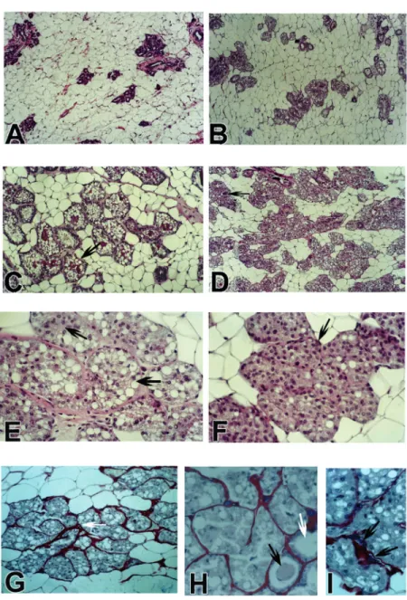
aci-Figure 1. Different morphologic aspects of the mammary gland after 6-month exposure of
female rats to testosterone. A, Low power view of the mammary gland of a control rat
(100X). B, Testosterone, 30 days. Acinotubular differentiation with secretory activity (100X).
C, Testosterone 60 days. Dilated acini (arrow) with secretory activity and with secretion in
the lumen (hematoxylin and eosin, H&E, 200X). D, Testosterone 120 days. Persistence of
the acinotubular hyperplasia, with the acinar cells filling the acinar lumen. Few acini (arrow)
with secretion accumulated in the lumen (H&E, 200X). E, Acini with vacuolated (secretory)
epithelial cells filling the lumen (arrows). In some acini there is loss of secretory differentia-tion evidenced by the absence of vacuoles. The nuclear pattern is not altered: there are
dark and clear nuclei (H&E, 400X) F, Testosterone 180 days. Acini with epithelial cells filling
the lumen, without vacuolization (arrow). In some acini there are vacuolated epithelial cells
evidencing persistent secretory activity (H&E, 400X). G, H and I, Testosterone 180 days.
Picrosirius red and hematoxylin staining for collagen. In G the white arrow indicates
increased intralobular collagen stained in red by sirius red. Epithelial cells with and without
secretory activity are filling the acini (200X). In H some acini are dilated, with (black arrow)
or without (white arrow) secretory material in the lumen and with hypotrophic acinar
epithelial cells (400X). In I the arrows indicate prominent basophilic mast cells stained by
nar epithelial cells without secretory activity were frequently present (Figure 1D-F). Most of the acini presented a solid pattern, with vacuolated acinar cells filling the lumen. The nuclear pattern observed in proliferat-ing acinar cells, with dark and clear nuclei, was similar to that observed in normal mam-mary acini. Epithelial proliferation filling the duct lumen was not observed and the myoepithelial cells apparently were unaf-fected. There was increased intralobular deposition of collagen after 90 days of an-drogen exposure (Figure 1H). Mast cells were frequently prominent in the intralobu-lar stroma with basophilic granules in the cytoplasm (Figure 1I).
The proliferation and secretory effects induced by testosterone exposure were simi-lar in all mammary gland pairs. No dysplas-tic lesions with nuclear pleomorphism or neoplastic lesions were observed in any treated animal.
Discussion
Adult, non-castrated, female rats were used because androgens have been used in combination with estrogens in hormonal re-placement therapy during perimenopause (6), when the ovaries still have some degree of activity.
The present study clearly demonstrated the potent proliferative effect of testosterone on the mammary glands of adult, non-cas-trated, female Wistar rats. There was ductal hyperplasia with differentiation of acinotu-bular structures and prominent secretory ac-tivity. Our observations up to 90 days of
exposure are similar to those reported for non-castrated adult female rats treated with testosterone propionate for 90 days (13) or castrated female rats treated with dehydroe-piandrosterone for 12 months (14). The histomorphologic pattern observed up to 60-90 days of testosterone exposure resembles those seen during pregnancy and lactation and could be defined as pseudo-gestational hyperplasia. After 90 days there was a pro-gressive loss of secretory differentiation, with increased deposition of collagen in the in-tralobular stroma. However, epithelial hy-perplasia was not seen in the ducts and nuclear pleomorphism was not observed in the aci-nar or ductal epithelial cells. In addition, proliferative lesions resembling those de-scribed by Xie et al. (7,8) in female Noble rats treated with testosterone were not ob-served. Thus, it appears that testosterone, although it induces a secretory acinotubular hyperplasia in non-castrated female Wistar rats, does not induce apparent preneoplastic lesions after 6 months of hormonal treat-ment.
The progressive loss of secretory differ-entiation after 90 days of testosterone treat-ment has not been reported by other investi-gators who studied the effects of testoster-one on non-castrated adult female rats. This may be due to differences in the time when treatment was started (11) or to the short period of observation (13). Sourla et al. (14) did not report alterations in the secretory pattern of the mammary acini of castrated rats up to 12 months of treatment with dehy-droepiandrosterone.
We have no explanation for the effect of testosterone on the mammary gland, induc-ing a strong proliferative and secretory activ-ity, followed by a reduction in secretory differentiation. Clinical observations in women indicate that testosterone induces atrophy of the mammary gland. However, in experimental animals (rats and mice) the hormone induces acinotubular differentia-tion and hyperplasia. In castrated female rats
Volume density (%)
70 123 123 123 123 12 12 12 12 12 12 12 12 123 123 123 123 123 123 123 123 123 12 12 12 12 12 12 12 12 12 12 12 12 123 123 123 123 123 123 123 123 123 123 123 123 12 12 12 12 12 12 12 12 12 12 12 12 * * *+ *+ *+
Co 30 60 90 120 180
Days of treatment 60 50 40 30 20 10 0 Figure 2. Volume density of the
mammary gland (acini and ducts) of adult female rats that received a silicone implant with 50 mg of testosterone. Co = control group consisting of 120-day-old female adult rats that re-ceived an empty silicone tube. The animals were killed 30, 60, 90, 120, and 180 days after im-plant of a silicone tube with tes-tosterone. Data are reported as the mean ± SD for five animals per group. *P < 0.05 compared to the control group
(Kruskal-Wallis test). +P < 0.05 compared
the induction of mammary acinotubular pro-liferation and secretion was blocked by flutamine, indicating a direct action of tes-tosterone on androgen receptors in acinar and tubular cells (14). In non-castrated fe-male rats the presence of an intact ovary would modify some effects of testosterone because this hormone induces increased ex-pression of prolactin receptors in mammary epithelial cells (19). In addition, the prolif-erative effect of exogenous testosterone may be related to the increased estrogen levels as a consequence of two other possible mechan-isms: i) mammary glands have a high aromatase activity that transforms testoster-one to estrogen (6); ii) corpus luteum was not observed in the ovaries of androgen-treated rats (data not shown), that probably were not cycling, with persistent production of estrogen in follicles. However, in these experiments plasma estrogen levels were not determined in testosterone-treated rats.
The increased collagen deposition in the intralobular stroma has not been reported in
castrated female rats treated with dihydro-testosterone or dehydroepiandrosterone af-ter 12 months of exposure (14) or in non-castrated animals after 90 days of treatment with testosterone propionate (13). Only Xie et al. (7,8) reported a proliferative effect of androgens on mammary periacinar connec-tive tissue of Noble rats, and these investiga-tors proposed that the carcinogenic effect of testosterone may be mediated by paracrine interrelations between stromal and epithelial cells.
Our data demonstrate that long-term tes-tosterone treatment of non-castrated, adult, female Wistar rats induced proliferation and secretory activity of the mammary glands with a progressive loss of secretory differen-tiation after 120 days, a result that could be defined as pseudo-gestational hyperplasia. The lack of evidence of epithelial dysplasia, as reported by Xie et al. (7,8), may reflect the differences in expression of androgens and estrogen receptors between Noble and Wistar rats (8,20,21).
References
1. Secreto G, Toniolo P, Berrino F, Recchione C, Cavalleri A, Pisani P, Totis A, Fariselli G & Di Pietro S (1991). Serum and urinary
andro-gens and risk of breast cancer in postmenopausal women. Cancer
Research, 51: 2572-2576.
2. Liao DJ & Dickson RB (2002). Roles of androgens in the
develop-ment, growth, and carcinogenesis of the mammary gland. Journal
of Steroid Biochemistry and Molecular Biology, 80: 175-189. 3. Yu H, Shu XO, Shi R, Dai Q, Jin F, Gao YT, Li BD & Zheng W (2003).
Plasma sex steroid hormones and breast cancer risk in Chinese
women. International Journal of Cancer, 105: 92-97.
4. Dorgan JF, Longcope C, Stephensosn HE, Falk RT, Franz C, Kahle L, Campbell WS, Tangrea JA & Scatzkin A (1997). Serum sex hormone levels are related to breast cancer risk in postmenopausal women.
Environmental Health Perspectives, 105 (Suppl): 583-585. 5. Dimitrakakis C, Zhou J & Bondy CA (2002). Androgens and
mam-mary growth and neoplasia. Fertility and Sterility, 77 (Suppl 4):
26-33.
6. Somboonporn W & Davis SR (2004). Testosterone effects on the
breast: implications for testosterone therapy for women. Endocrine
Reviews, 25: 374-388.
7. Xie B, Tsao SW & Wong YC (1999). Induction of high incidence of mammary tumor in female Noble rats with combination of
17ß-oestradiol and testosterone. Carcinogenesis, 20: 1069-1078.
8. Xie B, Tsao SW & Wong YC (1999). Sex hormone-induced mam-mary carcinogenesis in female Noble rats: the role of androgens.
Carcinogenesis, 20: 1597-1606.
9. Li S, Yan X, Bélanger A & Labrie F (1993). Prevention by dehydroepi-androsterone of the development of mammary carcinoma induced
by 7,12-dimethylbenz(a)anthracene (DMBA) in the rat. Breast
Can-cer Research and Treatment, 29: 203-217.
10. Dauvois S, Li S, Martel C & Labrie F (1989). Inhibitory effect of
androgens on DMBA-induced mammary carcinoma in the rat. Breast
Cancer Research and Treatment, 14: 299-306.
11. Welsch CW (1985). Host factors affecting the growth of carcinogen-induced rat mammary carcinomas: a review and tribute to Charles
Brenton Huggins. Cancer Research, 45: 3415-3443.
12. Tomooka Y & Bern HA (1982). Growth of mouse mammary glands
after neonate sex hormone treatment. Journal of the National
Can-cer Institute, 69: 1347-1352.
13. Laqueur GL & Fluhmann CF (1942). Effects of testosterone
propi-onate in immature and adult female rats. Endocrinology, 30: 93-101.
14. Sourla A, Martel C, Labrie C & Labie F (1998). Almost exclusive androgenic action of dihydroepiandrosterone in the rat mammary
gland. Endocrinology, 39: 753-764.
15. Casson PR & Carson SA (1996). Androgen replacement therapy in
Menopausal Studies, 41: 412-422.
16. Wang C & Swerdloff RS (1997). Androgen replacement therapy.
Annals of Medicine, 29: 365-370.
17. Burd ID & Bachmann GA (2001). Androgen replacement in
meno-pause. Current Women’s Health Reports, 1: 202-205.
18. Baak JPA & Oort J (1983). A Manual of Morphometry in Diagnostic
Pathology. Springer-Verlag, Berlim, Germany, 7-21.
19. Baratta M, Grolli S, Poletti A, Ramoni R, Motta M & TamaninI C (2000). Role of androgens in proliferation and differentiation of
mouse mammary epithelial cell line HC11. Journal of
Endocrinol-ogy, 167: 53-60.
20. Liao DJ, Pantazis CG, Hou X & Li AAS (1998). Promotion of estro-gen-induced mammary gland carcinogenesis by androgen in the
male Noble rat: probably mediated by steroid receptors.
Carcino-genesis, 19: 2173-2180.
21. Wong YC & Xie B (2001). The role of androgens in mammary
carcinogenesis. Italian Journal of Anatomy and Embryology, 106