on the somatic maturation of wistar rat offspring fed
a regional basic diet during pregnancy
Anselmo, CWSF.
a*, Silva, TL.
b, Holanda, TG.
b, Prado, LVS.
b,
Cabral-Filho, JE.
b, Catanho, MTJA.
cand Medeiros, MC.
baDepartamento de Fisioterapia, Universidade Federal de Pernambuco – UFPE,
Av. Prof. Moraes Rego, 1235, Cidade Universitária, CEP 50670-901, Recife, PE, Brazil
bDepartamento de Nutrição, Universidade Federal de Pernambuco – UFPE,
Av. Prof. Moraes Rego, 1235, Cidade Universitária, CEP 50670-901, Recife, PE, Brazil
cDepartamento de Biofísica, Universidade Federal de Pernambuco-UFPE,
Av. Prof. Moraes Rego, 1235, Cidade Universitária, CEP 50670-901, Recife, PE, Brazil *e-mail: [email protected], [email protected]
Received November 20, 2006 – Accepted March 19, 2007 – Distributed August 31, 2008
Abstract
The aim of the present study was to observe how the exposition of pregnant rats to an electromagnetic field (EMF),
with frequency of 60 Hz, and a magnetic field of 3 MT for 2 hours per day and/or using the so-called Regional Basic
Diet (RBD), influenced the somatic maturation in their offspring. Four groups were formed: Group A (casein), B (casein and EMF), C (RBD) and D (RBD and EMF). The diet manipulation occurred during pregnancy. The somatic maturation indexes - assessed daily between 12:00 AM and 2:00 PM - were: Eye Opening (EO), Auricle Opening (AO), Auditory Canal Opening (ACO), Low Incisor Eruption (LIE), and Upper Incisor Eruption (UIE). The associa-tion between EMF and deficient diet caused a delay in all Somatic Maturaassocia-tion Indexes (SMI) and the RBD caused delay only in the AO. Furthermore, the EMF caused delay in AO, ACO, LIE. In relation to the body weight, the EMF associated with the deficient diet caused change in the twenty-first day of life. The RBD, during pregnancy, caused lower body weight in the offspring in the first and third day of life. The body weight of the offspring whose mothers were fed casein and exposed to the EMF during pregnancy was lower in the third and sixth day of life. In conclusion, the EMF associated with under-nutrition caused delay in all SMI. In relation to the body weight, the EMF associated with under-nutrition caused a decrease in the body weight at the sixth day of life.
Keywords: casein, electromagnetic field, neurodevelopment, somatic maturation, regional basic diet.
Influência do campo eletromagnético de 60 Hz, 3 µT, na maturação somática de
filhotes de ratos wistar alimentados pela dieta básica regional durante a prenhez
Resumo
O objetivo deste estudo foi observar a influência do campo eletromagnético (CEM), com freqüência de 60Hz, campo
magnético de 3 MT, durante 2 horas por dia, associado ou não à dieta básica regional (DBR) no desenvolvimento
so-mático da prole. Quatro grupos foram formados: Grupo A (caseína), B (caseína e CEM), C (DBR) e D (DBR e CEM). A manipulação dietética ocorreu durante a prenhez. Os índices de maturação somática – Abertura dos Olhos (AO), Abertura do Pavilhão Auditivo (APA), Abertura do Conduto Auditivo (ACA), Erupção do Incisivo Inferior (EII), e Erupção do Incisivo Superior (EIS) - foram avaliados diariamente entre 12 e 14 horas. A associação entre o CEM e a dieta deficiente causou retardo em todos os índices de maturação somática (IMS) e a DBR causou retardo somente na APA. O CEM causou retardo na APA, ACA, EII. Em relação ao peso corporal, o CEM associado à dieta deficiente causou mudanças no 21° dia de vida. A DBR, durante a prenhez, causou diminuição do peso corporal dos filhotes no 1° e no 3° dia de vida. O peso corporal dos filhotes, cujas mães foram alimentadas pela caseína e expostas ao CEM, durante a prenhez, apresentaram uma diminuição no 3° e 6° dia de vida. Conclusão: o CEM, associado com a desnu-trição, causou retardo em todos os IMS. Em relação ao peso corporal, o CEM, associado à desnudesnu-trição, causou uma diminuição no peso corporal no 6° dia de vida.
Palavras-chave: caseína, campo eletromagnético, desenvolvimento nervoso, maturação somática, dieta básica
1. Introduction
Epidemiological studies have implicated maternal protein-calorie deficiency as an important public health problem in developing countries (Pissaia et al., 1980; Olubodun, 1992). In North-Eastern Brazil the diet that is consumed by the population living in the area of sugar-cane cultivation in coastal Pernambuco is known as the Regional Basic Diet or RBD (Teodósio et al., 1990).
The RBD was prepared by Teodósio et al. (1981) ac-cording to data from food consumption surveys in the Pernambuco coastal forest strip. The RBD is made from the most common foods and in the same proportion as that consumed by the population, as detected by surveys. Pioneer studies have indicated that this experimental diet, RBD, produces in rats a type of under-nutrition sim-ilar to that prevalent among children from this region of Brazil, which is associated with nutritional dwarfism and some clinical signs of marasmus (Silva, 1987). When this diet is compared with the standard one, it is seen to be deficient in proteins (content and quality), calories, fat, vitamins and minerals (Pessoa et al., 2000). From the obtained results, the RBD has been consolidated as a malnourishment experimental model (Guedes et al., 1987; Teodósio et al., 1990; Rocha de Melo and Guedes, 1997; Pessoa et al., 2000).
The development of the nervous system (NS) in-volves epigenetic processes that activate the genes in a sequential manner at different times (Jessel, 1995). These processes have many environmental and non cellular fac-tors that can change, modulate and direct the next stage of development (Nishi, 1994). Hence, only the genetic information in a living being may be insufficient to spec-ify the whole neuronal interconnections (Jessel, 1995). Morgane et al. (1993) showed that the NS development in mammals happens according to ‘time planning’ in which the different phases follow a predetermined chro-nology. This fast growth period of the NS is named the ‘critical or vulnerable’ period, being considered as the unique opportunity for its development. Therefore, its beginning and duration are different for different spe-cies. In humans this ‘critical period’ begins in the womb (the last three months of pregnancy) and lasts until the beginning of the postnatal phase (two to four years old). In rats this happens after birth (breast-feeding period). The series of events presented by the NS during the pre-natal and postpre-natal development determines the neuro-chemical composition and the definite morphofunctional structure, present in adults (Morgane et al., 1993).
Much experimental and clinical evidence has shown that aggression during those critical periods can change the ontogenesis sequence of events with diverse and per-sistent effects on the NS (Morgane et al., 1993; Dobbing, 1970; Lynch et al., 1975; Manhães de Castro et al., 2001). Experimental studies have shown that pre- or postnatal nutritional manipulation may programme adult size, metabolism, blood lipids, diabetes, obesity, behav-iour, and learning (Lucas, 1998). In trying to understand
these effects and living beings’ capability to adapt to dif-ferent forms of aggression, many researchers have been investigating the effects of different kinds of nutritional injury to the nervous system in the early phases of its development. These studies have shown morphologic, neurochemical, endocrine and functional alterations (Morgane et al. 1993; Del-Prado et al., 1997).
Over recent decades a remarkable diffusion of elec-trical equipment and an increased level in electromag-netic fields (EMF) in the environment characterize our society. Recent epidemiological studies of occupational and residential exposure to EMF are concerned with the biological effects of the 50-60 Hz fields (extremely low frequencies or ELF), particularly in relation to de-termining an increase in cancer incidence in individuals exposed to these types of radiation (Galloni and Marino, 2000). In accordance with Marino (2005), in order to un-derstand the relationship between environmental factors and disease in terms of an internal state variable called stress (for instance, death of a loved one, loss of job, an unhappy marital situation, poor diet, etc.), it is helpful to understand the influence of environmental EMFs.
Experiments investigating the possible effects of EMF associated with under-nutrition during pregnancy on development and somatic maturation of the offspring are scarce. The aim of the present study was to observe
how the exposition of pregnant rats to a 60 Hz, 3 MT,
EMF for two hours per day and/or using a RBD influ-enced the somatic maturation indexes in their offspring.
2. Materials and Methods
2.1. Animals
In this experiment 66 male, newly born rats from the Wistar strain, whose mothers were submitted to four different conditions, were used. The female rats were 90 days old when they mated. Fertilization was detected by the presence of sperm in the vaginal wash-ing of the mated females. Fertilized females were then immediately transferred to cages, two per cage, which were 60 cm long, 50 cm wide and 22 cm high, and put on supports made from polystyrene 35 cm wide, 50 cm long and 35 cm high (Lucena et al., 2002).The pregnant rats were divided in four Groups: Group A, composed of rats that consumed casein without being exposed to EMF; Group B, composed of rats that consumed casein and were exposed to EMF; Group C, composed of rats that consumed the RBD and were not exposed to EMF; and Group D, composed of rats that consumed RBD and were exposed to EMF. They were kept in conditions of
constant temperature (23 p 2 °C) and a light/dark cycle
(12/12 hours) with a background magnetic field of less than 0.3 MT.
form new groups. In this way, the genetic factor did not interfere with the experimental results. Every group from each female was reduced to six individuals on the second day after birth. Only male rats were used. Animal use was approved by the Federal University of Pernambuco Committee on Animal Research.
2.2. Diets
The ingredients of the multideficient diet used in this
experiment were beans (Phaseolus vulgaris), manioc
flour (Manihot esculenta), dried and salted meat and
sweet potato (Ipomaea batatas) (Teodósio et al., 1981).
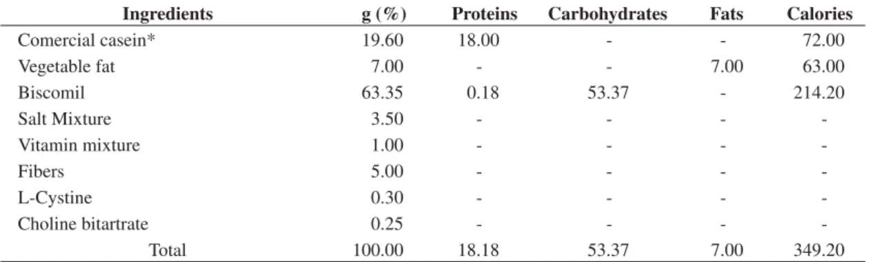
The diet was prepared in our laboratory as follows: all in-gredients (except manioc flour) were cooked, dehydrat-ed for 24-60 hours (according the type of ingrdehydrat-edient) at 60 °C and pulverized. Each component was mixed with manioc flour by humidifying. Meat fat was then added, and the mixture was shaped into balls which were dehy-drated for 24 hours at 60 °C. The centesimal composition of the RBD, which was determined by the Department of Nutrition, the Federal University of Pernambuco, is giv-en in Table 1. The caloric adequacy of the RBD was cal-culated to be about 316 Kcal per 100 g. The control diet provided 18% of protein (commercial casein) and was balanced according to recommendations for pregnant rats (AIN – 93), as shown in Table 2. The diet during the
mating period was maintenance pellet food (Purina® do
Brasil Ltd., São Paulo, SP, Brazil).
2.3. Feeding protocol
During the mating period all female and male rats were fed a standard balanced diet provided by Purina. In this investigation the diets (RDB and casein) were only used when the rats presented a positive test for pregnancy. The animals had food and water ad libittum. Two Groups were fed RBD (Groups C and D) and two Groups were fed casein (Groups A and B).
2.4. Exposure to EMF
The groups of pregnant rats fed casein and RBD, Groups B and D respectively, were exposed to a 60 Hz
senoidal, of 3 MT EMF, measured by a gauss meter, on
the scale of 0-100 mG (0-10 MT) for two hours per day
for twenty-one consecutive days, one hour in the morn-ing from 8:00 to 9:00 AM hours and one hour in the af-ternoon from 2:00 to 3:00 PM hours.
They were exposed when pregnancy was detected and removed from exposure when they gave birth. As the radiation source, transformers of 220/110 V of 500 W working with opened secondary and control-led by an electronic timer were used and placed under the polystyrene supports. Control groups of animals, Groups A and C, fed with casein and RBD respectively, were obtained by simply not placing the transformers under the polystyrene supports. The transformers were previously examined and tested to confirm their working
Table 1. Centesimal composition of the Regional Basic Diet (RBD).
Ingredients g
(%)
Proteins (%)
Carbohydrates (%)
Fats (%)
Fibers (%)
Kcal (%)
Beans 18.34 3.99 10.66 0.24 1.09 60.76
Manioc flour 64.81 0.84 48.59 0.12 5.64 198.80
Poor fat dried and salted meat fat 3.74 2.74 - 0.21 - 12.85
Dried and salted meat fat 0.35 - - 0.35 - 3.15
Sweet potato 12.76 0.30 9.99 0.03 0.48 41.43
Total 100.00 7.87 69.24 0.95 7.21 316.99
Table 2. Centesimal composition of the Control Diet (Casein).
Ingredients g (%) Proteins Carbohydrates Fats Calories
Comercial casein* 19.60 18.00 - - 72.00
Vegetable fat 7.00 - - 7.00 63.00
Biscomil 63.35 0.18 53.37 - 214.20
Salt Mixture 3.50 - - -
-Vitamin mixture 1.00 - - -
-Fibers 5.00 - - -
-L-Cystine 0.30 - - -
-Choline bitartrate 0.25 - - -
-Total 100.00 18.18 53.37 7.00 349.20
parameters at the Biomedical Engineering Department of the Biophysics and Radiobiology Department of the Federal University of Pernambuco. The EMF was measured inside the cage and it remained uniform ir-respective of its position. It should be noted that, except for the weekly cage cleaning and the weekly measur-ing of their weight, the rats were not moved or handled during this experiment. Exposed animals should be compared with control groups that have been derived from the same source and simultaneously handled and assayed in the same way, except for their exposure to the fields.
2.5. Somatic maturation indexes
The following somatic maturation indexes were investigated on a daily basis, between 12:00 AM and 2:00 PM hours from the second day after birth until when somatic maturation had occurred, namely Eye Opening (EO), Auricle Opening (AO), Auditory Canal Opening (ACO), Low Incisor Eruption (LIE) and Upper Incisor Eruption (UIE) (Smart and Dobbing, 1971). They were considered positive when the two eyes, the two auricles and the two auditory canals were opened and the two lower and upper incisors had appeared.
2.6. Somatic growth
2.6.1. Pup body weight
The animals were weighed on the 1st, 3rd, 6th, 9th,
12th, 15th and 21st day of life. The scale used was a digital
model, Quimis, with 0.1 g sensitivity.
2.7. Statistical analysis
The results of the somatic maturation development were evaluated by the Kruskal-Wallis analysis of vari-ance, followed by Dunn’s test for multiple comparisons among groups, with the significant level considered to be p < 0.05.
The Variance Analysis for Repeated Measures fol-lowed by the Tukey test was employed for comparisons of the body weight. When the normality test had been passed, the One Way Analysis of Variance (ANOVA)
was used followed by the Tukey Test for multiple com-parisons among groups. When the normality test had been failed, the Kruskal-Wallis analysis of variance was used followed by Dunn’s test for multiple comparisons among groups, considering the significant level consid-ered to be p < 0.05.
3. Results
3.1. Somatic maturation indexes
Comparing the suckling group from mothers ex-posed to EMF and malnourished, during pregnancy (Group D) to the suckling group from mothers fed casein (Group A), a delay was observed in the day (in median) that the following somatic maturation indexes appeared: AO, ACO, EO, UIE and LIE, (p < 0.05) (Table 3).
When the different diets were considered, the off-spring whose mothers were fed with a deficient diet and exposed to the EMF during pregnancy (Group D) showed a delay in the AO and EO indexes when compared to Group B whose mothers were fed casein and exposed to the EMF during pregnancy (p < 0.05) (Table 3).
When comparison was made between the offspring whose mothers were fed RBD, one group of which was exposed and the other group not exposed to (Groups D and C), during pregnancy, only a delay in the AO somatic index was noticed (p < 0.05) (Table 3). When compar-ing the groups whose mothers were fed casein diet, one group of which was exposed to EMF and the other group not during pregnancy (Groups B and A), Group B pre-sented a delay in the following somatic maturation in-dexes: AO, ACO and LIE (p < 0.05) (Table 3).
3.2. Body weight
After performing the statistical analysis of the body weight of the suckling over the first twenty-one days of live, there was no statistical difference observed between the weights measured from the first to the third day. However, from the fourth day a statistical weight gain was observed in all the groups.
Table 3. Somatic maturation times (age in days) of offspring from rats subjected to four different treatments during preg-nancy.
Groups*/SMI** A (n = 12) B (n = 24) C (n = 18) D (n = 12)
Md (Q1 – Q3)
EO 12.5 (10-13)a 13 (11-14)b 13 (13-14) 14 (14-14)ab
AO 1.5 (1-2)cde 3 (3-3)cf 3 (3-4)dg 4 (4-4)efg
ACO 10.5 (10-11)hi 13 (12-13)h 12 (12-13) 13 (13-13.5)i
LIE 10 (9-10)jk 12 (12-13)j 10.5 (10-14) 12 (12-12)k
UIE 8 (8-8.5)l 9 (8-13) 9 (7-11) 11 (10-11)l
Table 4 shows the suckling average weight p stand-ard error for the Groups A, B, C and D during the 1st, 3rd,
6th, 9th, 12th, 15th and 21st days after birth. At the twenty
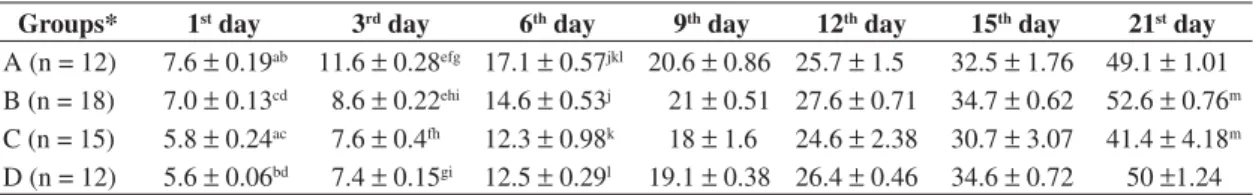
first day of life it was observed that the diet associated with the EMF, taken together, changed the body weight of offspring, where as Group C (formed from offspring of rats fed RBD but not subjected to EMF) showed lower body weight than Group B (formed from offspring of rats fed casein and subjected to EMF during pregnancy).
When the diet was considered, the offspring whose mothers were fed RBD during pregnancy (Groups C and D) had lower weight on the first and third day of life than the offspring whose mothers were fed casein (Groups A and B).
The EMF caused a decrease in the mean weight of offspring from rats fed casein and exposed to the EMF, during pregnancy (Group B), at the third and sixth day of life when compared with offspring of rats fed casein during pregnancy (Group A).
4. Discussion
The choice of intensity of 3 MT was due to the
ne-cessity of using a higher intensity of EMF than in resi-dences and most work places. In those places the average 50/60 Hz magnetic fields, as determined by the National Academy of Science (National Academy of Science,
1996), are between 0.1 and 0.3 MT. In this study, a value
ten times higher than the maximum expected value was used. Depending on the distance between the conduc-tors and the ground, the magnetic average flux densities
can reach 22 MT resulting from the current load in the
line (Simon, 1992). This value may also depend on the geographic location and the nature of the magnetic ma-terial near the subject area (Repacholi and Greenebaum, 1999).
The RBD was chosen as the experimental diet in this study because it is not only deficient in protein, but also in lipids, vitamins, sodium and other minerals (Teodósio et al., 1990; Pessoa et al., 2000; Monteiro et al., 2001). The pregnant rats were fed casein or RBD when they presented a positive test for pregnancy. According to Frazer and Huggett (1970), the fetus competes with the mother for the nutrients, but only for those that are ingested and stored throughout the pregnancy. They do
not compete for those already absorbed by the mother’s tissues at the moment of conception.
In this study, the somatic maturation indexes were in-vestigated from the second day after birth until when so-matic maturation had occurred, because this period cor-responds to the breast-feeding period and is considered by Dobbing (1970) as the critical period for development. The observation of the normal sequence of the stages of development in the ontogenesis of the reflexes and acqui-sition of the mature standard of the locomotive activities can be valuable indexes of the maturation and develop-ment of the nervous system (Smart and Dobbing, 1971; Walton et al., 1992; Morgane et al., 1993; Deiró et al., 2005). In rats, these patterns are established in the first three weeks after birth (Gramsbergen, 1998).
The offspring from rats exposed to EMF and RDB, taken together, presented a delay in all somatic matura-tion indexes. In accordance with Anselmo (2005),
preg-nant rats fed RBD and exposed to a 60Hz, 3 MT, EMF
for two hours per day showed a low dosage of serum
T3, which is in agreement with the results found in this
study. According to Porterfield and Hendrich (1993), Reyns et al. (2005), thyroid hormones are very important for the embryonic development in all vertebrates. In hu-mans, this is more evident in the central nervous system where the thyroid hormone deficit during the fetal and the neo-natal periods results in the syndrome of cretin-ism, which is characterized by hearing impairment, atax-ia, mental disability, and abnormal growth. According to Vara et al. (2002), the hypothyroidism which manifests during the development of rats, results in learning diffi-culties, delay in locomotion and cerebral skills and short-ened interneuron connectivity.
In this study offspring from rats fed casein showed delay in the auricle opening, auditory canal opening and lower incisor eruption when exposed to EMF. These results do not match with the results of Zusman et al. (1990), in which they observed a delay in the eye open-ing of offspropen-ing whose mothers were exposed to EMF of 20, 50, and 100 Hz, during pregnancy. On the other hand, the results obtained in this study agree with the results of Sokolova (1998) and Sienkiewicz et al. (1994). Sokolova (1998) reported that exposure to 2 or 20 mT fields did not exert any effect on eye opening in mice, and identical results were presented by Sienkiewicz et al.
Table 4. Offspring weight (in grams) born from rats subjected to four different treatments during pregnancy, on the 1st, 3rd,
6th, 9th, 12th, 15th, 21st days of life.
Groups* 1st day 3rd day 6th day 9th day 12th day 15th day 21st day
A (n = 12) 7.6p 0.19ab 11.6p 0.28efg 17.1p 0.57jkl 20.6p 0.86 25.7p 1.5 32.5p 1.76 49.1p 1.01
B (n = 18) 7.0p 0.13cd 8.6p 0.22ehi 14.6p 0.53j 21p 0.51 27.6p 0.71 34.7p 0.62 52.6p 0.76m
C (n = 15) 5.8p 0.24ac 7.6p 0.4fh 12.3p 0.98k 18p 1.6 24.6p 2.38 30.7p 3.07 41.4p 4.18m
D (n = 12) 5.6p 0.06bd 7.4p 0.15gi 12.5p 0.29l 19.1p 0.38 26.4p 0.46 34.6p 0.72 50p1.24
*A = casein; B = casein + electromagnetic field; C = regional basic diet; D = regional diet + electromagnetic field; The data show the offspring weight, in grams, mean p standard error, on the 1st, 3rd, 6th, 9th, 12th, 15th, 21st days of life. Equal
(1994) for CD1 mice exposed to 20 mT fields. However, Sienkiewicz et al. (1994) suggest that prenatal exposure to a 50 Hz, 20 mT magnetic field does not engender any gross impairment in postnatal development.
In our experiments we observed that the EMF caused a decrease in the mean weight in Group B (formed from offspring of rats fed casein and subjected to EMF) when compared to Group A (formed from offspring of rats fed by casein) at the third and sixth days of life. This re-sult agrees in part with Nishikawa et al. (1986). They observed that suckling mice exposed to pulsed fields of 1.6 mT during pregnancy suffered a decrease in their body weight between the second and the fifth days of life and an increase between the eighth and the twenty-first days of life. Zusman et al. (1990), showed that the body weight of rats, exposed to the EMF, 20 or 100 Hz dur-ing pregnancy was lower on the first day of life, while animals exposed to EMF of 50 Hz suffered a decrease in their body weight between the first and twenty-sixth days of life.
Our findings did not match the results obtained by Sokolova (1998), Matos et al. (2001) and Sienkwicz et al. (1994). Matos et al. (2001) observed that rats exposed to an EMF of 60 Hz during the breast-feeding period did not present changes in weight evolution when compared to the control group, but our rats were exposed during preg-nancy. This difference makes the comparison difficult. Sokolova (1998) and Sienkwicz et al. (1994) observed that after exposition to the EMF (50 Hz; 2 and 20 mT) during pregnancy and the postnatal (6 to 90 days) pe-riod, there were no changes in the body weight in mice. Rivas et al. (1985) exposed Swiss mice to a 50 Hz pulsed
field at either 2.3 mT or 83 MT from conception until
120 days of age. Male and female mice exposed at the higher field strength were significantly lower in weight at 120 days, but only males were reduced after exposure to the weaker field. The changes in weight were reported to increase throughout the duration of the experiment.
Groups that were fed RBD had a mean body weight lower than the groups fed casein at the first and third days of life. This can be explained by the fact that the suckling depends on its mother for a broad range of physiologi-cal functions, i.e. body temperature maintenance, urine excretion and feeding (Friedman, 1975). In approximate terms sucklings have their mother as the only source of food until the fourteenth day of life. The milk availabil-ity varies according to the presence of the mother in the nest and her emotional and nutritional states (Friedman, 1975). According to Teodósio et al. (1990), the RBD presents low vegetal protein content. These proteins are considered to be low in quality when compared with animal proteins, because they do not provide essential amino acid levels necessary for normal body develop-ment (Teodósio et al., 1990). Another important factor is that when the eye opening occurs (around the fourteenth day of life), the offspring are stimulated to eat solid food when the mother brings it to the nest (Galef and Clark, 1971).
What was done is this experiment can be called pro-gramming, because according to Lucas (1994), the nutri-tion and metabolic changes that act in the early stages of survival of individuals will permanently affect changes in the animal’s physiology. Experimental studies have shown that nutritional manipulation pre-and post-birth can program the size of the adult individuals, their me-tabolism, blood lipid levels, diabetes, obesity, blood pressure, behavior and learning skills (Lucas, 1998; Waterland and Garza, 1999).
5. Conclusion
The total Somatic Maturation Indexes studied in this research were delayed by the association of the EMF and malnutrition during pregnancy. In relation to the body weight, the association between the EMF and malnu-trition during pregnancy caused a decrease in the body weight at the sixth day of life.
References
ANSELMO, CWSF., 2005. Efeitos do Campo Eletromagnético de 60 Hz, 3 MT, na regulação hormonal e metabólica de ratas prenhas Recife. Pernambuco: Universidade Federal de Pernambuco, CCS Nutrição. [Tese de Doutorado].
DEIRÓ, TCBJ., MANHÃES-DE-CASTRO, R., CABRAL-FILHO, JE., BARRETO-MEDEIROS, JM., SOUZA, SL., MARINHO, SMOC., CASTRO, FMM., TOSCANO, AE., JESUS-DEIRÓ, RS. and BARROS, KMFT., 2005. Sertraline delays the somatic grown and reflex ontogeny in neonate rats. Physiology and behavior, vol. 87, no. 2, p. 338-344.
DEL-PRADO, M., DELGADO, G. and VILLALPANDO, S., 1997. Maternal Lipid Intake during Pregnancy and Production and Litter Growth in Rats. J. Nutr., vol. 127, no. 3, p. 458-462. DOBBING, J., 1970. Undernutrition and the developing brain. The relevance of animal models to the human problem. Am. J. Dis. Child., vol. 120, no. 5, p. 411-415.
FRAZER, JFD. and HUGGETT, AAG., 1970. The partition of nutrients between mother and conceptus in the pregnant rat. J. Physiol, vol. 207, p. 783-788.
FRIEDMAN, MI., 1975. Some Determinants of Milk Ingestion in Suckling Rats. J. Comp. Physiol. Psychol., vol.89, no. 6, p. 636-647.
GALEF, BG. and CLARK, MM., 1971. Parent-offspring interactions determine time and place of first ingestion of solid food by wild rat pups. Psychon. sci., vol. 25, p. 15-16.
GALLONI, P. and MARINO, C., 2000. Effects of 50 Hz Magnetic Field Exposure on Tumour Experimental Models. Bioelectromagnetics, vol. 21, no. 8, p. 608-614.
GRAMSBERGEN, A., 1998. Posture and locomotion in the rat: independent or interdependent development? Neurosci. Biobehav. Rev., vol. 22, no. 4, p. 547-553.
JESSEL, TM., 1995. Development of the Nervous System. In KANDEL, ER., SCHWARTZ, JH., JESSELL, TM. (Eds.). Essentials of Neural Science and Behavior. Stamford: Appleton & Lange, 2001.
LUCAS, A., 1994. Role of nutritional programming in determining adult morbidity. Arch. Dis. Child., vol. 71, no. 4, p. 288-290.
-, 1998. Programming by early nutrition: an experimental approach.J. Nutr., vol. 128, no. 2, p. 401S-406S.
LUCENA, ACT., ANSELMO, CWSF., OLIVEIRA, IM., BERNARDO-FILHO, M. and CATANHO, MTJA., 2002. Effects of 60 Hz Electric Magnetic Field on the Immune System in the Wistar Rats. In: Biological Effects of EMFs 2nd International Workshop, 2002, Rhodes, Greece. Annals Biological Effects of EMFs 2nd International Workshop. Rhodes, Greece, v. II, 837-845.
LYNCH, G., SMART, JL. and DOBBING, J., 1975. Motor coordination and cerebellar size in adults undernourished in early life. Brain Res., vol. 83, p. 249-259.
MANHÃES-DE-CASTRO, R., MEDEIROS, JMB., MENDES-DA-SILVA, C. FERREIRA, LMP., GUEDES, RCA., CABRAL-FILHO, JE. and COSTA, JA., 2001. Reduction of intraspecific aggression in adult rats by neonatal treatment with a selective serotonin reuptake inhibitor. Braz. J. Med. Biol. Res., vol. 34, no. 1, p. 121-124.
MARINO, AA., 2005. Environmental Electromagnetic Energy and public Health. Shreveport, Louisiana: Department of Orthopaedic Surgery, Louisiana State University School of Medicine in Shreveport. [September 3, 2005 ]. Available from: http://www.ortho.lsuhsc.edu/Faculty/Marino/Papers/79MBch27. pdf.
MATOS, RJB., MONTENEGRO, EJN., BARROS, KMFT., CASTRO, CMMB. and MANHÃES-DE-CASTRO, R., 2001. Campos eletromagnéticos não-ionizantes não alteram o desenvolvimento sensório-motor em ratos. An. Fac. Med. Univ. Fed. Pernamb. vol. 46, no. 2, p. 132-136.
MONTEIRO, FMF., LAHLOU, S., ALBUQUERQUE, JA. and CABRAL, AMS., 2001. Influence of a multideficient diet from north-eastern Brazil on resting blood pressure and baroreflex sensitivity in conscious, freely moving rats. Braz. J. Med. Biol. Res., vol. 34, no. 2, p. 271-280.
MORGANE, PJ., AUSTIN-LAFRANCE, RJ., BRONZINO, J., TONKISS, J., DIAZ-CINTRA, S., CINTRA, L., KEMPER, T. and GALLER, JR., 1993. Prenatal malnutrition and development of the brain. Neurosci. Biobehav. Rev., vol. 17, no. 1, p. 91-128.
NATIONAL ACADEMY OF SCIENCE, NATIONAL RESEARCH COUNCIL, 1996. Possible health effects of exposure to residential electric and magnetic fields. Washington: National Academy Press.
NISHI, R., 1994. Neurotrofic factors: two are better than one. Science, vol. 265, no. 5175, p. 1052-1053.
NISHIKAWA, U., HIROTANI, H. and TANAKA, O., 1986. Study on postnatal development in mice exposed to electromagnetic fields (PEMFs) during their prenatal period. Teratology, vol. 34, no. 3, p. 442-443.
OLUBODUN, JOB., 1992. Nutritional factors and heart failure in Nigerians with hypertensive heart disease. Int. J. Cardiol., vol. 35, no. 1, p. 71-76.
PESSOA, DPCN., LAGO, ES., TEODÓSIO, NR. and BION, FM., 2000. Dietary proteins on reproductive performance in three consecutives generations of rats. Arch. Latinoam. Nutr., vol. 50, no. 1, p. 55-61.
PISSAIA, O., ROSSI, MA. and OLIVEIRA, JSM., 1980. The heart in protein-calorie malnutrition in rats: morphological, electrophysiological and biochemical changes. J. Nutr., vol. 110, no. 10, p. 2035-2044.
PORTERFIELD, SP. and HENDRICH, CE., 1993. The role of thyroid hormones in prenatal and neonatal neurological development – current perspectives. Endocr. Rev., vol. 14, no. 1, p. 94-106.
REPACHOLI, MH. and GREENEBAUM, B., 1999. Interaction of static and extremely low frequency electric and magnetic fields with living systems: health effects and research needs. Bioelectromagnetics, vol. 20, no. 3, p. 133-160.
REYNS, GE., VERHOELST, CHJ., KÜHN, ER., DARRAS, VM. and VAN DER GEYTEN, S., 2005. Regulation of thyroid hormone availability in liver and brain by glucocorticoids. General and Comparative Endocrinology., vol. 140, no. 2, p. 101-108.
RIVAS, L., RIUS, C., TELLO, I. and OROZA, MA., 1985. Effects of chronic exposure to weak electromagnetic fields in mice.IRCS Med. Sci., vol. 13, p. 661-662.
ROCHA-DE-MELO, AP. and GUEDES, RCA., 1997. Spreading depression is facilitated in adult rats previously submitted to short episodes of malnutrition during the lactation period. Braz. J. Med. Biol. Res., vol. 30, no. 5, p. 663-669.
SIENKIEWICZ, aj., ROBBINS, L., HAYLOCK, RGE. and SAUNDERS, RD., 1994. Effects of Prenatal Exposure to 50 Hz Magnetic Fields on Development in Mice: II Postnatal Development and Behavior. Bioelectromagnetics, vol. 15, no. 4, p. 363-375.
SILVA, AT., 1987. Nerve conduction velocity of malnourished rats fed the human “basic regional diet” of the northeast of Brazil.Braz. J. Med. Biol. Res., vol. 20, no. 3-4, p. 383-392. SIMON, NJ., 1992. Biological effects of static magnetic fields: a review. Boulder, Colorado: International Cryogenic Materials Comission. p. 284.
SMART, JL. and DOBBING, J., 1971. Vulnerability of developing brain. II. Effects of early nutritional deprivation on reflex ontogeny and development of behaviour in the rat. Brain Res., vol. 28, no. 1, p. 85-95.
SOKOLOVA, IP., 1998. The state of reproductive function in female mice exposed to a 50 Hz magnetic field. In: Mechanisms of Biological Effects of Non ionizing Electromagnetic Radiation, vol. 12, 92 p. Abstracts of Reports.
TEODÓSIO, NR., VARELA, RM., BION, FM., SIQUEIRA-CAMPOS, FAC., LIRA, RAB., FLORES, H., 1981. Protein deficiency and calorie deficiency in etiology of early malnutrition in rats. In: International Congress of Nutrition, XII. San Diego. Abstract n. 1013.
TEODÓSIO, NR., LAGO, ES., ROMANI, SAM. and GUEDES, RCA., 1990. A regional basic diet from Northeast Brazil as a dietary model of experimental malnutrition. Arch. Latinoam. Nutr., vol. 40, no. 4, p. 533-547.
WATERLAND, RA., GARZA, C., 1999. Potential mechanisms of metabolic imprinting that lead to chronic disease. Am. J. Clin. Nutr., vol. 69, no.2, p. 179-197.
ZUSMAN, I., YAFFE, P., PINUS, H. and OMOY, A., 1990. Effects of pulsing electromagnetic fields on the prenatal and postnatal development in mice and rats: in vivo and in vitro studies.Teratology, vol. 42, no. 2, p. 157-170.
in neonatal rat hippocampus. Neuroscience, vol. 110, no. 1, p. 19-28.