HEMATOLOGICAL AND PHYSIOLOGICAL CHANGES
INDUCED BY SHORT-TERM EXPOSURE TO COPPER IN
THE FRESHWATER FISH,
Prochilodus scrofa
MAZON, A. F., MONTEIRO, E. A. S., PINHEIRO, G. H. D. and FERNANDES, M. N. Departamento de Ciências Fisiológicas, Universidade Federal de São Carlos, C. P. 676,
CEP 13565-905, São Carlos, SP, Brazil
Correspondence to: Marisa N. Fernandes, Departamento de Ciências Fisiológicas, Universidade Federal de São Carlos, C. P. 676, CEP 13565-905, São Carlos, SP, Brazil, e-mail: [email protected]
Received October 4, 2001 – Accepted April 9, 2002 – Distributed November 30, 2002 (With 5 figures)
ABSTRACT
Hematological and physiological changes in the blood of juveniles of the freshwater fish, Prochilodus scrofa were determined after acute exposure to 20, 25, and 29 µgCu L–1 in water (pH 7.5; hardness
24.5 mg L–1 as CaCO
3) for 96 h. Copper exposure to 25 and 29 µgCu L
–1 caused significant increase
in the hematocrit and red blood cell values. The increase in red blood cells was associated with in-crease in whole blood hemoglobin only in fish exposed to 29 µgCu L–1. Leukocytes increased
fol-lowing copper exposure and were significantly higher in fish exposed to 29 µgCu L–1. Differential
leukocyte percentage displayed significant reduction in lymphocytes and an increase in neutrophils in fish exposed to 25 and 29 µgCu L–1. The percentage of monocytes remained unchanged after copper
exposure. The thrombocytes did not change. There was a significant decrease in plasma [Na+] and
[Cl–] and a significant drop in blood pH in fish exposed to 25 and 29 µgCu L–1 while [K+] showed
significant increase in fish exposed to 29 µgCu L–1. Copper exposure led to ionoregulatory
impair-ment, although chloride cell hypertrophy was induced. The changes in red blood cells suggest a compensatory response to respiratory surface reduction of gills (tissue damage and cell proliferation) in order to maintain oxygen transference from water to the tissues, allowing the fish to survive during the so-called shock phase of LC50 exposure, at least while at rest.
Key words: copper, hematological parameters, plasma ions, gill histopathology, Prochilodus scrofa.
RESUMO
Alterações hematológicas e fisiológicas em Prochilodus scrofa induzidas durante exposição aguda ao cobre
As alterações hematológicas e fisiológicas em Prochilodus scrofa juvenis foram determinadas após exposição aguda a 20, 25 e 29 µgCu L–1 no meio aquático (pH 7,5; dureza 24,5 mg L–1 como CaCO3)
durante 96 h. A exposição a 25 e 29 µgCu L–1 causou aumento significativo nos valores de hematócrito
e número de eritrócitos. O aumento no número de eritrócitos foi associado a um aumento na porcentagem de hemoglobina somente nos peixes expostos a 29 µgCu L–1. O aumento nos leucócitos
após exposição ao cobre foi significativamente maior nos peixes expostos a 29 µgCu L–1. A
porcentagem diferencial de leucócitos apresentou redução significativa nos linfócitos e aumento nos neutrófilos nos peixes expostos a 25 e 29 µgCu L–1, entretanto nenhuma modificação ocorreu na
porcentagem de monócitos e trombócitos após a exposição ao cobre. Houve decréscimo significativo na [Na+] e [Cl–] plasmática e redução significativa no pH sangüíneo em peixes expostos a 25 e 29
µgCu L–1, enquanto a [K+] mostrou aumento significativo em peixes expostos a 29 µgCu L–1. A
tenha sido induzida, e as mudanças nos parâmetros hematológicos sugerem resposta compensatória à redução da superfície respiratória das brânquias (lesões no tecido branquial e proliferação celular) de forma a manter a transferência do oxigênio da água para o sangue, permitindo a sobrevivência dos peixes durante a fase de choque da exposição a CL50, pelo menos, sob condições de repouso.
Palavras-chave: cobre, parâmetros hematológicos, íons plasmáticos, histopatologia branquial,
Prochilodus scrofa.
INTRODUCTION
Water pollution has become a global problem. Some essential metal trace elements for animal life, such as copper, are continuously increasing in water which may result in toxic effects on aquatic organisms, including fish (Heath, 1995). In Brazil, as a result of increases in industrial development the Southeast Brazilian rivers have experienced increasing copper concentrations, a situation aggravated by the ocurrence of episodic ecological accidents. Previously, the copper concentration in these environments was usually lower than 5 µg L–1 but
is has increased during the last decade reaching occasionally, 50 µg L–1 (CETESB, 1992-2000)
although the Brazilian Environmental Bureau has adopted the copper limits recommended by the U.S. EPA (US EPA, 1984) for the protection of aquatic life (20 µgCu.L–1). However, no toxicological studies
have been done on the effects of copper on native fish of these environments.
The gill is the primary target organ for the toxic action of copper. Impairment of the respiratory and the ionoregulatory functions may occur due to the structural changes and an increased the ion permeability of the gill epithelia (Laurén & McDonald, 1985; Wilson & Taylor, 1993), and inhibition of the Na+/K+-ATPase activity (Li et al.,
1998). Such toxic effects may result in biochemical and physiological changes in fish blood (Nussey
et al., 1995a, b, c). These changes can be an indicator of the physiological state of fish, as it is well known that the blood’s function is to maintain tissue stability by keeping the internal environment of the body constant (Banerjee & Homechaudhuri, 1990; Heath, 1995).
Prochilodus scrofa is an active species living in Southeastern Brazilian rivers. Juvenile specimens show high sensitivity to copper and can be a potential vertebrate bio-indicator organism for environmental monitoring in this region of Brazil (Mazon & Fernandes, 1999). Gill and kidneys
accumulate high amounts of copper during acute exposure, and preliminary morphological examination of these organs detected pathological changes, even at low concentration of copper in water (Mazon, 1997; Mazon et al., 2002), suggesting possible respiratory and ion-osmoregulatory impairment. Thus, the purpose of this study wasto determine the hematological and physiological changes of the blood of P. scrofa, as well as to examine gill tissue after exposure to different copper concentrations in water in order to evaluate the homeostatic status of the fish and possible adaptive responses to environmental copper exposure.
MATERIAL AND METHODS
Animals
Juvenile Prochilodus scrofa, Steindachner 1881, weighing 15-25 g were obtained from the Hydrobiology and Aquaculture Station of Furnas Hydroelectric Power Plant, Furnas, MG, Brazil. Following their transfer to the Zoophysiology and Comparative Biochemistry Laboratory, Federal University of São Carlos, São Carlos, SP, the fish were maintained at 25 ± 1oC in tanks (1,000 L)
with continuously aerated and flowing dechlorinated tap water (pH 7.0 ± 0.22, hardness 24.5 ± 0.3 mg L–1 as CaCO
3; alkalinity 23.7 ± 1.9
mg L–1 as CaCO
3) at least one month prior to the
experiments. Fish were fed ad libitum with balanced fish food for this species provided by the Aquaculture Research and Training Center CEPTA/IBAMA. Feeding was suspended 24 h before experiments. The laboratory photoperiod was 12D:12L.
Experiment protocol
Groups of 10 fish were exposed (96 h) to 20
µgCu L–1 (copper limit for the protection of aquatic
life), 29 µgCu L–1, LC50 of copper calculated for
intermediate copper concentration (25 µgCu L–1) in a
200 L glass aquarium, not exceeding 1 g fish.L–1
(with replicate), using a static test system. Each aquarium was continuously aerated (water PO2 > 130 mmHg) and the same physical and chemical characteristics of the water as those in laboratory acclimation were maintained. The copper agent was CuSO4.5H2O and its concentration in the water was measured using an atomic absorption spectrophotometer. Control fish were maintained under the same conditions in water devoid of copper detectable. Dead fish were removed from the aquarium. After 96 h, 10 control fish and 10 fish from each copper concentration exposure were randomly sampled, anaesthetized with 0.01% benzocaine (ethyl p-aminobenzoate), and in less than 1 minute their blood was withdrawn, from the caudal vein into heparinized plastic tubes. Sub-samples were used for hematological and ion analyses. The gills of each fish were rapidly excised and fixed for histological processing.
Blood analysis
Analyses of blood pH, hematocrit (Hct), red blood cell count (RBC), and hemoglobin concentration [Hb] were conducted immediately. The pH was measured using a Micronal B375 pHmeter (São Paulo, Brazil) and the electrode was adjusted with high precision buffer. Hct was determined by spinning the blood sample contained in heparinized capillary tubes in a microhematocrit centrifuge. The RBC count was carried out in a modified Neubauer chamber after saline (0.9% NaCl solution) dilution of the blood and the [Hb] was determined by the cyanomethaemoglobin method. The blood indices, mean corpuscular volume (MCV), mean corpuscular hemoglobin (MCH), and mean corpuscular hemoglobin concentration (MCHC) were then calculated using the blood measurements above. Blood smears were fixed with methanol and stained with Leishman solution for immature red blood cell counts, and thrombocytes and leukocytes by 5,000 cell count according to the method described by McKnight (1966). To prevent errors arising from uneven distribution of cells, the slides were divided into four segments and cells were counted in fields in a parallel row commencing from the outside edge of the slide to the inside. Differential leukocyte
counts were made by identifying 200 leukocytes in each slide (Dick & Dixon, 1985). The leukocytes were classified according to their general form and affinity to the dye (Takashima & Hibiya, 1995). Plasma samples were obtained by blood centrifugation and cooled at –20ºC until ion analyses were done. Plasma sodium [Na+] and
potassium [K+] concentrations were determined
using a ZEISS M4Q2 flamephotometer and the plasma chloride concentration [Cl–] was determined
by the thiocyanate method using a commercial kit (SIGMA 461).
Gill morphology analysis
To assess the effects of copper on gill morphology, 20 random samples contained 5-7 filament pairs from the gill arches of the right side of each fish were fixed in 1% glutaraldehyde and 4% paraformaldehyde buffered to pH 7.3 with 0.1 M phosphate buffer and processed for light microscopy. Gill samples were dehydrated in graded ethanol solutions and embedded in historesin (LEICA). Sagittal sections were stained with toluidine blue which permits colored localization of mucous (pink), chloride cells (light blue), pavement cells, and nucleus (dark blue). Gill tissue and cell morphology were analyzed under an Olympus-Micronal CBA-K photomicroscope. In brief, every 10 sections were used and, at least 5 fields from each section were selected at random for analysis of histopathological changes.
Statistical analysis
The analysis of variance (ANOVA) was used to determine the significance of the data and the Tukey test with 95% confidence limit was applied to compare the means whenever the data were significant. All the tests were done using the software program InStat for Windows (GraphPads Software, San Diego, CA).
RESULTS
Hematological parameters
The P. scrofa exposed to 29 µgCu L–1 (LC50)
increase in Hct and RBC (p < 0.05) but no change was found in [Hb]. Lower copper concentration (20 µgCu L–1) showedonly a slight change of blood
parameters that were maintained within the control range (Fig. 1). With increasing copper concentration in water, MCH tended to decrease but the change was not significant (Fig. 1). Circulating immature as opposed to mature red blood cells were very low (0.47 ± 0.02%) in control P. scrofa and did not increase in fish exposed to copper.
Total leukocyte number tended to increase in fish exposed to copper and was significantly higher following exposure to 29 µgCu L–1 (24.65 ±
0.23 103 mm3 for controls and 28.93 ± 0.10,
31.63 ± 0.11, and 53.99 ± 0.16 103 mm3 for fish
exposed to 20, 25, and 29 µgCu L–1, respectively).
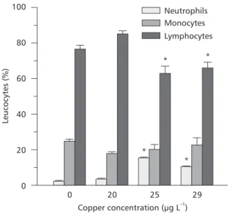
Differential leukocyte counts (Fig. 2) showed that lymphocytes were the most frequent white blood cells in control P. scrofa (75 ± 5%), and the proportion of these cells was reduced to 62% and 66% respectively in fish exposed to 25 and 29
µgCu L–1. The percentage of neutrophils was low
compared to that of monocytes. After copper exposure, the monocyte percentage showed a slight increase but resulted in a nonsignificant change, while neutrophils increased significantly in fish exposed to 25 and 29 µgCu L–1. Basophils were
not found in the prepared smears and eosinophils were very rare (less than 0.33%).
Thrombocytes were easily identified and did not show significant change following copper exposure. The mean thrombocytes number was 45.94 ± 2.18 103 mm3.
500
400
300
200
100
0
RBC
x
10
(mm
)
43
*
* 150
100
50
0
MCV
(
m
)
µ
3
50
40
30
20
10
0
Hct
(%)
* * 60
40
20
0
MCH
(pg
cell
)
–1
20
15
10
5
0
Hb
(g/100
ml)
*
45
30
15
0
MCHC
(%)
0 20 25 29
0 20 25 29
Cooper concentration ( g L )µ –1
Fig. 2 — Changes in the percentage of differential leukocyte counts of P. scrofa blood after exposure to different copper concentrations. Points are means ± SEM. * Indicates significant difference (p < 0.05) from controls.
Physiological parameters
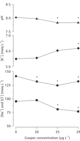
Copper exposure induced ionoregulatory disturbances in P. scrofa. Plasma [Na+] and [Cl–]
decreased significantly (p < 0.05) in fish exposed to lethal and sublethal copper concentration (Fig. 3). The reduction in plasma [Cl–] was 44% higher
than the corresponding fall in plasma [Na+] and the
Na/Cl ratio consequently increased significantly in fish exposed to 25 and 29 µgCu L–1 (p < 0.05).
Plas-ma [K+] increased with increasing copper in the
water, reaching significant values at 29 µgCu L–1
exposure (p < 0.05) (Fig. 3). The percentage of increased plasma [K+] was similar to the percentage
of plasma [Na+] lost (approximately 13%). Blood
pH decreased significantly (p < 0.05) in fish exposed to 25 and 29 µgCu L–1.
Gill histophology
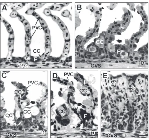
When compared with gills of control P. scrofa
(Fig. 4A), several distinct histopathologies were observed in fish exposed to copper on both the epithelia and blood vessels, even at the low copper concentrations in water recommended by the US EPA (1984). The exposure to copper induced intense proliferation of pavement cells and hypertrophy of both pavement and chloride cells (Fig. 4A-E).
Fila-ment epithelium height of fish exposed to copper was higher than that of control fish (3-5 cell layers, 0.0134 to 0.0280 mm in height), usually consisting of 5-15 hypertrophied cells layer (0.0284 to 0.0408 mm in height) and evidenced a dose-dependent response increasing with copper concentration in the water (Fig. 4B-D). Cell proliferation resulted in incomplete fusion of several lamellae in 28%, 46%, and 58% of examined samples of fish exposed to 20, 25, and 29 µgCu L–1 (LC50) respectively, and in complete
lamellar fusion (Fig. 4E) in 1%-4% of samples of fish exposed to 20 and 25 µgCu L–1, reaching 27%
in fish exposed to 29 µgCu L–1. Detachment of
lamellar epithelium and necrosis were common and increased with increasing copper in the water.
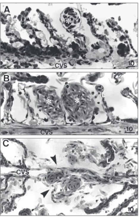
In addition to cell changes in the filament and lamellar epithelium, several histopathologies in the vascular system were identified in the gills (Fig. 5). Erythrocyte congestion was common in the marginal channel (telangiectasis) (Fig. 5A) in fish exposed to 20, 25, and 29 µgCu L–1.
Erythrocyte congestion throughout the entire lamella (aneurysm) (Fig. 5B) was usually observed in fish exposed to 25 and 29 µgCu L–1, and rupture
of the lamellar epithelium and the pillar cell system indicating hemorrhage foci was mainly observed in fish exposed to 29 µgCu L–1 (Fig. 5C).
Neutrophils Monocytes Lymphocytes
Leucocytes
(%)
100
80
60
40
20
0
0 20 25 29
Copper concentration ( g L )µ –1
* *
DISCUSSION
Heavy metal exposure is known to induce changes in blood parameters in fish (see review Heath, 1995). The direct effects of copper on blood parameters are usually associated with increased erythrocytes disintegration or, in the case of more sensitive species, damage of the hemopoietic system (Svobodová et al., 1994). In P. scrofa the increase of Hct, RBC, and [Hb] may indicate a compensatory response of this species to increase the blood’s O2 carrying capacity. The changes in gill epithelia of
P. scrofa caused by copper, such as cell hypertrophy, cell proliferation, and epithelial lifting may represent a defense response, as pointed out by Mallatt (1985), because these changes increase the distance across which copper must diffuse to reach the bloodstream.
150
125
100
75
50
0 20 25 29
7.0
6.0
5.0 8.5
8.0
7.5
Cooper concentration ( g L )µ –1
[K
]
(meq
L
)
+–
1
[Na
]
and
[Cl
]
(meq
L
)
+–
–
1
pH * *
*
* *
*
* *
Fig. 3 — Blood pH (F) and plasma ions Na+ (l), K+ (u), Cl– (n) of P. scrofa after exposure to different copper concen-trations. Points are means ± SEM. * Indicates significant difference (p < 0.05) from controls.
Fig. 4 — Representative sections of gill filament of P. scrofa. A – Control fish. B – Fish exposed to 20 µgCu L–1. Note filament epithelium (F) hypertrophy (arrow). C – Fish exposed to 25 µgCu L–1. Note lamellar epithelium and pavement (PVC) and chloride (CC) cell hypertrophy. D and E – Fish exposed to 29 µgCu L–1. Note CC hypertrophy (arrows) in D and total fu-sion of several lamellae (*) due to intense cell proliferation. CC chloride cell, CVS central venous sinus, L lamella, PVC pavement cell. Scale bar in µm.
Nevertheless, the increase in Hct coupled with the increase in the RBC and [Hb], with no significant changes in the MCH and MCHC blood indices, cell size, and circulating immature red blood cells suggest the body’s attempts to absorb more oxygen from the external environment so as to supply the oxygen requirement of tissue.
Leukocytopenia, an overall reduction in leukocytes, has been demonstrated in teleosts exposed to copper (Mishra & Srivastava, 1980; Dick & Dixon, 1985; Svobodová et al., 1994) and other heavy metals (Srivastava & Agrawal, 1979; Mishra & Srivastava, 1980; Gill & Pant, 1987). Leukocytopenia is a nonspecific response to a variety of stressors mediated by corticosteroid
hormones (Ellis, 1981) and cannot be considered a specific cytotoxic action of copper (Dick & Dixon, 1985). However, the leukocytosis reported in O. mossambicus afteracute exposure to copper (Nussey et al., 1995a, b) and found in P. scrofa in the present study may be attributed to increased leukocyte mobilization to protect the body against infections in copper-damaged tissue.
Neutrophils and monocytes are important white blood cells to protect the body, through their elevated phagocytic activity, against bacterial infection in damaged tissue. The percentage of these cell types generally decreases during acute exposure to copper (Nussey et al., 1995b; Svobodová et al., 1994), and in situations of chronic copper exposure, the neutrophil percentage has been reported to increase (Dick & Dixon, 1985). In P. scrofa, the monocyte percentage did not change following acute copper exposure but neutrophil percentage increased significantly which may also be related to gill tissue damage. This would reflect such direct deleterious effects of copper on gill epithelia as cell degeneration and the intense rupture and peeling of lamellar epithelial after 96 h exposure to 25 and 29 µgCu L–1. Basophils and
eosinophils were not found in P. scrofa although they have been identified in some fish species (Takashima & Hibiya, 1995). In O. mossambicus
increased counts of eosinophils were found during copper exposure (Nussey et al., 1995b).
Thrombocytes are comparable to mammal blood platelets and play an important role in the blood clotting which prevents blood loss from hemorrhaging. A high number of thrombocytes reduces clotting time (Srivastava, 1969), by as much as 50% in cases where the number of circulating thrombocytes was found to be 1 to 2 times higher than normal (Cassilas & Smith, 1977). Some species exposed to copper displayed a high increase in the thrombocyte percentage (Mishra & Srivastava, 1980; Dick & Dixon, 1985) although, the thrombocytes increase did not entail clotting time reduction in fish exposed to copper due to thrombocyte malfunction (Nussey et al., 1995c). In P. scrofa, the thrombocytes did not change following copper exposure, although several hemorrhage foci identified as vessel ruptures in the lamellae were found in gills of fish exposed to 29 µgCu L–1.
Copper causes serious ion imbalance in P. scrofa. Chloride cell hypertrophy on the gills of
P. scrofa exposed to copper appear to result from a failure to compensate for ion losses. Copper concentrations as low as 20 µg L–1 decreased the
concentration of sodium in the plasma and at a concentration of 29 µg L–1 (96 h LC50 (Mazon &
Fernandes, 1999)), plasma sodium and chloride were significantly lower than the control, with a
concentration. Previous investigations have also found ion imbalance in Salvelinius fontinalis
(McKim et al., 1970), Ictalurus nebulosus
(Christensen et al., 1972), Lepomis macrochirus
(Heath, 1991), and O. mossambicus (Nussey et al., 1995a; Pelgrom et al., 1995). Ionregulatory disruption induced by copper is related to inhibition of branchial Na+-K+-ATPase and ion uptake with
concomitant stimulation of ion efflux in freshwater fish (Laurén & McDonald, 1985; Pelgrom et al., 1995). The increased membrane permeability favoring ion efflux may be due to disruption of the membrane integrity of gill cells (Stagg & Shuttleworth, 1982; McDonald & Wood, 1993). In general, plasma ions increased in marine teleosts (Stagg & Shuttleworth, 1982) and decreased in freshwater fish (Christensen et al., 1972; McKim
et al., 1970). The imbalance of Na/Cl ratio may also reflect disturbed acid-base regulation (McDonald & Wood, 1993).
The reason for K+ increases in plasma (K+/
Na+ ratio from 0.03 in controls to 0.05 in fish
exposed to 29 µg L–1) may be due to increased cell
membrane permeability, as pointed out by Laurén & McDonald (1985), allowing the intracellular K+
to diffuse passively to extracellular fluid (Perry & Laurent, 1993). The significant blood acidosis found in P. scrofa has also been reported in O. mykiss (Wilson & Taylor, 1993) and O. mossambicus (Pelgrom et al., 1995). It is generally related to increase in lactic acid or other acid production as a result of metabolism increase. In
P. scrofa the acid-base imbalance may be related to decreased H+ excretion due to gill cell damage
or other acid production since no change in blood lactate was reported by Mazon et al. (2000) in this species exposed to 29 µgCu L–1 for 96 h in the same
water conditions and temperature as those of the present study. Blood lactate increase was found only during the first 24 h- copper exposure and returned to control value in 48 hours.
the other hand, the changes in red blood cell parameters suggest a compensatory response to the disruption of structural integrity of gills with consequent reduction of respiratory surface, in order to increase O2-carrying capacity and maintain the level of oxygen transference from water to tissues, allowing fish to survive during the nominated shock phase of LC50 exposure, at least under restful conditions. However, the blood changes found in the present study may be more drastic for mature
P. scrofa, particularly during upstream migration for breeding which requires more energy expenditure.
Acknowledgments — This study was supported by grants from FAPESP and CNPq. The authors thank the Hydrobiology and Aquaculture Station of Furnas Hydroelectric Powerplant, Furnas, Minas Gerais, Brazil for supplying the fish. A. F. Mazon, E. A. S. Monteiro, and G. H. D. Pinheiro thank CNPq for scholarships.
REFERENCES
BANERJEE, S. & HOMECHAUDHURI, S., 1990, Hematological monitoring of a bio-indicator fish,
Heteropneustes fossilis, on exposure to copper toxicity. Israel J. Aquacult., Bamiggeh, 42: 46-51.
CASSILAS, E. & SMITH, L. S., 1977, Effect of stress on blood coagulation and haematology in rainbow trout (Salmo gairdneri). J. Fish Biol., 10: 481-491. CETESB, 1992-2000, Relatório de qualidade das águas
interiores do Estado de São Paulo. São Paulo, Brasil. CHRISTENSEN, G. M., McKIM, J. M., BRUNGS, W. A. &
HUNT, E. P., 1972, Changes in the blood of Brown bullhead (Ictalurus nebulosus, Le Sueur) following short and long term exposure to copper (II). Toxicol. Appl. Pharmacol., 23: 417-427.
DICK, P. T. & DIXON, D. G., 1985, Changes in circulating blood cell levels of rainbow trout, Salmo gairdneri
Richardson, following acute and chronic exposure to copper. J. Fish Biol., 26: 475-484.
ELLIS, A. E., 1981, Stress and the modulation of defense mechanisms in fish. In: A. D. Pickering (ed.), Stress and Fish. Academic Press, London.
GILL, T. S. & PANT, J. C., 1987, Haematological and pathological effects of chromium toxicosis in the freshwater fish, Barbus conchonius Ham. Water, Air Soil Poll., 35: 241-250.
HEATH, A. G., 1991, Effect of water-borne copper on physiological responses of bluegill (Lepomis macrochirus) to acute hypoxic stress and subsequent recovery. Comp. Biochem. Physiol., 100(C): 559-564. HEATH, A. G., 1995, Water pollution and fish physiology.
CRC Press, Boca Raton, Fl.
LAURÉN, D. J. & McDONALD, D. G., 1985, Effects of copper on branchial ionoregulation in the rainbow trout,
Salmo gairdneri Richardson. J. Comp. Physiol., 155(B): 636-644.
LI, J., QUABIUS, E. S., WENDELAAR BONGA, S. E. & LOCK, R. A. C., 1998, Effects of waterborne copper on branchial Na+-transport in Mozambique tilapia (Oreochromis mossambicus). Aquat. Toxicol., 43: 1-11. MALLATT, J., 1985, Fish gill structural changes induced by toxicants and other irritants: a statistical review. Can. J. Fish. Aquat. Sc., 42: 630-648.
MAZON, A. F., 1997, Efeitos do íon cobre sobre o curimbatá,
Prochilodus scrofa (Steindachner, 1881). Dissertação de Mestrado em Ecologia e Recursos Naturais, UFSCar, São Carlos, 160p.
MAZON, A. F. & FERNANDES, M. N., 1999, Toxicity and differential tissue accumulation of copper in the tropical freshwater fish, Prochilodus scrofa (Prochilodontidae).
Bull. Environ. Contam. Toxicol., 63: 797-804. MAZON, A. F., CERQUEIRA, C. C. C. & FERNANDES, M.
N., 2002, Gill cellular changes induced by copper exposure in the South American tropical freshwater fish
Prochilodus scrofa. Environm. Res. A, 88: 52-63. MAZON, A. F., PINHEIRO, G. H. D. & FERNANDES, M.
N., 2000, Contaminação dos ecosistemas aquáticos pelo cobre e risco potencial à biodiversidade. Estudo da toxicidade do cobre em curimbatá, P. scrofa (Teleostei, Prochilodontidae). In: E. L. G. Espíndola, C. M. R. B. Paschoal, O. Rocha, M. B. C. Bohrer & A. L. Oliveira Neto (eds.), Ecotoxicologia:Perspectivas para o Século XXI. RiMa Editora, São Carlos.
MAZON, A. F., FERNANDES, M. N., NOLASCO, M. & SEVERI, W., 1998, Functional morphology of gills and respiratory area of two active rheophilic fish species,
Plagioscion squamosissimus and Prochilodus scrofa. J. Fish Biol., 52: 50-61.
McDONALD, D. G. & WOOD, C. M., 1993, Branchial mechanisms of acclimation to metals in freshwater fish.
In: J. C. Rankin & F. B. Jensen (eds.), Fish Ecophysiology. Chapman & Hall, London.
McKIM, J. M., CHRISTENSEN, G. M. & HUNT, E. P., 1970, Changes in the blood of the brook trout Salvelinus fontinalis after short-term and log-term exposure to copper. J. Fish. Res. B. Can., 27: 1883-1889. McKNIGHT, I. M., 1966, A hematological study on the
mountain whitefish, Prosopium williamsoni. J. Fish. Res. B. Can., 23: 45-64.
MISHRA, S. & SRIVASTAVA, A. K., 1980, The acute toxic effects of copper on the blood of a teleost. Ecotoxicol. Environ. Safety,4: 191-194.
NUSSEY, G., VAN VUREN, J. H. J. & DU PREEZ, H. H., 1995a, Effect of copper on haematology and osmoregulation of the Mozambique tilapia, Oreochromis mossambicus (Cichlidae). Comp. Biochem. Physiol.,
111(C): 369-380.
1995b, Effect of copper on the differential white blood cell counts of the Mozambique tilapia (Oreochromis mossambicus). Comp. Biochem. Physiol., 111(C): 381-388. NUSSEY, G., VAN VUREN, J. H. J. & DU PREEZ, H. H., 1995c, Effect of copper on blood coagulation of
Oreochromis mossambicus (Cichlidae). Comp. Biochem. Physiol., 111(C): 359-367.
PELGROM, S. M. G. J., LOCK, R. A. C., BALM, P. H. M. & WENDELAAR BONGA, S. E., 1995, Integrated physiological response of tilapia, Oreochromis mossambicus, to sublethal copper exposure. Aquat. Toxicol., 32: 302-320.
PERRY, S. F. & LAURENT, P., 1993, Environmental effects on fish gill structure and function. In: J. C. Rankin & F. B. Jensen (eds.), Fish Ecophysiology. Chapman & Hall, London.
PILGAARD, L., MALTE, H. & JENSEN, F. B., 1994, Physiological effects and tissue accumulation of copper in freshwater rainbow trout (Oncorhynchus mykiss) under normoxic and hypoxic conditions. Aquat. Toxicol., 29: 197-212.
SOIVIO, A. & NIKINMAA, A., 1981, The swelling of erythrocytes in relation to the oxygen affinity of the blood of the rainbow trout, Salmo gairdneri Richardson. In: A. D. Pickering (ed.), Stress and Fish. Academic Press, London.
SRIVASTAVA, A. K., 1969, Studies on the haematology of certain freshwater teleosts-V. Thrombocytes and the clotting of blood. Anat. Anz B., 124: 368-174. SRIVASTAVA, A. K. & AGRAWAL, S. J., 1979,
Haematological anomalies in a freshwater teleost, Colisa fasciatus, on acute exposure to cobalt. Acta Pharmacol. Toxicol., 44: 197-199.
STAGG, R. M. & SHUTTLEWORTH, T. J., 1982, The accumulation of copper in Platichthys flesusL. and its effects on plasma electrolyte concentrations. J. Fish Biol.,
20: 491-500.
SVOBODOVÁ, Z., VYKUSOVÁ, B. & MÁCHOVÁ, J., 1994, The effects of pollutants on selected haematological and biochemical parameters in fish. In: R. Müller & R. Lloyd (eds.), Sublethal and Chronic Effects of Pollutants on Freshwater Fish. Fishing New Books, London. TAKASHIMA, F. & HIBIYA, T., 1995, An atlas of fish
histology: normal and pathological features. 2. ed. Kodansha, Tokyo.
US EPA, 1984, Ambient water quality criteria for copper.
United States Environmental Protection Agency, Washington, D.C.
![Fig. 1 — Changes in hematocrit (Hct), red blood cells (RBC), whole blood hemoglobin concentration [Hb], mean cell volume (MCV), mean corpuscular hemoglobin (MCH) and mean corpuscular hemoglobin content (MCHC) of P](https://thumb-eu.123doks.com/thumbv2/123dok_br/15826984.654866/4.892.222.658.509.980/changes-hematocrit-hemoglobin-concentration-corpuscular-hemoglobin-corpuscular-hemoglobin.webp)