Elevated amniotic fluid amino acid
levels in fetuses with gastroschisis
1Department of Obstetrics and Gynecology, 2Department of Clinical Biochemistry,
Dicle University School of Medicine, Diyarbakir, Turkey A. Kale1, E. Kale2,
N. Akdeniz1
and N. Canoruc2
Abstract
Our objective was to measure maternal plasma and amniotic fluid amino acid concentrations in pregnant women diagnosed as having fetuses with gastroschisis in the second trimester of pregnancy. Twenty-one pregnant women who had fetuses with gastroschisis detected by ultrasonography (gastroschisis group) in the second trimester and 32 women who had abnormal triple screenings indicating an increased risk for Down syndrome but had healthy fetuses (control group) were enrolled in the study. Amniotic fluid was obtained by amniocentesis, and maternal plasma samples were taken simultaneously. The chrom-osomal analysis of the study and control groups was normal. Levels of free amino acids and non-essential amino acids were measured in plasma and amniotic fluid samples using EZ:fast kits (EZ:fast GC/FID free (physiological) amino acid kit) by gas chromatography (Focus GC AI 3000 Thermo Finnigan analyzer). The mean levels of essential amino acids (histidine, isoleucine, leucine, lysine, methionine, phen-ylalanine, threonine, tryptophan, and valine) and non-essential amino acids (alanine, glycine, proline, and tyrosine) in amniotic fluid were found to be significantly higher in fetuses with gastroschisis than in the control group (P < 0.05). A significant positive correlation be-tween maternal plasma and amniotic fluid concentrations of essential and nonessential amino acids was found only in the gastroschisis group (P < 0.05). The detection of significantly higher amino acid concentrations in the amniotic fluid of fetuses with a gastroschisis defect than in healthy fetuses suggests the occurrence of amino acid malabsorption or of amino acid leakage from the fetus into amniotic fluid.
Correspondence
A. Kale
Department of Obstetrics and Gynecology
Dicle University School of Medicine 21280 Diyarbakir
Turkey
E-mail: [email protected]
Received November 24, 2005 Accepted April 26, 2006
Key words
•Amino acids •Gastroschisis •Amniotic fluid
Introduction
Gastroschisis is a full thickness abdomi-nal wall defect in which the viscera herniate through the abdominal wall lateral to the umbilicus. This condition occurs in 1 to 3 per 10,000 live births and, unlike exomphalos, the viscera are not surrounded by a
membra-nous sac (1). The pathophysiologic mechan-ism is presumed to be occlusion of the ompha-lomesenteric artery, leading to disruption of the umbilical ring with subsequent hernia-tion of the abdominal contents (2).
pregnancy and infants with gastroschisis al-most universally experience problems with absorptive and motility functions of the in-testine which are the major cause of mortal-ity and morbidmortal-ity (3).
Bowel loops may become dilated due to partial lymphatic and venous obstruction caused by obstruction on the side wall of the defect. Severe complications, such as volvu-lus, atresia, ischemia, increased mucosal permeability, and intestinal villous atrophy, result in increased mortality and morbidity (4). Furthermore, animal models of gas-troschisis have shown that there is a signifi-cant deficiency in nutrient absorption and protein concentration in the small bowel after prolonged exposure to amniotic fluid (3,5).
The aim of the present study was to de-termine the concentrations of amino acids in amniotic fluid and maternal serum of preg-nant women whose fetuses were diagnosed to have gastroschisis in the second trimester of pregnancy. We hypothesized that the con-centrations of amino acids may be higher in fetuses with gastroschisis due to deficiency in nutrient absorption, intestinal dysfunction and increased mucosal permeability of the intestines.
Patients, Material and Methods
The study was performed at the Prenatal
Diagnosis Unit of Dicle University Hospital between January 2002 and June 2005. The study was approved by the institutional re-view board and Ethics Committee of the university hospital, and written informed consent was obtained from all participants. All pregnant women who had a fetus with gastroschisis (N = 21) in the second trimes-ter were included in the study. The first 32 women who attended our clinic and had abnormal triple screens indicating an creased risk for Down syndrome were in-cluded in the study as the control group. Mean maternal age was 25.5 ± 2.01 years for the gastroschisis group and 24.7 ± 3.1 years for the study group. The mean gestational age at sampling was 19.1 ± 1.1 weeks for the gastroschisis group and 18.6 ± 1.0 weeks for the study group. Maternal body mass index was 26.2 ± 1.0 kg/m2 in gastroschisis group
and 25.9 ± 1.1 kg/m2 in the study group.
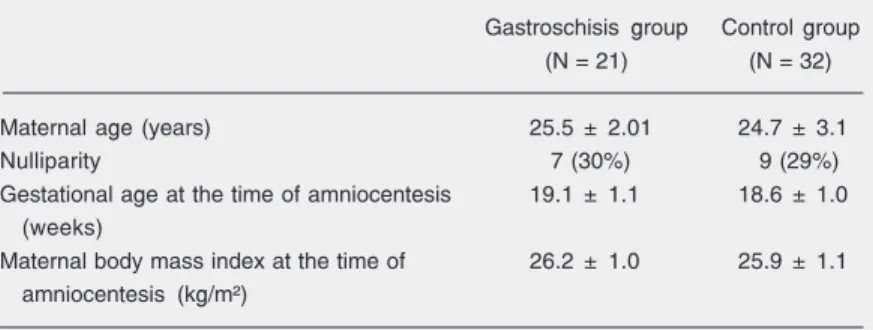
Seven women in the gastroschisis group and 9 in the control group were nulliparous (Table 1).
Obese patients and those with any sys-temic or endocrine disorder were excluded from the study. All pregnancies were accu-rately dated by the last menstrual period and by first-trimester ultrasonographic investi-gation. Amniotic fluid samples were ob-tained by routine transabdominal amniocen-tesis and collected into 10-mL dry tubes. All amniotic fluid samples were free of blood contamination. Venous blood samples were taken within 10 min after amniocentesis from the pregnant women and collected into EDTA-containing tubes. All samples were immediately centrifuged at 3000 g for 10 min and stored at -20ºC until assayed. Lev-els of free amino acids (essential amino acids: histidine, leucine, lysine, isoleucine, methi-onine, phenylalanine, thremethi-onine, tryptophan, and valine) and non-essential amino acids (alanine, asparagine, aspartic acid, cys-tathionine, cysteine, glutamic acid, gluta-mine, glycine, ornithine, and proline) were measured in plasma and amniotic fluid Table 1. Demographic characteristics of the study and control groups.
Gastroschisis group Control group
(N = 21) (N = 32)
Maternal age (years) 25.5 ± 2.01 24.7 ± 3.1
Nulliparity 7 (30%) 9 (29%)
Gestational age at the time of amniocentesis 19.1 ± 1.1 18.6 ± 1.0 (weeks)
Maternal body mass index at the time of 26.2 ± 1.0 25.9 ± 1.1 amniocentesis (kg/m²)
samples using EZ:fast kits (EZ:fast GC/FID free (physiological) amino acid kit) by gas chromatography (Focus GC AI 3000 Thermo Finnigan analyzer, Milan, Italy; injection: Split 1:15 at 250ºC, 2.5 µ; carrier gas: helium 1.5 mL/min (60 kPa) at 110ºC; pressure rise: 6 kPa/min; oven program: 30ºC/min from 110º to 320ºC, hold at 320º for 1 min; Detec-tor: FID at 320ºC; intravariability: 2.4%; intervariability: 3.2%).
The results are reported as means ± SD. A t-test was performed for statistical analy-sis. The statistical relationship between the two variables was checked by Pearson cor-relation coefficients. A P value of less than 0.05 was considered to be statistically sig-nificant.
Results
Twenty-one women who had fetuses with gastroschisis were included in the study (gas-troschisis group, N = 21). Gas(gas-troschisis was diagnosed by ultrasonography and confirmed after delivery. The chromosomal analysis of the gastroschisis group was normal. In the gastroschisis group there was one intrauter-ine death at 34 weeks, whereas the obstetri-cal outcome of the other affected fetuses was good. The control group consisted of 32 women submitted to amniocentesis per-formed because of abnormal triple screens indicating an increased risk for Down syn-drome (control group, N = 32). None of the control group fetuses showed structural ab-normalities in ultrasonography at the time of amniocentesis and none had chromosome abnormalities. All patients in the control group gave birth to a healthy child. The characteristics of both groups of patients are shown in Table 1. The rates of nulliparity, the mean maternal and gestational ages and body mass index at the time of amniocente-sis did not differ significantly between the two groups (P < 0.05).
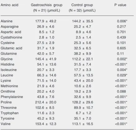
The mean concentrations of amino acids in the gastroschisis and control groups are
given in Table 2. The mean concentrations of essential and non-essential amino acids were significantly higher in the gastroschisis group than in the control group (P < 0.05), whereas the mean concentrations of acidic amino acids (glutamine, glutamic acid, as-partic acid, asparagine), ornithine and cys-tathionine did not differ statistically between groups (P < 0.05). There were significant positive correlations between maternal plasma and amniotic fluid concentrations of ala-nine, cysteine, glutamine, glycine, ornithine, proline, tyrosine, and essential amino acids in the gastroschisis group (P < 0.05, Table 3). None of the amino acids showed a statis-tically significant difference between mater-nal serum and amniotic fluid in the control group (P < 0.05).
Table 2. Concentrations of twenty amino acids in amniotic fluid samples of fetuses with gastroschisis and controls.
Amino acid Gastroschisis group Control group P value (N = 21) (µmol/L) (N = 32) (µmol/L)
Alanine 177.9 ± 49.2 144.2 ± 35.5 0.006*
Asparagine 26.9 ± 4.6 25.2 ± 4.7 0.217
Aspartic acid 8.5 ± 1.2 8.9 ± 4.6 0.701
Cystathionine 2.8 ± 1.0 2.5 ± 1.4 0.439
Cysteine 27.5 ± 2.9 25.3 ± 5.6 0.101
Glutamic acid 31.7 ± 1.9 32.5 ± 6.5 0.605
Glutamine 42.0 ± 5.7 38.2 ± 9.9 0.11
Glycine 145.4 ± 41.9 112.2 ± 22.1 0.002*
Histidine 54.1 ± 13.6 31.5 ± 7.4 <0.001*
Isoleucine 20.7 ± 3.3 17.7 ± 3.3 0.004*
Leucine 66.3 ± 14.6 57.5 ± 13.5 0.029*
Lysine 71.5 ± 14.0 43.4 ± 20.0 <0.001*
Methionine 21.9 ± 4.6 10.6 ± 2.6 <0.001*
Ornithine 20.2 ± 4.0 19.2 ± 2.9 0.088
Phenylalanine 43.8 ± 7.6 32.6 ± 9.9 <0.001*
Proline 212.4 ± 20.0 128.2 ± 29.6 <0.001*
Threonine 102.6 ± 8.5 89.9 ± 10.7 <0.001*
Tryptophan 11.6 ± 2.3 6.7 ± 1.2 <0.001*
Tyrosine 45.2 ± 9.3 35.1 ± 7.0 <0.001*
Valine 153.4 ± 12.3 113.1 ± 16.5 <0.001*
Discussion
The results of the present study demon-strate that fetuses with gastroschisis have higher amniotic fluid concentrations of es-sential and non-eses-sential amino acids than controls (P < 0.05). This suggests that there is malabsorption of these amino acids or loss of essential amino acids from the fetus to the amniotic fluid. A similar phenomenon has been observed in studies on rabbit fetuses demonstrating that fetuses with iatrogenic gastroschisis are smaller and have a reduced uptake of amino acids and other nutrients compared to controls (5,6).
Carroll et al. (3) reported that fetuses with gastroschisis have lower serum protein concentrations and higher amniotic fluid to-tal protein levels than do cases of exomphalos or controls. Pathologic changes in the small intestine associated with gastroschisis, such as villous atrophy, increased mucosal
per-meability, inflammatory bowel wall changes, and ischemia may contribute to the leakage of amino acids from the fetus to the amniotic fluid (3-5).
Midrio et al. (7) reported delayed matu-ration of intestinal pacemaker cells and smooth muscle cells in the rat model of gastroschisis which might explain the intes-tinal malfunction and reduced uptake of amino acids.
Blakelock et al. (8) studied 112 babies born with gastroschisis to explore the hypo-thesis that normal fetal gut function is needed for normal growth in late gestation and found that normal growth is dependent on a nor-mally functioning gastrointestinal tract dur-ing this phase.
We found a significant positive correla-tion between maternal serum and amniotic fluid concentrations of essential and non-essential amino acids (P < 0.05). Our hypo-thesis is that the changes in maternal serum
Table 3. Correlations between maternal serum (MS) and amniotic fluid (AF) amino acid levels in the gastroschisis group.
Amino acid Maternal serum Amniotic fluid MS vs AF
levels (N = 21) (µmol/L) levels (N = 21) (µmol/L)
r value P value
Alanine 304.1 ± 35.1 177.9 ± 49.2 0.69 <0.001*
Asparagine 29.7 ± 5.2 26.9 ± 4.6 0.08 NS
Aspartic acid 9.1 ± 2.3 8.5 ± 1.2 0.02 NS
Cystathionine 4.0 ± 2.5 2.8 ± 1.0 0.09 NS
Cysteine 62.14 ± 13.2 27.5 ± 2.9 0.55 <0.001*
Glutamic acid 33.9 ± 5.1 31.7 ± 1.9 0.07 NS
Glutamine 488.5 ± 21.1 42.0 ± 5.7 0.85 <0.001*
Glycine 168.9 ± 29.3 145.4 ± 41.9 0.09 <0.05*
Histidine 100.3 ± 23.4 54.1 ± 13.6 0.60 <0.001*
Isoleucine 41.4 ± 2.9 20.7 ± 3.3 0.90 <0.001*
Leucine 98.2 ± 6.6 66.3 ± 14.6 0.67 <0.001*
Lysine 147.0 ± 22.5 71.5 ± 14.0 0.80 <0.001*
Methionine 33.3 ± 4.6 21.9 ± 4.6 0.61 <0.001*
Ornithine 31.2 ± 5.4 20.2 ± 4.0 0.58 <0.001*
Phenylalanine 55.1 ± 10.0 43.8 ± 7.6 0.29 <0.001*
Proline 278.0 ± 23.9 212.4 ± 20.0 0.04 <0.001*
Threonine 121.8 ± 22.5 102.6 ± 8.5 0.24 <0.001*
Tryptophan 37.5 ± 2.0 11.6 ± 2.3 0.97 <0.001*
Tyrosine 60.4 ± 12.9 45.2 ± 9.3 0.32 <0.001*
Valine 191.6 ± 37.9 153.4 ± 12.3 0.32 <0.001*
Data are reported as mean ± SD. NS = not significant.
References
1. Kazaura MR, Lie RT, Irgens LM, Didriksen A, Kapstad M, Egenaes J, et al. Increasing risk of gastroschisis in Norway: an age-period-cohort analysis. Am J Epidemiol 2004; 159: 358-363.
2. Hoyme HE, Higginbottom MC, Jones KL. The vascular pathogen-esis of gastroschisis: intrauterine interruption of the omphalomesen-teric artery. J Pediatr 1981; 98: 228-231.
3. Carroll SG, Kuo PY, Kyle PM, Soothill PW. Fetal protein loss in gastroschisis as an explanation of associated morbidity. Am J Obstet Gynecol 2001; 184: 1297-1301.
4. Dykes EH. Prenatal diagnosis and management of abdominal wall defects. Semin Pediatr Surg 1996; 5: 90-94.
5. Shaw K, Buchmiller TL, Curr M, Lam MM, Habib R, Chopourian HL, et al. Impairment of nutrient uptake in a rabbit model of gastroschi-sis. J Pediatr Surg 1994; 29: 376-378.
6. Guo W, Swaniker F, Fonkalsrud EW, Vo K, Karamanoukian R.
Effect of intraamniotic dexamethasone administration on intestinal absorption in a rabbit gastroschisis model. J Pediatr Surg 1995; 30: 983-986.
7. Midrio P, Faussone-Pellegrini MS, Vannucchi MG, Flake AW. Gas-troschisis in the rat model is associated with a delayed maturation of intestinal pacemaker cells and smooth muscle cells. J Pediatr Surg
2004; 39: 1541-1547.
8. Blakelock R, Upadhyay V, Kimble R, Pease P, Kolbe A, Harding J. Is a normally functioning gastrointestinal tract necessary for normal growth in late gestation? Pediatr Surg Int 1998; 13: 17-20. 9. Harding J. Nutritional causes of impaired fetal growth and their
treatment. J R Soc Med 1999; 92: 612-615.
10. Regnault TR, Friedman JE, Wilkening RB, Anthony RV, Hay WJ. Fetoplacental transport and utilization of amino acids in IUGR - a review. Placenta 2005; 26 (Suppl A): S52-S62.
concentrations of essential and non-essen-tial amino acids might cause changes in amniotic fluid amino acid concentrations in pregnancies involving fetuses with gas-troschisis. This might be the result of leak-age from the intestinal fluid via a defective abdominal wall, intestinal dysfunction, in-creased mucosal permeability, or intestinal villous atrophy. Furthermore, prolonged ex-posure of the bowels to amniotic fluid might explain amino acid deficiency in fetus and increased concentrations of amino acids in amniotic fluid.
Human studies and experimental models have shown that amino acid uptake is de-creased in fetuses with intrauterine growth restriction and that intrauterine growth re-striction is likely to be associated with a shift in amino acid transport capacity and meta-bolic pathways within the fetoplacental unit (9,10).
We found higher levels of amniotic fluid essential and non-essential amino acids in the gastroschisis group than in the control group which might explain intrauterine
growth restriction in fetuses with gastroschi-sis.
The causes of growth restriction, fetal distress, and death in fetuses with gastroschi-sis may be complicated by amino acid defi-ciency as a result of malabsorption or loss, which might partly explain fetal morbidity and mortality. We speculate that replace-ment of essential and non-essential amino acids might improve the outcome of infants with gastroschisis during the neonatal pe-riod.