Humoral and cellular immune responses
to
Blomia tropicalis and concanavalin
A-binding fractions in atopic patients
R. Alves
1, D.A.O. Silva
1, J.F.C. Fernandes
1,
K.C. Almeida
1, L.H. Ynoue
1,
C.T.V. Bernardes
1,
P.F.S. Moreira
1, M.L. Gennari-Cardoso
2, S.J. Sung
3and
E.A. Taketomi
11
Laboratório de Alergia e Imunologia Clínica, Instituto de Ciências Biomédicas, Universidade Federal de
Uberlândia, Uberlândia, MG, Brasil
2
Divisão de Imunologia e Microbiologia, Departamento de Ciências Biológicas, Universidade Estadual de
Santa Cruz, Ilhéus, BA, Brasil
3
Department of Medicine, Center for Immunity, Inflammation and Regenerative Medicine, University of
Virginia, Charlottesville, VA, USA
Correspondence to: E.A. Taketomi, Laboratório de Alergia e Imunologia Clínica, ICBIM, UFU, Av. Pará,
1720, Bloco 4C, Campus Umuarama, 38400-902 Uberlândia, MG, Brasil
Fax: +55-34-3232-8620. E-mail: eat4y@yahoo.com.br
Blomia tropicalis, Dermatophagoides pteronyssinus and D. farinae are prevalent house dust mites. Concanavalin A-binding components derived from B. tropicalis (Bt-ConA extract) are highly immunogenic in allergic diseases. The aim of the present study was to evaluate the humoral and cellular immune responses to B. tropicalis in mite-sensitized patients. A total of 137 patients with allergic rhinitis with/without asthma and 109 non-atopic subjects were selected and analyzed by the skin prick test, and for total serum IgE and specific IgE levels to both Bt-total and Bt-ConA extracts, their proliferative response and cytokine (IFN-γ and IL-5) production by peripheral blood mononuclear cells (PBMC) stimulated with both extracts. Skin prick test showed that 70% of the patients were sensitized to Bt (Bt+) and similar levels of specific IgE to Bt-total and Bt-ConA extracts were demonstrable in Bt+ patients. Significant PBMC proliferation was observed in response to total extract in Bt+, but not in Bt-patients and non-atopic subjects (P < 0.001). Bt-ConA extract induced increased proliferative responses in all patient groups compared to medium alone (P < 0.05), but these responses were significantly decreased in the presence of the mannopyrano-side ConA inhibitor (P < 0.05). Significant IFN-γ production was observed after Bt-ConA stimulation of Bt+ patients (P < 0.05), while Bt-total extract had no effect. IL-5 production was consistently detected in Bt+ patients after allergen-specific stimulation or with no stimulus, indicating that PBMC from allergic patients are prone to produce Th2 profile cytokines, spontaneously or inductively by allergen restimulation. These data showed that ConA-binding components isolated from B. tropicalis may contain relevant antigens that are involved in both humoral and cellular immune responses. However, without an additional purification procedure to eliminate the residual contamination with ConA, its use in immunotherapeutic procedures cannot be recommended.
Key words: Blomia tropicalis; Concanavalin A; Cellular proliferation; IFN-γ; IL-5; Immunoglobulin E
Research supported by CAPES (#503-313/COIPA), CNPq (#479577/2004-8), and FAPEMIG (#CDS 1605/05).
Received January 31, 2008. Accepted August 26, 2008
Introduction
In tropical and subtropical climates, Blomia tropicalis (Bt), Dermatophagoides pteronyssinus (Dp) and D. farinae (Df) are prevalent house dust mites (HDMs) (1,2). HDMs
antibodies to HDMs are demonstrable in allergic patients (4). Glycosylated antigens play a crucial role in different pathologies and are highly immunogenic (5-7). Mannan-rich glycosylated components derived from Candida albi-cans have been recognized by IgE antibodies in patients with allergic manifestations including asthma, rhinitis, and atopic dermatitis (8). Recently, we reported IgE, IgG1 and IgG4 responses to B. tropicalis in patients with respiratory allergy using Bt-total extract and concanavalin A-binding components derived from B. tropicalis (Bt-ConA extract) (9). The results of that study showed that these patients seem to be more frequently sensitized to B. tropicalis high-molecular weight (54-104 kDa) components, mostly pres-ent in Bt-ConA extract than to the low-molecular weight (11-15 kDa) allergens, which have been demonstrated primarily in other reports (10,11).
Several investigators have shown consistently in both human and mouse models that Th2 cytokines (IL-4, IL-5 and IL-13) are the main contributors to the development of allergic diseases by stimulating B cells to produce IgE antibodies, recruitment and activation of eosinophils, and mucus production. Interferon-γ (IFN-γ), a Th1 cytokine profile, acts in conjunction with Th2 cytokines to maintain chronic allergic inflammation (12-14).
The aims of the present study were to evaluate the humoral and cellular immune responses to B. tropicalis in mite-sensitized patients by determining the levels of total serum IgE and specific IgE to Bt-total and Bt-ConA ex-tracts, the proliferative response and the cytokine (IFN-γ and IL-5) production by peripheral blood mononuclear cells (PBMC) stimulated with both allergen extracts.
Subjects and Methods
A total of 137 patients (54 males and 83 females; mean age ± SD: 27.6 ± 9.4 years) with perennial allergic rhinitis with/without intermittent or persistent, mild-to-moderate asthma were selected for the study at the Allergy and Clinical Immunology Unit, Federal University of Uberlândia, Uberlândia, MG, Brazil, based on their history of respira-tory symptoms related to house dust exposure and physi-cal examination (15-17). The presence of upper airway infections in the last 30 days prior to the study, the use of antihistaminic drugs earlier in the week, and the use of oral or topic corticosteroids in the previous 4 weeks were used as exclusion criteria. As the control group, 109 healthy subjects (37 males and 72 females; mean age ± SD: 31.1 ± 11.7 years), with no history of allergic diseases and a negative SPT to all aeroallergen extracts tested were included. The study was approved by the Ethics Commit-tee in Human Research of the Federal University of
Uberlândia and written informed consent was obtained from all participants.
Skin prick test and serum samples
All individuals underwent SPT with the following aller-gen extracts: mite (B. tropicalis, D. pteronyssinus and D. farinae) extracts prepared as described in Ref. 18, and commercial extracts of cockroach (Blattella germanica and Periplaneta americana), molds (Cladosporium spp) and pet danders (Felis domesticus and Canis familiaris) obtained from IPI/ASAC Brasil, Brazil. A mean wheal 3 mm in diameter larger than the negative control (diluent) was considered to be positive. In parallel, blood samples (5 mL) were collected from all individuals and the serum was stored at -20°C.
Mite extracts
Total extracts of Bt, Dp, and Df were obtained from mite bodies and feces as described elsewhere (18). Briefly, mite powder was triturated in liquid nitrogen and allergens were extracted in 5 mM borate-buffered saline containing protease inhibitors. After centrifugation (20,000 g for 45 min at 4°C), the supernatant was dialyzed and protein concentration was determined by the method of Lowry et al. (19).
ConA-binding components from total extracts of
B. tropicalis
ConA-binding components from the Bt-total extract were obtained by affinity chromatography using ConA-Sepharose (Amersham Pharmacia Biotech, Sweden) as described (9). After elution with 50 mM α -D-methylmanno-pyranoside (Ferro Pfanstiehl Laboratories Inc., USA), ConA-bound (Bt-ConA) fractions were concentrated, dialyzed and the protein content was determined.
Measurement of specific IgE to B. tropicalis
Levels of specific IgE to B. tropicalis and its ConA-binding fractions were measured by ELISA as described (9). Briefly, plates were coated with Bt-total or Bt-ConA extracts (1 µg/well), blocked with PBS containing 0.05% Tween 20 and 1% bovine serum albumin (PBST-BSA), and incubated with serum samples diluted 1:2 in PBST-BSA. After washing, plates were incubated with biotinyl-ated anti-human IgE (Kierkegaard and Perry Lab., USA) and subsequently with streptavidin-peroxidase (Sigma, USA). The assay was developed with 10 mM 2,2'-azino-bis-(3-ethyl-benzthiazoline) sulfonic acid (ABTS; Sigma, USA) and 0.03% H2O2.Absorbance was determined in a
sera. Antibody titers were reported as ELISA index (EI) and determined as follows: EI = absorbance test sample/ cutoff, where cutoff was calculated as the mean absorb-ance of 3 negative control sera plus 5 standard deviations, as described elsewhere (9). EI values >1.2 were consid-ered to be positive in order to exclude borderline reactivity values close to EI = 1.0.
Measurement of total serum IgE
Total serum IgE was measured with a monoclonal antibody-based ELISA (20). Briefly, plates were coated with monoclonal anti-human IgE (1:5000; Sigma), blocked with PBST-BSA and subsequently incubated with serum (1:5, 1:50 and 1:500) and biotinylated goat anti-human IgE (1:4000). Subsequent steps were similar to specific IgE-ELISA. Results are reported as international units per milliliter (IU/mL) and calculated on the basis of a standard curve constructed with a reference serum that contained 3000 IU/mL of total IgE.
Cellular immune response
Twenty-six atopic patients with positive SPT to Bt, Dp, and Df (Bt+ group), 19 atopic patients with negative SPT to Bt, but positive SPT to Dp and Df (Bt- group), and 24 non-atopic subjects with negative SPT to all tested aeroaller-gens (NA group) were selected to evaluate the cellular immune response to B. tropicalis.
Proliferation assays
PBMC were freshly isolated from 30 mL heparinized blood samples by density gradient centrifugation over Ficoll-Hypaque solution (Amersham Pharmacia). PBMC were cultured in triplicate (2 x 10⁵ cells/200 µL per well) in
96-well plates in RPMI 1640 medium supplemented with 10% human AB serum (Sigma) and stimulated with Bt-total (10 µg/mL) or Bt-ConA (5 µg/mL) extracts. Control cultures were incubated with medium alone, or mitogens (phytohe-magglutinin (PHA) at 10 µg/mL or ConA at 5 µg/mL), or non-allergen control (tetanus toxoid (TT) at 10 µg/mL). Cells were incubated at 37°C and 5% CO2 for 3 or 5 days
when stimulated with mitogens or antigens, respectively. Cells were pulsed with 0.5 µCi/well of tritiated thymidine (New England Nuclear, USA) for the final 8 h of culture, harvested on glass fiber filters, and counted in a liquid scintillation ß-counter (Packard Tri-Carb 2100TR, USA) and results are reported as counts per minute (cpm).
Proliferative response in the presence of ConA inhibitor
To evaluate the interference of residual ConA in the Bt-ConA extract on the proliferative response, PBMC were obtained from 14 patients (6 from Bt+ group, 4 from
Bt-group and 4 from NA Bt-group) and cultured with stimuli as described above. In parallel, the Bt-ConA extract was added in the presence of a ConA inhibitor, α -D-methyl-mannopyranoside at 50 mM as previously established in preliminary experiments. As control, ConA mitogen was also added in the presence of the ConA inhibitor. All subsequent steps were performed as described above.
Cytokine production assays
PBMC (1 x 10⁵ cells/250 µL per well) were cultured in
48-well plates and stimulated with allergen extracts (Bt-total and Bt-ConA), mitogens (PHA, ConA), non-allergen control (TT) or medium alone, at the same concentrations used for the proliferation assays. Supernatant solutions were collected from independent replicate wells after 3 and 7 days of culture, and stored in aliquots at -70°C until determination of cytokine levels. IFN-γ and IL-5 levels were measured in supernatants from cell cultures with an ELISA sandwich assay according to manufacturer instruc-tions (R&D Systems, USA). The sensitivity of the assays was 7.8 pg/mL for IFN-γ and 11.7 pg/mL for IL-5. The intra-and inter-assay coefficients of variations for these cyto-kines were below 10 and 15%, respectively.
Statistical analysis
Statistical analysis was performed using the GraphPad Prism 4.0 software (GraphPad Software, Inc., USA). Lev-els of total serum IgE and specific IgE to B. tropicalis were compared using the Kruskal-Wallis and the Dunn multiple comparison tests. Differences between proportions were analyzed by the chi-square test. Proliferation responses and cytokine levels were compared by the Kruskal-Wallis analysis. Differences were considered to be statistically significant when P < 0.05.
Results
Subject characteristics
The demographic and clinical characteristics of the study subjects are reported in Table 1. Subjects were distributed into three groups according to SPT positivity to Bt-total extract: 1) Bt+ group: patients with positive SPT to Bt-total; 2) Bt- group: patients with negative SPT to Bt-total, but positive SPT to Dp and/or Df extract; 3) NA group: non-atopic subjects with negative SPT to all aeroallergens tested.
although no significant difference was found in positive SPT results (90-100%). All NA subjects showed a negative SPT to mite extracts in addition to other aeroallergens tested (data not shown) according to the selection criteria used.
SPT to B. tropicalis, D. pteronyssinus and D. farinae
SPT results of 137 patients with allergic rhinitis with/ without asthma showed that 96 (70.1%) patients had posi-tive SPT to B. tropicalis, 95 (69.4%) were simultaneously positive to 3 mites (Bt+/Dp+/Df+), and only 1 patient (0.7%) was positive to both Bt and Df extracts (Bt+/Dp-/Df+). No patient was monosensitized to B. tropicalis (Bt+/Dp-/Df-). On the other hand, of 41 (29.9%) patients with negative SPT to B. tropicalis, 37 (27%) were reactive to both Dp and Df extracts (Bt-/Dp+/Df+) and only 4 (2.9%) showed reac-tivity to Dp extract alone (Bt-/Dp+/Df-).
Levels of specific IgE to B. tropicalis and total serum IgE
Levels of specific IgE to both Bt-total and Bt-ConA extracts (Figure 1A) were significantly higher in the Bt+ group (EI = 1.73 and 1.20, respectively) than in the Bt-group (EI = 0.86 and 0.84, respectively) or the NA Bt-group (EI
= 0.78 and 0.78, respectively; P < 0.001). From all Bt+ patients, 73% had specific serum IgE to Bt-total extract and 50% to Bt-ConA extract by ELISA, with a significant difference between the seropositivity rates (P = 0.0013). In contrast, Bt- patients and NA subjects had no serum IgE reactivity to either Bt-total or Bt-ConA extracts. Sera as-sayed in plates previously coated with Con-A (negative control for the Bt-ConA extract) did not show any reactivity, with EI values below 1.2 (data not shown).
Levels of total serum IgE measured by ELISA in Bt+ (196.2 IU/mL) and Bt- (105.8 IU/mL) atopic patients were significantly higher than those detected in non-atopic sub-jects (21.7 IU/mL; P < 0.001), but with no significant differ-ence between the atopic patients (P > 0.05).
Proliferative responses
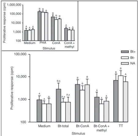
Bt-total allergen-induced cellular proliferative responses were significantly higher compared to negative control (medium alone) only for Bt+ patients, while Bt-ConA ex-tract induced increased proliferative responses in all groups (P < 0.05; Figure 2). TT antigen-induced proliferative re-sponses were significantly elevated for both groups of atopic patients (P < 0.05). Positive controls (PHA and Con-A) showed 30- to 50-fold higher proliferation rates in rela-tion to medium alone (Figure 2, inset). Comparing the groups, Bt-total extract induced a significantly higher proliferative response in Bt+ patients than in and NA subjects (P < 0.001). In contrast, Bt-ConA showed significantly increased cellular re-sponse in the Bt+ group in relation to the NA group (P < 0.01). Both Bt+ and Bt- atopic patients showed significantly elevated proliferative responses after TT stimulation compared to NA subjects (P < 0.05).
Proliferative response in the presence of ConA inhibitor
The Bt-total extract again induced a significant proliferative response in Bt+ patients compared to medium alone, while the Bt-ConA extract once more induced increased proliferative responses in all groups (P < 0.05; Figure 3). However, in the pres-ence of ConA inhibitor, the Bt-ConA extract-induced responses were significantly decreased (P < 0.05). When comparing the groups, only the Bt-total ex-tract induced significantly increased proliferative re-sponses in Bt+ patients compared to Bt- and NA subjects (P < 0.05). TT antigen-induced proliferative responses were also reproducible and significantly increased in both atopic patients (P < 0.05). Positive controls (PHA and ConA) showed significantly higher proliferation rates compared to medium while a
sig-Table 1. Table 1.Table 1. Table 1.
Table 1. Demographic and clinical characteristics of the study subjects.
Characteristics Groups
Bt+ Bt- NA
Number of subjects (N) 96 41 109
Age (years, mean ± SD) 27.3 ± 9.5 28.3 ± 9.2 31.1 ± 11.7
Gender (male/female) 40/56 14/27 37/72
Clinical diagnosis (N, %)
Rhinitis 78 (81.3%)a 37 (90.2%)a 0
Rhinitis + asthma 18 (18.7%)b 4 (9.8%)b 0
Positive SPT (mean wheal size, mm) and positivity (%)
B. tropicalis (Bt) 7.4 ± 3.0a 0 0
100%a 0 0
D. pteronyssinus (Dp) 9.8 ± 3.4b 7.5 ± 2.7a 0
99%a 100%a 0
D. farinae (Df) 7.9 ± 2.8a 7.3 ± 2.6a 0
100%a 90%a 0
Figure 1. Figure 1.Figure 1. Figure 1.
Figure 1. Levels of specific IgE (A) to Blomia tropicalis total extract (Bt-total) and concanavalin A-binding fraction (Bt-ConA), and total IgE (B) in serum samples from patients with positive (Bt+, N = 96) or negative (Bt-, N = 41) skin prick test to B. tropicalis and from non-atopic subjects (NA, N = 109). The dashed line indicates the cutoff of the assay (ELISA index, EI = 1.2). *P < 0.001 for comparisons indicated by the horizontal lines (Kruskal-Wallis and Dunn multiple comparison tests). The scales of the ordinates are logarithmic.
nificant decreased proliferative response was seen only for the ConA mitogen in the presence of its specific inhib-itor (Figure 3, inset).
Cytokine assays
No significant difference was observed in IFN-γ
pro-duction after any stimulus among the patient groups, ex-cept for the Bt-ConA extract that significantly induced increased IFN-γ levels in Bt+ patients compared to medi-um alone (P < 0.05; Figure 4). Positive controls increased IFN-γ production significantly in relation to medium in all subject groups (P < 0.05; Figure 4, inset).
Figure 2. Figure 2.Figure 2. Figure 2.
Figure 3 Figure 3Figure 3 Figure 3
Figure 3... Proliferative response of periph-eral blood mononuclear cells from 6 atopic patients with positive skin prick test (SPT) to Blomia tropicalis (Bt+), 4 atopic patients with negative SPT to B. tropicalis (Bt-) and 4 non-atopic (NA) subjects. Cells were stim-ulated with B. tropicalis total extract (Bt-total), or concanavalin A-binding fraction (Bt-ConA), or Bt-ConA extract in the pres-ence of methyl-α-D-mannopyranoside (Bt-ConA + methyl), or tetanus toxoid (TT) as non-allergen control. Positive and negative controls included mitogens (phytohemag-glutinin, PHA or ConA) and medium alone, respectively (inset). Data are reported as means ± SD. Different uppercase letters indicate significant differences between the stimuli for each group; different lowercase letters indicate significant differences be-tween the groups for each stimulus (P < 0.05, Kruskal-Wallis and Dunn multiple comparison tests). The scales of the ordi-nates are logarithmic.
Figure 4. Figure 4.Figure 4. Figure 4.
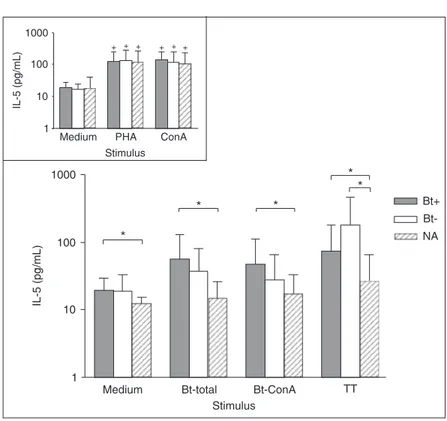
IL-5 levels were not significantly different after any stimulus in relation to medium alone, but significantly higher IL-5 levels were detected in the Bt+ patients compared to the NA subjects for any stimulus analyzed (P < 0.05; Figure 5). IL-5 levels were also significantly higher in Bt- patients after stimulation with the non-allergen control (TT) in rela-tion to NA group (P < 0.01; Figure 5). As expected, positive controls increased IL-5 production significantly in relation to medium in all subject groups (P < 0.05; Figure 5, inset).
Discussion
In the present study, we investigated the cellular and humoral immune responses to B. tropicalis allergens and their ConA-binding fractions in mite-sensitized patients. As expected, the great majority (70%) of patients with allergic rhinitis with/without asthma was sensitized to B. tropicalis, with nearly all patients (69%) presenting positive SPT concomitantly to the three mites (B. tropicalis, D. pteronyssinus, and D. farinae) and no patient was monosensitized to B. tropicalis. These results were similar to those of our previous studies carried out with another group of atopic patients from the same region (9,18), confirming the low sensitization to B. tropicalis only and that a concurrent sensitization seems to occur with the three mites, even though a need for previous sensitization
to other mite allergens cannot be ruled out. Therefore, this concomitant sensitization has complicated the evalu-ation of the role of B. tropicalis in these patients, particu-larly in this geographic region in which these mites have commonly been found. In addition, the sensitization to Dermatophagoides spp seems to be a major mechan-ism in the pathogenesis of respiratory allergic diseases in Brazil.
Similar levels of specific IgE to Bt-total and Bt-ConA extracts were found among Bt+ patients, supporting our previous reports (9) that Bt-ConA extract contains clini-cally relevant allergens responsible for the induction of IgE antibodies. In addition, patients non-sensitized to B. tropi-calis or non-atopic subjects showed no reactivity to both extracts, confirming the high specificity of the assay. De-spite the similar levels of specific IgE, seropositivity rates to Bt-total and Bt-ConA extracts were significantly differ-ent, indicating that the glycosylated components are not uniformly recognized by atopic patients and that the frac-tionation of the total extract could have contributed to the lack of some relevant sensitizing epitopes. As expected, the total serum IgE levels were found in both groups of atopic patients, regardless of the mite sensitizing agents, B. tropicalis and/or Dermatophagoides spp. Thus, despite the detection of total serum IgE, which is not able to discriminate among different atopic patients, IgE has been
Figure 5. Figure 5.Figure 5. Figure 5.
shown to be a useful laboratory parameter for differentiat-ing atopic from non-atopic subjects. Taken together, ELISA for the detection of B. tropicalis-specific IgE antibodies and IgG subclasses might be used to obtain consistent and reproducible results and it can be considered as an additional method for evaluating the humoral immune response to B. tropicalis in allergic patients.
Significant PBMC proliferation, a measure of the cel-lular immune response, was observed in response to Bt-total allergen extract in B. tropicalis-sensitized (Bt+), but not in non-sensitized (Bt-) patients and non-atopic indi-viduals. This indicates that the allergic response involves not only specific IgE production to allergens with hista-mine release, but also cellular proliferation, particularly of allergen-specific T lymphocytes and cytokine secretion (21). In contrast, the Bt-ConA allergen extract induced increased proliferative responses in all groups of pa-tients, even though significantly only in Bt+ atopic patients compared to non-atopic subjects. These findings sug-gest a role for mannose-enriched glycosylated compo-nents present in this extract, as potential inducers of the cell proliferation. Alternatively, these data could reflect a residual effect of ConA present in the Bt-ConA extract. To clarify this latter hypothesis, cellular proliferation assays using Bt-ConA in the presence of specific ConA inhibitor showed that these responses were significantly decreased, reinforcing the presence of ConA residues in the Bt-ConA extract. It is noteworthy that the Bt-ConA extract was probed with polyclonal mouse anti-ConA antibodies in ELISA and no specific reactivity was observed (data not shown). Therefore, further studies are required for improving the ConA-Sepharose column cross-linking in order to remove residual ConA and subsequently to evaluate the actual role of these mannose-enriched glycosylated antigens.
Little is known about the cellular immune response to B. tropicalis. Recently, Lozano et al. (22) evaluated the
PBMC proliferative response to Bt allergen extract and BtM recombinant protein, demonstrating that recombinant allergens are also able to induce cellular immune re-sponses similar to native allergen-induced rere-sponses, with potential applications in immunotherapeutic procedures. In the present study, a significant IFN-γ production, a Th1 typical cytokine, was observed only after Bt-ConA stimula-tion in B. tropicalis-sensitized patients while Bt-total aller-gen extract showed no significant change in this cytokine production. On the other hand, significant IL-5 production, a Th2 typical cytokine, was consistently seen in B. tropica-lis allergic patients under specific allergen stimulation (Bt-total and Bt-ConA) as well as unrelated antigen (TT) and even with no stimulus (medium alone), indicating that PBMC from allergic patients are prone to produce Th2 profile cytokines, spontaneously or inductively by allergen restimulation. In our previous study (9), Bt-ConA extract components were recognized by specific IgG1 antibodies (Th1 profile) rather than IgG4 antibodies (Th2 profile) in serum samples from B. tropicalis-sensitized patients, prob-ably reflecting its ability to induce a protective immune response through the development of Th1 cells. Alto-gether, it can be concluded that ConA-binding compo-nents isolated from B. tropicalis may contain relevant antigens that are involved in both humoral and cellular immune responses. However, without an additional purifi-cation procedure to eliminate the residual contamination with ConA, its use in immunotherapeutic procedures can-not be recommended.
Acknowledgments
We thank Dr. Federico Montealegre-Golcher, Ponce School of Medicine, Ponce, USA, for providing dried samples of B. tropicalis and D. pteronyssinus.
References
1. Arlian LG, Bernstein D, Bernstein IL, Friedman S, Grant A, Lieberman P, et al. Prevalence of dust mites in the homes of people with asthma living in eight different geographic areas of the United States. J Allergy Clin Immunol 1992; 90: 292-300.
2. Fernandez-Caldas E, Puerta L, Mercado D, Lockey RF, Caraballo LR. Mite fauna, Der p I, Der f I and Blomia tropica-lis allergen levels in a tropical environment. Clin Exp Allergy 1993; 23: 292-297.
3. Arruda LK, Rizzo MC, Chapman MD, Fernandez-Caldas E, Baggio D, Platts-Mills TA, et al. Exposure and sensitization to dust mite allergens among asthmatic children in São
Paulo, Brazil. Clin Exp Allergy 1991; 21: 433-439.
4. Arruda LK, Vailes LD, Platts-Mills TA, Fernandez-Caldas E, Montealegre F, Lin KL, et al. Sensitization to Blomia tropica-lis in patients with asthma and identification of allergen Blo t 5. Am J Respir Crit Care Med 1997; 155: 343-350. 5. Puccia R, Travassos LR, Rodrigues EG, Carmona AK,
Identification of a 105 kilodalton Cryptococcus neoformans mannoprotein involved in human cell-mediated immune re-sponse. J Med Vet Mycol 1997; 35: 299-303.
7. Gomez BL, Figueroa JI, Hamilton AJ, Diez S, Rojas M, Tobon A, et al. Detection of the 70-kilodalton histoplasma capsulatum antigen in serum of histoplasmosis patients: correlation between antigenemia and therapy during follow-up. J Clin Microbiol 1999; 37: 675-680.
8. Savolainen J. A standardized densitometric immunoblotting analysis of Candida albicans protein allergens. Clin Exp Allergy 1995; 25: 357-363.
9. Almeida KC, Silva DA, Gennari-Cardoso ML, Cunha-Junior JP, Alves R, Ynoue LH, et al. Responses of IgE, IgG1, and IgG4 to concanavalin A-binding Blomia tropicalis antigens in allergic patients. Braz J Med Biol Res 2006; 39: 1445-1454.
10. Tsai JJ, Wu HH, Shen HD, Hsu EL, Wang SR. Sensitization to Blomia tropicalis among asthmatic patients in Taiwan. Int Arch Allergy Immunol 1998; 115: 144-149.
11. Caraballo L, Puerta L, Martinez B, Moreno L. Identification of allergens from the mite Blomia tropicalis. Clin Exp Allergy 1994; 24: 1056-1060.
12. Ngoc PL, Gold DR, Tzianabos AO, Weiss ST, Celedon JC. Cytokines, allergy, and asthma. Curr Opin Allergy Clin Immunol 2005; 5: 161-166.
13. Romagnani S. Lymphokine production by human T cells in disease states. Annu Rev Immunol 1994; 12: 227-257. 14. Akdis CA, Blaser K, Akdis M. Genes of tolerance. Allergy
2004; 59: 897-913.
15. Bousquet J, Khaltaev N, Cruz AA, Denburg J, Fokkens WJ,
Togias A, et al. Allergic Rhinitis and its Impact on Asthma (ARIA) 2008 update (in collaboration with the World Health Organization, GA(2)LEN and AllerGen). Allergy 2008; 63 (Suppl 86): 8-160.
16. IV Diretrizes Brasileiras para o Manejo da Asma. Rev Bras Alergia Imunopatol 2006; 29: 222-245.
17. Sociedade Brasileira de Pneumologia e Tisiologia. III Consenso Brasileiro de Manejo da Asma. J Pneumol 2002; 28: 1-28.
18. Pereira EA, Silva DA, Cunha-Junior JP, Almeida KC, Alves R, Sung SJ, et al. IgE, IgG1, and IgG4 antibody responses to Blomia tropicalis in atopic patients. Allergy 2005; 60: 401-406.
19. Lowry OH, Rosebrough NJ, Farr AL, Randall RJ. Protein measurement with the Folin phenol reagent. J Biol Chem 1951; 193: 265-275.
20. Silva DA, Gervasio AM, Sopelete MC, Arruda-Chaves E, Arruda LK, Chapman MD, et al. A sensitive reverse ELISA for the measurement of specific IgE to Der p 2, a major Dermatophagoides pteronyssinus allergen. Ann Allergy Asthma Immunol 2001; 86: 545-550.
21. Valenta R, Sperr WR, Ferreira F, Valent P, Sillaber C, Tejkl M, et al. Induction of specific histamine release from baso-phils with purified natural and recombinant birch pollen aller-gens. J Allergy Clin Immunol 1993; 91: 88-97.