Letter to Editor
305 Rev Bras Hematol Hemoter. 2012;34(4):305-6
Heterozygosis for hemoglobin Porto Alegre identiied by a combination of laboratory
diagnostic methodologies
Hemoglobin (Hb) Porto Alegre is a beta globin chain mutant [beta 9 (A6) Ser>Cys] that was initially described in a Caucasian Brazilian family in 1963(1). It was subsequently identiied in other families in Brazil and in other places such as Cuba and the Canary Islands. The origin of the mutation was reported as Portuguese by the co-inheritance of an intragenic polymorphism characteristic of the population(2).
The laboratory identiication of the variant by routine methods is often dificult because the mutant fraction migrates to the same position as Hb A in alkaline electrophoresis, the most widely used method for identifying hemoglobins in Brazil. In this case report, we describe the identiication of the globin variant by associating electrophoretic, chromatographic and molecular methods after suspicion of a hemoglobin variant by capillary electrophoresis.
The patient was a female, adult Caucasian. Blood tests did not identify anemia and the suspected variant was only investigated after her clinician requested hemoglobin electrophoresis.
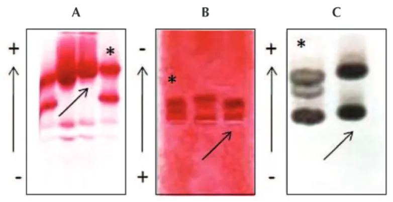
With the presence of a fraction close to Hb A and with 41.0% identiied as Hb Atlanta by capillary electrophoresis, an investigation was begun to conirm the suspicion. The fraction was not identiied by cation-exchange high-performance liquid chromatography (HPLC) as elution had the same retention time of hemoglobin A, but with unknown peaks close to the elution time of Hb C. Only a diffuse fraction with migration close to Hb A was identiied by electrophoresis in cellulose acetate at alkaline pH. The proile of Hb A was also conirmed by electrophoresis in agarose at an acid pH. Hb A2 was 4.2% by HPLC. Figure 1 shows the Hb proile for electrophoretic procedures at different pH, by capillary electrophoresis and HPLC.
The results of electrophoretic and chromatographic procedures were discordant with the initial suspicion of Atlanta Hb, because this Hb has an unstable component, and testing for thermal and isopropanol instability was negative. Given the inconsistency of indings of conventional laboratory methods, and how the mutant beta chain is suggested by the percentage of the fraction obtained by capillary electrophoresis, electrophoresis at alkaline pH was performed that showed the proile A alpha/A beta globin chain. The next step was to amplify the three exons of the gene for beta globin and perform nucleotide base sequencing. The result showed the presence of a mutation in the codon responsible for the amino acid number nine, with replacement of C by G (TCT > TGT).
Marcos José Cataldo1
Ana Carolina Bonini-Domingos2
Claudia Regina Bonini-Domingos2
1Labs D’or, Rio de Janeiro, RJ, Brazil 2Universidade Estadual Paulista “Júlio de Mesquita Filho” - UNESP, São José do Rio Preto, SP, Brazil
Conlict-of-interest disclosure:
The authors declare no competing inancial
interest
Submitted: 3/21/2012 Accepted: 5/2/2012
Corresponding author:
Claudia Regina Bonini-Domingos
Laboratory of Hemoglobin and Genetics of Hematological Diseases, Biology Department,
Universidade Estadual Paulista “Júlio de Mesquita Filho” – UNESP
Rua Cristóvão Colombo, 2265,
Jardim Nazareth
15054-000 São José do Rio Preto, SP, Brazil
www.rbhh.org or www.scielo.br/rbhh
DOI: 10.5581/1516-8484.20120077
A B C
306
Cataldo MJ, Bonini-Domingos AC, Bonini-Domingos CR
Rev Bras Hematol Hemoter. 2012;34(4):305-6 The mutation in the exon leads to the beta globin amino
acid substitution in the external portion of the molecule, which does not result in hematologic or clinical manifestations. The mutant fraction can be better seen by isoelectric focusing(3) due to the proximity of migration to other Hbs. Hb Porto Alegre presents normal Bohr effect, slightly decreased cooperativity and increased oxygen afinity(4). Hb Porto Alegre is also found in
association with beta thalassemia and other variants(5).
Conirmation of heterozygous Hb Porto Alegre and also the presence of an intragenic polymorphism at codon 27, reinforcing the contribution of Portuguese genetics for this mutant, were only possible with molecular analysis. Greater attention should be given to obtaining hemoglobin proiles using procedures with lower sensitivity, because the co-migration of fractions can hinder identiication.
Acknowledgments
The authors acknowledge the collaboration of Dr. Paula Juliana Antoniazzo Zamaro and Larissa Paola Rodrigues Venancio for the molecular analysis.
References
1. Tondo CV, Salzano FM, Rucknagel DL. Hemoglobin Porto Alegre, a possible polymer of normal hemoglobin in a Caucasian Brazilian family.
Am J Hum Genet. 1963;15:265–79.
2. Gonçalves MS, Sonati MF, Kimura EM, Arruda VR, Costa FF, Nechmen JF, et al . Association of Hb Santa Ana and [alpha 2 beta (2)88(F4) Leu- Pro] and Hb Porto Alegre [alpha 2 beta (2)9(A6)Ser-Cys] in a Brazilian female. Hemoglobin. 1994;18(03):235-9.
3. Hardison R, Chui DH, Riemer CR, Miller W, Carver MF, Molchanova TP, et al. Access to a syllabus of human hemoglobin variants (1996) via
the World Wide Web. Hemoglobin. 1998;22(2):113-27.
4. Tondo C, Bonaventura J, Bonaventura C, Brunori M, Amiconi G, Antonini E. Functional properties of hemoglobin Porto Alegre (alpha2A beta2 9Ser leads to Cys) and the reactivity of its extra cysteinyl residue. Biochim Biophys Acta. 1974;342(1):15-20.
5. Baudin-Creuza V, Fablet C, Zal F, Green BN, Promé D, Marden MC, et al. Hemoglobin Porto Alegre forms a tetramer of tetramers superstructure. Protein Sci. 2002,11(1):129–36.
xxx
Figure 1 - The arrows identify the patient; (D) electrophoretic proile by capillary electrophoresis systems show the Hb fraction close to Hb A; (E) Chromatographic proile obtained by high-performance liquid chromatography; Standard (*) – hemoglobin AS.