(1) Hepatology Branch, Discipline of Clinical Gastroenterology, Department of Gastroenterology; University of São Paulo School of Medicine(FMUSP), São Paulo, SP, Brazil (2) Discipline of Digestive Tract Surgery, Department of Gastroenterology; FMUSP, São Paulo, SP, Brazil.
(3) Department of Pathology, FMUSP, São Paulo, SP, Brazil.
Correspondence to: Marcelo Eidi Nita, Rua Capote Valente 154/13, 05409-000 São Paulo, SP, Brazil. Email: marcelo_nita@uol.com.br
REVIEW
MOLECULAR ASPECTS OF HEPATIC CARCINOGENESIS
Marcelo Eidi NITA(1,2), Venâncio Avancini Ferreira ALVES(3), Flair José CARRILHO(1), Suzane Kioko ONO-NITA(1), Evandro Sobroza de MELLO(3) & Joaquim J. GAMA-RODRIGUES(2)
SUMMARY
Exogenous agents correlated with hepatocellular carcinoma (HCC) have been identified and well characterized. These agents, including the different viruses that cause chronic hepatitis and cirrhosis, can lead to regenerative nodules and dysplastic nodules/ adenomatous hyperplasia. These conditions associated with several molecular alterations of hepatocyte ultimately culminate in hepatocellular carcinoma. Recently, there has been a great progress in the identification of somatic and germinative mutations that may be correlated with the development of HCC, justifying a review on the subject. Hence, the factors involved in the process of hepatic carcinogenesis, such as infection by the hepatitis B and C viruses, with a special focus in the molecular alterations described in recent years are discussed herein, pointing out areas potentially relevant for clinical development.
KEYWORDS: Liver cancer; Carcinogenesis; Hepatitis B; Hepatitis C; p53; Cell cycle; Apoptosis.
INTRODUCTION
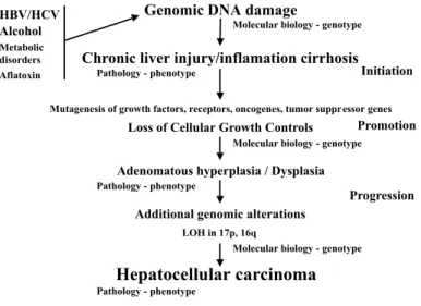
The carcinogenesis is a complex event, involving alterations in several genes and, depending also of external factors, such as infection by hepatitis B or C and chronic alcohol intake, can lead to a pre-neoplastic lesion, like the low and high grade displastic nodule / adenomatous hyperplasia105. Subsequently, this lesion can progress to early cancer and finally, culminate with advanced hepatocellular carcinoma (HCC)4. The knowledge about the development and progression to HCC have increased in later years with the increasing using of several molecular biology techniques71,80. Thus, one of the most frequent molecular events in cancer is an aberrant gene expression. This can be due to both genetic alterations - like point mutations or deletions in segments of genes - or to epigenetic modifications that modulate the gene function, like gene methylation, without changing the DNA sequence but altering the resulting protein. The activation of oncogenes and inactivation of tumor suppressor genes (TSG) have an important role in the mechanisms of cancer development. These genes can induce malignant transformation when inappropriately expressed as a result of mutation, deletion, amplification or rearrangement. Overexpression of oncogenes leads to constitutive activity of key regulatory molecules that are normally modulated during the cell cycle. In the case of HCC, the activation of oncogenes has been seen with a low frequency, leading some authors to suggest a secondary role of this genetic event. On the other hand, reports of inactivation of TSG are increasing seen in the process of hepatic
carcinogenesis. Thus, the advances of molecular genetics of HCC as summarized in Fig. 1 and Table 1, already justify a review on the theme.
Fig. 1 - Molecular model for hepatocarcinogenesis (see the text). Figure adapted from CHAN,
Furthermore, the progress generated by the publication of the first draft of the human genome, the start up of several projects post-genome sequencing to identify human genes and the knowledge acquired with molecular biology will soon make an impact in the routine of the physicians management of several diseases. We will, thus, try to discuss several factors involved in the process of hepatic carcinogenesis, such as infection by the hepatitis B and C viruses, the related molecular alterations recently discovered and their relevance for clinical management.
HEPATITIS B VIRUS: Hepatitis B virus (HBV) has a circular DNA
genome, with about 3.2 kb, which can originate an acute and chronic necroinflammatory response from the liver85. In fact, HBV is not directly cytopathic to the hepatocyte, and it is believed that the hepatic disease is due to an immunologic response to the viral antigens expressed by the infected hepatocytes. The virus is composed of several forms of proteins: large envelope protein, transactivating protein that contains the HBV X, core-pre core proteins and viral polymerase. The HBV infection during the adult life is often subclinical, and most of the adult patients with acute hepatitis B recovers completely and eliminate the virus15. About 5% of the patients infected during adult life, however, will have persistent infection and will develop chronic hepatic disease. In contrast, more than 90% of the patients with neonatal transmission of the HBV develop chronic disease. Vertical transmission of HBV is common in areas of Africa and Asia with high demographic density, and because of that hundreds of thousands of people are chronically infected5,79. Many of these individuals will develop chronic hepatic diseases, and even cirrhosis and HCC still in young ages. In Taiwan there are about 433 cases of HCC per 100,000 HBV infected male, compared to 5 cases per 100,000 persons without infection by HBV2. In Japan, where neonatal transmission of HBV is rare, the risk of developing HCC in patients with diagnosis of chronic infection by HBV is 7 fold higher the in non-infected individuals97.
HCC-induced by HBV is likely the result of a variety of mechanisms71. (I) The viral integration into the host genome, that would lead to an alteration in genes regulating cell cycle; (II) Chronic hepatitis or hepatic cirrhosis could lead to a hipermutagenic status affecting cell cycle genes by somatic mutation and/or epigenetic alteration, promoting deregulated proliferation; (III) Chronic liver inflammation could lead to a mutagenesis prone environment; (IV) The viral transactivating protein
HBV X, after host genome integration, could cause deregulation of genes involved in the cell cycle, such as p53 and p21Waf1, among others.
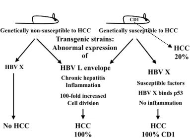
All of these mechanisms have been investigated in patients infected with HBV and in several animal models for studying hepatitis. One of the first used models was the woodchuck88. Captive woodchucks infected at birth with woodchuck hepatitis virus (WHV), a DNA-virus very similar to the HBV, developed HCC after 17 to 36 months of persistent infection in 97% of the cases; WHV infection occurring during adult life carries a far lower risk of HCC development. This pattern of infection is somewhat analogous to the HCC incidence within HBV infection in humans. Integrated WHV genomes are found in most cases of HCC in woodchucks infected at birth or while young, as it is the case for human patients with chronic HBV infection. Integration of WHV causes deletions, translocations and inversions of host chromosomes. However, no predominant transforming oncogenes and no characteristic chromosomal abnormality have been identified. Integration events leading to the direct activation of oncogenes and inactivation of tumor-suppressor genes, therefore, seem to be rare and random in both woodchucks and humans, and only in a few cases can be correlated with unordered cellular proliferation. Alterations in the expression of the insulin growth factor type II detected in the pre-neoplastic lesion in the liver of woodchucks infected with WHV are potentially important because the gene of IGF-II is deleted in several human HCC samples118. Nonetheless the integration of HBV can lead to rearrangements of chromosome 11p, and this may also account for the deletions observed in this chromosome in HCC patients. However, differences between this animal model and cancer in humans have also been observed; the activation of mutation in the oncogenes n-myc and c-myc is frequently found in woodchucks67, but these alterations have not been found in HCC associated with hepatitis. Recently several models of infection by HBV have been studied in transgenic mice8. These transgenic mice have been constructed containing only HBV-derived regulatory sequences that express all of the viral gene products and replicate the virus in the hepatocyte. Therefore, they are considered to be excellent models for dissecting the human disease. One potential problem with this methodology is that the expression of HBV genes and the process of viral replication are not per se cytotoxic for the hepatocytes. On the other hand, the abnormal expression of two products of viral genome, the large envelope protein and the protein HVB X can lead to HCC development (Fig. 2). Transgenic mice expressing large Table 1
Molecular alterations in hepatocellular carcinoma (Modified from OZTURK80)
Gene Mutation(%) Other alterations References
p53 20 – 70 Interaction with HBx gene, aflatoxin 14,22,32,70
Cyclin D 11 Amplification 73
p16Ink4 0 – 50 Hypermethylation, decreased expression 7,39
p21Waf1/Cip1 0 Decreased expression 29,37
Rb 15 Homozygous deletion 57,120
Beta-catenin 16 – 26 Increased expression 64
M6P/IGF2R 18-33 Homozygous deletion 17,84
E-cadherin Methylation 42,43
COX-2 Increased expression 53,95
amounts of the HBV large envelope protein show severe hepatitis, and
the degree of hepatocellular injury is directly proportional to the amount of HBsAg maintained in the endoplasmic reticulum. Prolonged storage of the long subviral particles in the endoplasmic reticulum in large amounts can lead the hepatocytes to die spontaneously. This chronic hepatocellular necrosis causes a secondary inflammatory and regenerative response, which leads to HCC in all transgenic mouse strains overexpressing the large envelope protein8,9. Hepatocellular turnover in these mice, relative to nontransgenic controls, is increased 100-fold for at least a year before the onset of HCC36. Moreover, there is a dramatic increase in the oxidative damage of the hepatocellular genomic DNA. It is reasonable to assume that these events could lead to and are fundamental for the HCC formation in these animals, and can serve as reference in the human hepatocarcinogenesis.
A high level of expression of the HBV X protein in transgenic mice results in almost ubiquitous HCC in some mouse strains49,54. The transgenic mouse strains that develop HCC upon overexpression of the HBV X protein have a CD-1 genetic background. They are naturally susceptible to HCC and as such have been used extensively to study hepatic carcinogenesis. The transgenic mouse strains that express the X protein in a genetic background other than CD-1 do not develop HCC. These findings suggest that the X protein may function as a cofactor in promoting HCC, besides host genetic susceptibility. The mechanism by which these mice develop HCC is not fully understood22,48,108. It has been shown that the HBV X protein interacts with the wild-type human p53 protein and inhibits its sequence-specific DNA binding and transcriptional activity in vitro. Furthermore, the HBV X protein can transcriptionally activate several cellular genes in vitro, including c-myc and c-jun, which are associated with growth control, what could also be transforming.
HEPATITIS C VIRUS: Hepatitis C virus (HCV) is a RNA virus,
of approximately 10 kb, belonging to the Flaviridae family10,85 and is acquired mainly by parenteral exposure5; it has been considered the main agent causing chronic hepatitis, cirrhosis and HCC15,66,96,103. Due to the great sequence heterogeneity of the viral genome, HCV is classified into various subtypes, with different epidemiological characteristics98,99. The viral replication, from a RNA strand into a new RNA strand, does
not involve a DNA synthesis step and, thus, no viral integration in the host genome. Some scientists propose a cytopathic action of HCV, due to the structural analogy with several known cytopathic virus, such as flavivirus and pestivirus. However, this has been vigorously contested by those who believe in the existence of “asymptomatic carrier of HCV”. The HCV genome codes for a polyprotein of 3,010 to 3,033 aminoacids11. This polyprotein is processed into at least 10 proteins, of which four are structural (core, E1, E2 and p7) and 6 non-structural (NS2, NS3, NS4A, NS4B, NS5A and NS5B). KATO et al., working at Omata’s lab, showed that the HCV core and NS4B proteins45,46 work as an induction agent in the activation of intracellular signal, specially in the system NF-kB, AP1 and SRE, contributing for cellular proliferation and production of inflammatory cytokines, potentially relevant in the formation of HCC. MORIYA et al. accepted that transgenic mice for the HCV core gene developed sequentially: hepatic steatosis, adenomas and hepatocarcinomas66. Altogether, these results imply that HCV can have an important role in hepatocarcinogenesis.
p53: The p53 gene is one of the most important tumor suppressor genes, being altered in more than half of all different kinds of tumor and HCC is not an exception to this rule86,106. This gene has characteristics of tumor suppressor in its wild-type form and can function as a dominant oncogene when mutated. Usually, the altered protein of a tumor suppressor gene is due to the loss of heterozigosity of one allele and another mutation in the remaining one, according to the “two-hits theory” proposed by KNUDSON Jr. et al.52. Exceptionally, p53 is a transcription factor that requires alteration of only one of its alleles to have its function altered, since the mutant protein prevails and inhibits the function of the wild type protein112. The 20 kb-gene is located in the short arm of chromosome 17 and contains 11 exons (protein-coding nucleotides), and 10 introns (non-coding nucleotides). The messenger RNA codes for a protein of 393 amino acids, which is expressed at low levels in all different tissues of the body. The p53 gene product is a phosphoprotein of 53 kDa involved in several functions to maintain the cellular homeostasis. Cell cycle and apoptosis are two key determinants of the cell fate controlled by p53. Cell cycle arrest at the “checkpoints” in G1 and G2 (Fig. 3) allows the process of DNA repair and prevent mutation or other genetic alterations to be transmitted to the next cellular
Fig. 2 - Transgenic mice and hepatocellular carcinoma (see the text).
generation. When DNA damage is severe with no possible repair, the cell can undergo a programmed death, named apoptosis, in which specific genes are activated and lead the cell to commit suicide. Upon DNA damage, the p53 gene is activated and acts in vital regulatory functions through, for instance, p21Waf1/Cip1 - arresting the cell cycle (see cell cycle below) - and Bcl-2 and Bax proteins leading to apoptosis. This is why p53 was called the “guardian of the genome”59, explaining the high frequency of alterations in this gene in many different kinds of cancer. In liver cancer, p53 is closely associated to aflatoxins - which are toxins produced by the fungus Aspergillus flavus and related species, that grow in food like peanuts, corn and rice when stored in inadequate conditions of high temperature and humidity, creating the perfect environment for the fungus reproduction. The HCC found in patients from areas with high exposure to aflatoxin, like China, Senegal and other African countries, has been associated with a specific mutation in the p53 gene35,81. About 20 to 75% of the HCC-patients from these regions have a p53 mutation, which in most of the cases is a G to T transversion in the third nucleotide of codon 249 - resulting in substitution of arginine by serine14,70,115. This mutation has been found in individuals with intense exposure to aflatoxin B1, but with an apparently normal liver, suggesting that this is an early genetic event in the process of hepatocarcinogenesis. On the other hand, this mutation has not been found in regions with high incidence of HCC and HBV; thus it is likely that the hepatitis B virus is not associated with this particular type of p53 mutation. In areas with low levels of aflatoxin, like Japan and some European countries, the frequency of this type of p53 alteration is much lower, about 10 to 20% depending on the area30,33,44,113,115.
CELL CYCLE: Cancer cells are characterized by unlimited and
unordered growth due to disequilibrium between cell proliferation and death. The intimate relation between cancer and some genes that control cell cycle has been elucidated by molecular biology74,76,107. Cell cycle is divided into four phases: G1 phase (gap1), S phase (DNA synthesis), G2 phase (gap2) and mitotic phase (Fig. 3). The progression through each phase is controlled by intracellular signals, that either negatively control the cycle, arresting the cell cycle, or positively control the cycle, allowing the advance into the next phase41,92. The G1 phase is well defined and frequently associated with carcinogenesis; at this point, the genes that positively control the cell cycle to proceed into the next phase code for cyclins and cyclin dependent kinases (CDK). The main substrate of cyclin and CDK is the retinoblastoma gene product (Rb); the wild-type form of the Rb gene inhibits the development of tumor. Phosphorylation of Rb by the cyclin/CDK complex allows progression of the cell cycle by freeing the E2F family of proteins. The inhibition of this machinery occurs through a class of small proteins called CDK inhibitors (CKI), such as p16INK4, p21Waf1/cip1 and p27Kip1, and their alteration have been associated with different types of cancers. Besides these, other genes, such as p53 and TGF-beta (transforming growth factor–beta), are involved in the G1 phase of the cell cycle by regulating the CKI.
Any alteration in any level of this pathway can break the regulatory machinery, resulting in unordered and continuous cellular growth–two paradigms of a cancer cell. That is why these genes are being intensely researched and the study of the alteration of the genes p16INK4, p21Waf1/cip1 and p27Kip1, Cyclin D1, p53, Rb, and TGF-beta receptor II are leading to a better understanding of the carcinogenesis process in several different neoplasias74,106,107, among them, the hepatocellular carcinoma38,72.
Cyclin D1 is one of the cyclins that when associated with CDK phosphorilates the Rb protein, allowing cellular proliferation. The chromosomal location of the Cyclin D1 gene is 11q13 and its protein expression is altered in HCC. Neoplastic hepatocytes have a 3 times increased expression of Cyclin D1 when compared with normal hepatocyte; this alteration was also confirmed at DNA level, in which amplification of the Cyclin D1–gene was found mainly in advanced cases73,121. Since this alteration may be associated with a more aggressive behavior of the tumors, it is believed that the amplification and overexpression of Cyclin D1 can deregulate cell proliferation.
The product of the gene p16INK4 was initially identified as a protein of 16 kDa that inhibits the Cyclin/CDK complex, not allowing phosphorylation of Rb and, consequently, stopping the cell cycle. The gene p16INK4 is located in the region 9p21 and is also called MTS1 (multiple tumor suppressor 1), because it is involved in multiple types of cancer, being melanoma the best studied. This gene has several ways of being inactivated, and some of them are different from the classical p53 and Rb genes that are inactivated by point mutations; the p16INK4 gene is inactivated by homozygous deletion in mesothelioma and bladder cancer considering that it is inactivated by point mutation in melanomas, esophageal and biliary tract cancer. Besides these genetic alterations, p16INK4 can be inactivated by an epigenetic modification, such as hypermethylation, like was observed in lung-carcinoma small cell and colon cancer. The product of the p16INK4 gene is altered in about 34% of hepatocellular carcinomas39 and in about 22% of early cases of HCC. These results suggest that p16INK4 can be involved in the early phases of the hepatocellular carcinogenesis. CHAUBERT et al.7,50 also suggest that the inactivation of this gene occur mainly by hypermethylation in 48% of the HCC cases, and their data were confirmed by WONG et al.114. The p21Waf1/cip1 gene is one of the main substrates for p53 and is the link between p53 and cell cycle. Furthermore, p21 is considered to be an universal inhibitor of the cyclin/CDK complex, thus also inhibiting the cell cycle. Several experiments have suggested that the p21Waf1/ cip1 could suppress tumoral growth, being then a potential tumor suppressor gene. However, the p21Waf1/cip1 gene is seldom mutated in most of the cancers, leading the researchers to look for possible alterations at the expression level29. HUI et al. found that this gene has low levels of mRNA in almost 40% of HCC; they also found a correlation between this low level of expression and mutation in p5337. Taking all in consideration, it is possible that the p21Waf1/cip1 gene contributes to the process of hepatocarcinogenesis93.
where this gene is located is frequently observed in about 30% of patients
with HCC, and it was associated with loss of protein function. Probably, such gene inactivation is involved in the process of hepatocarcino-genesis120. More recently, it was demonstrated that the E2F1 family of genes, that complex with Rb, is associated with adenomas and HCC in animal models of hepatocarcinogenesis13.
In summary, drawing all these studies together, it is clear that, although those alterations are not necessarily correlated between themselves, HCC patients have at least one of them present in their tumors. Thus, loss of cell cycle regulation in HCC is frequently found, and can be involved in the hepatic carcinogenesis, since it could lead to an unordered growth of the hepatocyte. The elucidation of the machinery governing the cell cycle may allow novel preventive and therapeutic approaches aiming to correct these alterations.
APOPTOSIS: Apoptosis, or programmed cell death, is a biologic
process essential to several physiologic and pathologic conditions47,74,78. The number of cells in a living organism depends basically on the balance between cellular proliferation and apoptosis, and breaking the homeostasis between these two processes can contribute to the appearance of a hepatic disease. High rate of apoptosis seems to be involved in viral hepatitis, cholestatic diseases, such as primary biliary cirrhosis, and hepatic disease induced by alcohol82. On the other hand, low rate of apoptosis in hepatocytes allows survival of cells that otherwise would die, leading to neoplastic transformation, due to clonal expansion of cells with molecular abnormalities. Alternatively, there would be an increase of apoptosis rate in the hepatocellular carcinoma and in the pre-neoplastic lesions, but not enough to compensate for the increased proliferation rate that features in cancer tissue31. Furthermore, inhibition of apoptosis is one of the main mechanisms of acquiring treatment resistance, such as chemoresistance, that characterize solid tumors; thus approaches that induce apoptosis can be a way to overcome resistance to solid-tumors treatment18,19,91. Among the genes controlling apoptosis, the emerging Bcl-2 family of apoptosis regulators56 was the first gene of this family to be identified, and thereafter a series of novel genes were isolated. These genes are now divided in two groups: those that block apoptosis, such as Bcl-2 and Bcl-XL; and those that induce apoptosis, such as Bax, Bak and Bcl-XS. In vitro studies have shown that Bcl-2 or Bcl-XL overexpression, as well as Bax deficiency, can confer resistance to antineoplastic drugs25,63. Conversely, the overexpression of Bcl-XS or Bax can sensitize neoplastic cells to radiotherapy or chemotherapy89.
The Bcl-2 gene product was immunohistochemically found in HCC, but not in pre-neoplastic lesions such as dysplasia122. This finding suggests that overexpressed Bcl-2 could be detected by immunohistochemistry and explored as a marker for neoplasia. There are, however, some recent reports describing the Bcl-XL gene, and not the Bcl-2, as the predominant gene in HCC116. Experimental work61 showed that hepatoblastoma cell lines have an increased sensitivity to the chemotherapy drugs when the expression of the Bcl-XL gene is reduced, or the opposite when the gene is fully expressed28. Similar results have been obtained by experimental work with colorectal cancer cell lines75,77. These works suggest that Bcl-XL can be a target for genetic therapy allowing the overcome of chemoresistance in digestive cancers. Another important protein that is involved in the interaction of the tumor with the immune system is the Fas (Apo-1, CD95), a member of the family of the nerve growth factor/tumoral necrosis factor that mediates apoptosis in response
to specific antibodies after ligation with Fas-L. In normal liver, Fas is weakly expressed in the hepatocyte membrane and cell of the biliary duct, while Fas-L is not expressed. In chronic hepatitis, Fas-L is expressed in areas of high interface activity, being variable in the case of HCC.
WNT SIGNALLING PATHWAY (APC/AXIN/BETA–
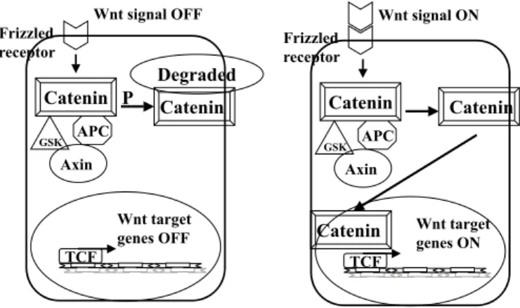
CATENIN/TCF): The Wnt signalling pathway plays a critical and
evolutionarily conserved role on regulating the cell fate in different phases of organogenesis and regulation of cell-cell interaction (Fig. 4). The genes involved in the Wnt signaling pathway are also involved in several types of cancer, including the HCC12. At the cell membrane, the Wnt signal links to the family of Frizzled receptors, a transmembrane cellular receptor. The signal triggered by Wnt goes down through several cytoplasmic molecules, such as APC, axin and GSK3beta, and stabilizes the beta–catenin protein65. This protein is then accumulated and enters the nucleus, where it forms a complex with Tcf/Lef (also known as lymphocyte enhancer-binding factor). Together, they function as a transcription factor activating the genes involved in several cellular functions, such as c-myc34. When Wnt signal is absent, the signal transduction pathway is off because beta–catenin is rapidly destroyed and can not enter the nucleus. Inactivation of APC or axin by mutations can result in permanent activation of the Wnt signaling pathway and accumulation of beta–catenin in the cytoplasm. Mutation in the CTNNB1 gene, that codes for the beta–catenin64, or in the APC gene (Adenomatous Polyposis Coli responsible for adenomatous familiar polyposis – FAP) have been reported in several types of cancer65. The hepatic cancer, however, shows a higher occurrence of abnormal distribution and accumulation of beta-catenin than it would be expected by the rate of mutation in the gene CTNNB1, raising the hypothesis that beta-catenin accumulation would be due to other reasons12. In the case of colorectal cancer the innapropriate activation of Tcf/Lef by beta-catenin can occur by means of APC inactivation, an important tumor suppressor gene involved in the colorectal carcinogenesis. Alterations, nevertheless, of APC are very rare in the case of HCC. SATO et al., working at the Nakamura’s lab, studied HCC derived cell lines and 100 primary liver tumors, and uncovered that the axin gene is mutated in HCC90. Axin has features of a tumor suppressor, and they reported that both alleles are inactivated in some tumors, by mutation or loss of heterozigosity (LOH), in accordance with the Knudson hypothesis51. Thus, it is possible that axin is a new tumor suppressor gene. Functional analysis of this gene
showed that all mutations detected in that study would yield a truncated protein missing the binding site for beta-catenin. Moreover, they reported that mutation on any of these genes, CTNNB1 (beta-catenin) ou Axin, leads to an increased interaction with TCF/LEF in the nucleus64,90. They also have shown that genetic therapy with transfection of the wild-type axin gene in liver or colon cancer cells can induce these cells to apoptosis in background where the beta-catenin was accumulated either by mutation on APC or CTNNB1 or axin. Taking together, these experiments not only give further insights to the understanding of hepatocarcinogenesis, but also indicate a possible way for treatment of both liver cancer and colon cancer.
ALLELOTYPE STUDIES – LOSS OF HETEROZIGOSITY:
Loss of heterozigosity (LOH) studies, in certain chromossomal regions, is a frequently employed strategy for the detection of alleles alteration in the human genome23,110. A LOH indicates a situation where there is loss of a second allele on one chromossome that had already lost the other one by a germinative alteration (two-hits theory by KNUDSON)51. Such alteration is preferably detected in a malignant tumor as compared with the heterozygous state in the normal tissue. For such study, the chromossomes can be conveniently divided into a few hundreds loci whose loss of integrity (LOH) can be indirectly pointed out by specific markers. The loci showing LOH suggest that the genes residing in these regions have been preferentially removed during the cancer development, and thus, can be considered candidates for tumor suppressor genes (TSG). Table 2 depicts some chromosome regions in which LOH were described as the likely candidates to TSG near this region and the occurrence of this fact in early stages of HCC.
Chromosome 1p allelic loss was studied in 104 cases of HCC and was detected in 38% of the early tumors (less than 2 cm) and in 33% of the well differentiated tumors, indicating that it might be an early event associated with HCC development58,104. The gene M6P/IGFR2 (mannose 6-phosphate/insulin-like growth factor-II receptor) reside on chromosome 6q, where was observed LOH in cancer tissues16 and in pre-neoplastic lesions of the liver117. Moreover, it was described in HCC a mutation in the other allele in about 18 to 32% of the cases17,84. The M6P/IGFR-2 gene interacts with IGFII, and activates the growth inhibitor TGF-beta, regulating cellular proliferation. Another chromosome described by researchers at the University of Tokyo as being altered in about 1/4 of the early cases of the studied HCC is 8p57. In this region DLC1 (Deleted
in Liver Cancer) was isolated, and it is a gene with low expression level in HCC, a potentially novel tumor suppressor gene. More recently, another gene with decreased expression in hepatic tumors was reported in this region and called LPTS – for liver-related putative tumor suppressor60. Functional studies, using gene transfection techniques, showed that LPTS is able to control the growth of cancer cells in vitro, suggesting that this gene may inhibit the growth of hepatocytes. The region of chromosome 13q is also important because it harbors the genes Rb and BRCA23,57. The 13q region showed allelic imbalance in about 15% of the early HCC and 36% of the cases of well differentiated HCC57. Loss of chromosome 16q appears to be one of the most common genetic defects in HCC119. One of the candidates to be the target is the E-cadherin gene – located on 16q22.1. Although no mutations or gross structural alterations of this gene have been reported in HCC, loss of E-cadherin expression has been observed in this type of cancer94. Recently, de novo methylation of the 5’CpG island of the E-cadherin gene has been found in 46% of liver tissue showing chronic hepatitis or cirrhosis and in 67% of the HCC analyzed42. Such epigenetic change correlated significantly with reduced E-cadherin expression. It is suspected that the inactivation of this gene by deletion or hypermethylation may play an important role in the development of HCC43. In the region of p53, 17p, it was also observed LOH in 17% of the early tumors and 35% of the well-differentiated tumors, being, thus, this gene a possible target for genetic therapy in the near future. For this reason, the wild-type p53 gene is transfected to the cancer cell in the hope that the correction of this abnormality leads to an effective treatment of HCC. It is important to note that the discrepancy between high LOH rates in the 17p region and the low frequency of p53 deletion in some reports may indicate the presence of another tumor suppressor gene in this region.
GENETIC ALTERATION OF THE ONCOGENES RAS AND
MYC:The oncogenes ras and myc are reported to be involved in the process of pancreas and colo-rectal carcinogenesis. Frequently, these oncogenes are targets of mutations, amplifications and deletions that can lead to an overexpression of their proteins, triggering a process of hyper activation of regulatory molecules involved in important cellular functions, such as cell cycle and apoptosis. Alteration of the ras genes has been described in animal models of hepatocarcinogenesis26, but this result is yet to be confirmed in human hepatocellular carcinoma88,100. Most authors, therefore, believe that they do not play an important role in the liver cancer development.
Table 2
Chromosomal location, frequency of LOH, the gene most likely to be in this region and if it is reported to be an early event or not
Chromosome Frequency of LOH1 Associated gene Early event
1p 32%58,104 p73 Yes in 38%58
4q 40 to 83%3,119 - Yes in 17%58
6q 35 to 80%3,17 IGF-II/M6p-receptor17 Yes16,117
8p 50 to 60%3,60 DLC-1? LPTS?60 Yes in 25%57
10q 25%27 PTEN/MMAC1 No
13q 25 to 50%3,68 Rb or BRCA-2 Yes in 15%57
16p 40%3,90 Axin No
16q 36 to 70%3,27,119 E-cadherin No
17p 36 to 54%3,27,58,68 p53 Yes in 17%58 and 50%68
COX2: Cyclooxygenase (Cox) is the key enzyme in the conversion
of arachidonic acid to prostaglandin and has been the focus of a series of research in the field of inflammatory diseases and carcinogenesis21. Two isoforms of this enzyme were recently identified: the COX-1 variant is constitutively expressed in most of the tissues, while its isoform, COX-2, is inducible by a variety of physiologic stimuli, including cytokines, growth factors, and tumor promoters. This inducible isoform was found to be upregulated in several human cancers, including colon cancer, gastric cancer, and squamous cell carcinoma20,87. A significant reduction in mortality from colorectal cancer has been reported for individuals taking nonsteroidal anti-inflammatory drugs (NSAIDs), attributed to the inhibition of the isoform 2 in the cancer cells. Recently, this COX-2 variant was also described to present an increased expression in hepatocellular carcinoma, mainly in those small and well differentiated tumors53. This finding led these authors to suggest that this change might be an early event in the development of HCC. Similar results were published by other groups1,95, raising the possibility that COX inhibitors may have a therapeutic or preventive role in liver cancer similarly to what has been seen for colorectal tumors1.
TELOMERASE: At the end of a chromosome there are long 5’
DNA repeats called telomeres. This structure consists of DNA and proteins, and, in eukaryotic cells, the telomeres DNA sequence cannot be completely copied during the replication mechanism. Therefore, telomeres undergo progressively shortening within each mitotic cycle as part of the normal ageing process. The length of telomeres seems to be determined by the enzymatic activity of telomerase. Upon an increased activity of this enzyme, a ribonucloprotein that is necessary to maintain the cancer cell tend to immortality, the telomere has its length increased and the cell extend its potential to continuous replication or even become resistant to the death62. The reactivation of telomerase activity and the change in the length of the telomeres have been described in most cases of HCC55,69,101,102, and at early phases of HCC development55. For this reason, monitoring telomerase activity has been assessed as a potential molecular marker for early cancer diagnosis, not only in HCC but in several other types of cancer. However, in a small percentage of the cases the altered telomerase activity has also been observed in normal cells. Although, these authors101 explain these changes in normal cells as a consequence of the presence of inflammatory cells, the confirmation of this finding in other series may jeopardize the utility of telomerase in the process of differential diagnosis of hepatic lesions.
CONCLUSION
Compared with other solid tumors, such as the digestive tract tumors109,111, the process of molecular carcinogenesis of HCC is still not clear. There are several genes possibly related to hepatocarcinogenesis but confirmation of these preliminary studies is yet to be provided. Figure 1 shows a model proposed for the process of hepatocarcinogenesis. There are about 30,000 to 50,000 genes in each human cell, and it is necessary to identify those that are involved in the process of hepatocarcinogenesis. We believe that the disclosure of such genes and their function will make it feasible to create new tools for diagnosis, follow up and treatment of HCC patients. We hope that with this review of molecular alteration of HCC can, by reviewing the current knowledge, stimulate new lines of research and open up the possibility of daily use in clinical setting of the genetic alteration described until now.
RESUMO
Aspectos moleculares da carcinogênese hepática
Agentes exógenos correlacionados com o carcinoma hepatocelular (HCC) têm sido identificados e bem caracterizados. Esses agentes, entre os quais se incluem os diferentes vírus que causam hepatite e cirrose hepática, podem provocar o aparecimento de nódulos regenerativos e nódulos displásicos/hiperplasia adenomatosa. Essas condições associadas com diversas alterações moleculares do hepatócito podem culminar com o aparecimento do HCC. Recentemente, grandes progressos têm ocorrido na identificação de mutações somáticas ou germinativas que estariam correlacionadas com o desenvolvimento do HCC, justificando ampla revisão do tema. Procuramos discutir nesta revisão os fatores envolvidos no processo de carcinogênese hepática, tal como a infecção pelos vírus das hepatites B e C, com ênfase nas alterações moleculares descritas nos últimos anos, assinalando áreas em que potenciais avanços na abordagem clínica poderão surgir em futuro próximo.
ACKNOWLEDGEMENTS
This work is part of the Hepatology – Hepatitis/Cancer Project partially supported by Alves de Queiroz Family Fund for Scientific Research. São Paulo, Brazil.
REFERENCES
1. BAE, S.H.; JUNG, E.S.; PARK, Y.M. et al. – Expression of cyclooxygenase-2 (COX-2) in hepatocellular carcinoma and growth inhibition of hepatoma cell lines by a COX-2 inhibitor, NS-398. Clin. Cancer Res., 7: 1410-1418, COX-2001.
2. BEASLEY, R.P.; HWANG, L.Y.; LIN, C.C. & CHIEN, C.S. Hepatocellular carcinoma and hepatitis B virus. A prospective study of 22 707 men in Taiwan. Lancet, 2(8256): 1129-1133, 1981.
3. BOIGE, V.; LAURENT-PUIG, P.; FOUCHET, P. et al. – Concerted nonsyntenic allelic losses in hyperploid hepatocellular carcinoma as determined by a high-resolution allelotype. Cancer Res., 57: 1986-1990, 1997.
4. CARRILHO, F.J.; ALVES, V.A.F.; GAYOTTO, L.C.C. & DA SILVA, L.C. – Carcinoma hepatocelular: aspectos etiopatogênicos, clínicos e diagnóstico. In: DA SILVA, L.C., ed. Hepatites agudas e crônicas. 2 ed. São Paulo, Sarvier, 1995. p. 299-309. 5. CARRILHO, F.J. & DA SILVA, L.C. – Epidemiologia. In: DA SILVA, L.C., ed. Hepatites
agudas e crônicas. 2 ed. São Paulo, Sarvier, 1995. p. 73-95.
6. CHAN, J.Y.H.; LO, K.W.; LI, H.M. & LIEW, C.T. – Molecular aspects. In: LEONG, A.S.Y.; LIEW, C.T.; LAU, J.W.Y. & JOHNSON, P.J., ed. Hepatocellular carcinoma: diagnosis, investigation and management. London, Arnold, 1999. p 131-145.
7. CHAUBERT, P.; GAYER, R.; ZIMMERMANN, A. et al. – Germ-line mutations of the p16INK4(MTS1) gene occur in a subset of patients with hepatocellular carcinoma. Hepatology, 25: 1376-1381, 1997.
8. CHISARI, F.V. – Hepatitis B virus transgenic mice: insights into the virus and the disease. Hepatology, 22: 1316-1325, 1995.
9. CHISARI, F.V. – Hepatitis B virus transgenic mice: models of viral immunobiology and pathogenesis. Curr. Top. Microbiol. Immunol., 206: 149-173, 1996.
8
11. CHOO, Q.L.; RICHMAN, K.H.; HAN, J.H. et al. – Genetic organization and diversity of the hepatitis C virus. Proc. nat. Acad. Sci. (Wash.), 88: 2451-2455, 1991. 12. CLEVERS, H. – Axin and hepatocellular carcinomas [news]. Nature Genet., 24:
206-208, 2000.
13. CONNER, E.A.; LEMMER, E.R.; OMORI, M. et al. – Dual functions of E2F-1 in a transgenic mouse model of liver carcinogenesis. Oncogene, 19: 5054-5062, 2000. 14. COURSAGET, P.; DEPRIL, N.; CHABAUD, M. et al. – High prevalence of mutations at
codon 249 of the p53 gene in hepatocellular carcinomas from Senegal. Brit. J. Cancer, 67: 1395-1397, 1993.
15. DA SILVA, L.C. – Hepatites agudas e crônicas. 2a. ed. São Paulo, Sarvier, 1995.
16. DE SOUZA, A.T.; HANKINS, G.R.; WASHINGTON, M.K. et al.– Frequent loss of heterozygosity on 6q at the mannose 6-phosphate/insulin-like growth factor II receptor locus in human hepatocellular tumors. Oncogene, 10: 1725-1729, 1995. 17. DE SOUZA, A.T.; HANKINS, G.R.; WASHINGTON, M.K.; ORTON, T.C. & JIRTLE,
R.L. – M6P/IGF2R gene is mutated in human hepatocellular carcinomas with loss of heterozygosity. Nature Genet., 11: 447-449, 1995.
18. DIVE, C. & HICKMAN, J.A. – Drug-target interactions: only the first step in the commitment to a programmed cell death? Brit. J. Cancer, 64: 192-196, 1991.
19. DOLE, M.G.; JASTY, R.; COOPER, M.J. et al. – Bcl-xL is expressed in neuroblastoma cells and modulates chemotherapy-induced apoptosis. Cancer Res., 55: 2576-2582, 1995.
20. EBERHART, C.E.; COFFEY, R.J.; RADHIKA, A. et al. – Up-regulation of cyclooxygenase 2 gene expression in human colorectal adenomas and adenocarcinomas. Gastroenterology, 107: 1183-1188, 1994.
21. EBERHART, C.E. & DUBOIS, R.N. – Eicosanoids and the gastrointestinal tract. Gastroenterology, 109: 285-301, 1995.
22. ELMORE, L.W.; HANCOCK, A.R.; CHANG, S.F. et al. – Hepatitis B virus X protein and p53 tumor suppressor interactions in the modulation of apoptosis. Proc. nat. Acad. Sci. (Wash.), 94: 14707-14712, 1997.
23. FEARON, E.R. & VOGELSTEIN, B. – A genetic model for colorectal tumorigenesis. Cell, 61: 759-767, 1990.
24. FIORENTINO, M.; ALTIMARI, A.; D’ERRICO, A. et al. – Acquired expression of p27 is a favorable prognostic indicator in patients with hepatocellular carcinoma. Clin. Cancer Res., 6: 3966-3972, 2000.
25. FISHER, T.C.; MILNER, A.E.; GREGORY, C.D. et al. – bcl-2 modulation of apoptosis induced by anticancer drugs: resistance to thymidylate stress is independent of classical resistance pathways. Cancer Res., 53: 3321-3326, 1993.
26. FREY, S.; BUCHMANN, A.; BURSCH, W. et al. – Suppression of apoptosis in C3H mouse liver tumors by activated Ha-ras oncogene. Carcinogenesis, 21: 161-166, 2000.
27. FUJIMORI, M.; TOKINO, T.; HINO, O. et al. – Allelotype study of primary hepatocellular carcinoma. Cancer Res., 51: 89-93, 1991.
28. FUKUDA, K. & YAMAMOTO, M. – Acquisition of resistance to apoptosis and necrosis by Bcl-xL over-expression in rat hepatoma McA-RH8994 cells. J. Gastroent. Hepat., 14: 682-690, 1999.
29. FURUTANI, M.; ARII, S.; TANAKA, H. et al. – Decreased expression and rare somatic mutation of the CIP1/WAF1 gene in human hepatocellular carcinoma. Cancer Lett., 111: 191-197, 1997.
30. GOLDBLUM, J.R.; BARTOS, R.E.; CARR, K.A. & FRANK, T.S. – Hepatitis B and alterations of the p53 tumor suppressor gene in hepatocellular carcinoma. Amer. J. Surg. Path., 17: 1244-1251, 1993.
31. GRASL-KRAUPP, B.; RUTTKAY-NEDECKY, B.; MULLAUER, L. et al. – Inherent increase of apoptosis in liver tumors: implications for carcinogenesis and tumor regression. Hepatology, 25: 906-912, 1997.
32. GREENBLATT, M.S.; FEITELSON, M.A.; ZHU, M. et al. – Integrity of p53 in hepatitis B x antigen-positive and -negative hepatocellular carcinomas. Cancer Res., 57: 426-432, 1997.
33. HAYASHI, H.; SUGIO, K.; MATSUMATA, T. et al. – The mutation of codon 249 in the p53 gene is not specific in Japanese hepatocellular carcinoma. Liver, 13: 279-281, 1993.
34. HE, T.C.; SPARKS, A.B.; RAGO, C. et al. – Identification of c-MYC as a target of the APC pathway. Science, 281: 1509-1512, 1998.
35. HSU, I.C.; METCALF, R.A.; SUN, T. et al. – Mutational hotspot in the p53 gene in human hepatocellular carcinomas. Nature (Lond.), 350: 427-428, 1991.
36. HUANG, S.N. & CHISARI, F.V. – Strong, sustained hepatocellular proliferation precedes hepatocarcinogenesis in hepatitis B surface antigen transgenic mice. Hepatology, 21: 620-626, 1995.
37. HUI, A.M.; KANAI, Y.; SAKAMOTO, M.; TSUDA, H. & HIROHASHI, S. – Reduced p21(WAF1/CIP1) expression and p53 mutation in hepatocellular carcinomas. Hepatology, 25: 575-579, 1997.
38. HUI, A.M.; MAKUUCHI, M. & LI, X. – Cell cycle regulators and human hepatocarcinogenesis. Hepatogastroenterology, 45: 1635-1642, 1998.
39. HUI, A.M.; SAKAMOTO, M.; KANAI, Y. et al. – Inactivation of p16INK4 in hepatocellular carcinoma. Hepatology, 24: 575-579, 1996.
40. HUI, A.M.; SUN, L.; KANAI, Y.; SAKAMOTO, M. & HIROHASHI, S. – Reduced p27Kip1 expression in hepatocellular carcinomas. Cancer Lett., 132: 67-73, 1998. 41. HUNTER, T. & PINES, J. – Cyclins and cancer. Cell, 66: 1071-1074, 1991. 42. KANAI, Y.; USHIJIMA, S.; HUI, A.M. et al. – The E-cadherin gene is silenced by CpG
methylation in human hepatocellular carcinomas. Int. J. Cancer, 71: 355-359, 1997.
43. KANAI, Y.; USHIJIMA, S.; TSUDA, H.; SAKAMOTO, M. & HIROHASHI, S. – Aberrant DNA methylation precedes loss of heterozygosity on chromosome 16 in chronic hepatitis and liver cirrhosis. Cancer Lett., 148: 73-80, 2000.
44. KAR, S.; JAFFE, R. & CARR, B.I. – Mutation at codon 249 of p53 gene in a human hepatoblastoma. Hepatology, 18: 566-569, 1993.
45. KATO, N.; LAN, K.H.; ONO-NITA, S.K.; SHIRATORI, Y. & OMATA, M. – Hepatitis C virus nonstructural region 5A protein is a potent transcriptional activator. J. Virol., 71: 8856-8859, 1997.
46. KATO, N.; YOSHIDA, H.; ONO-NITA, S.K. et al. – Activation of intracellular signaling by hepatitis B and C viruses: C-viral core is the most potent signal inducer. Hepatology, 32: 405-412, 2000.
47. KERR, J.F.; WINTERFORD, C.M. & HARMON, B.V. – Apoptosis. Its significance in cancer and cancer therapy. Cancer, 73: 2013-2026, 1994.
48. KEW, M.C. – Increasing evidence that hepatitis B virus X gene protein and p53 protein may interact in the pathogenesis of hepatocellular carcinoma. Hepatology, 25: 1037-1038, 1997.
49. KIM, C.M.; KOIKE, K.; SAITO, I.; MIYAMURA, T. & JAY, G. – HBx gene of hepatitis B virus induces liver cancer in transgenic mice. Nature (Lond.), 351: 317-320, 1991. 50. KITA, R.; NISHIDA, N.; FUKUDA, Y. et al. – Infrequent alterations of the p16INK4A
gene in liver cancer. Int. J. Cancer, 67: 176-180, 1996.
4 52. KNUDSON Jr., A.G.; HETHCOTE, H.W. & BROWN, B.W. – Mutation and childhood
cancer: a probabilistic model for the incidence of retinoblastoma. Proc. nat. Acad. Sci. (Wash.), 72: 5116-5120, 1975.
53. KOGA, H.; SAKISAKA, S.; OHISHI, M. et al. – Expression of cyclooxygenase-2 in human hepatocellular carcinoma: relevance to tumor dedifferentiation. Hepatology, 29: 688-696, 1999.
54. KOIKE, K.; MORIYA, K.; IINO, S. et al. – High-level expression of hepatitis B virus HBx gene and hepatocarcinogenesis in transgenic mice. Hepatology, 19: 810-819, 1994.
55. KOJIMA, H.; YOKOSUKA, O.; IMAZEKI, F.; SAISHO, H. & OMATA, M. – Telomerase activity and telomere length in hepatocellular carcinoma and chronic liver disease. Gastroenterology, 112: 493-500, 1997.
56. KORSMEYER, S.J. – Bcl-2 initiates a new category of oncogenes: regulators of cell death. Blood, 80: 879-886, 1992.
57. KUROKI, T.; FUJIWARA, Y.; NAKAMORI, S. et al. – Evidence for the presence of two tumour-suppressor genes for hepatocellular carcinoma on chromosome 13q. Brit. J. Cancer, 72: 383-385, 1995.
58. KUROKI, T.; FUJIWARA, Y.; TSUCHIYA, E. et al. – Accumulation of genetic changes during development and progression of hepatocellular carcinoma: loss of heterozygosity of chromosome arm 1p occurs at an early stage of hepatocarcinogenesis. Genes Chrom. Cancer, 13: 163-167, 1995.
59. LANE, D.P. – Cancer. p53, guardian of the genome. Nature (Lond.), 358: 15-16, 1992.
60. LIAO, C.; ZHAO, M.; SONG, H. et al. – Identification of the gene for a novel liver-related putative tumor suppressor at a high-frequency loss of heterozygosity region of chromosome 8p23 in human hepatocellular carcinoma. Hepatology, 32: 721-727, 2000.
61. LUO, D.; CHENG, S.C.; XIE, H. & XIE, Y. – Effects of Bcl-2 and Bcl-XL protein levels on chemoresistance of hepatoblastoma HepG2 cell line. Biochem. Cell Biol., 78: 119-126, 2000.
62. MEYERSON, M. – Role of telomerase in normal and cancer cells. J. clin. Oncol., 18: 2626-2634, 2000.
63. MINN, A.J.; RUDIN, C.M.; BOISE, L.H. & THOMPSON, C.B. – Expression of bcl-xL can confer a multidrug resistance phenotype. Blood, 86: 1903-1910, 1995. 64. MIYOSHI, Y.; IWAO, K.; NAGASAWA, Y. et al. – Activation of the beta-catenin gene in
primary hepatocellular carcinomas by somatic alterations involving exon 3. Cancer Res., 58: 2524-2527, 1998.
65. MORIN, P.J.; SPARKS, A.B.; KORINEK, V. et al. – Activation of beta-catenin-Tcf signaling in colon cancer by mutations in beta-catenin or APC. Science, 275: 1787-1790, 1997.
66. MORIYA, K.; FUJIE, H.; SHINTANI, Y. et al.– The core protein of hepatitis C virus induces hepatocellular carcinoma in transgenic mice. Nature Med., 4: 1065-1067, 1998.
67. MOROY, T.; MARCHIO, A.; ETIEMBLE, J. et al.– Rearrangement and enhanced expression of c-myc in hepatocellular carcinoma of hepatitis virus infected woodchucks. Nature (Lond.), 324: 276-279, 1986.
68. MURAKAMI, Y.; HAYASHI, K.; HIROHASHI, S. & SEKIYA, T. – Aberrations of the tumor suppressor p53 and retinoblastoma genes in human hepatocellular carcinomas. Cancer Res., 51: 5520-5525, 1991.
69. NAKAYAMA, J.; TAHARA, H.; TAHARA, E. et al. – Telomerase activation by hTRT in human normal fibroblasts and hepatocellular carcinomas. Nature Genet., 18: 65-68, 1998.
70. NG, I.O.; SRIVASTAVA, G.; CHUNG, L.P.; TSANG, S.W. & NG, M.M. - Overexpression and point mutations of p53 tumor suppressor gene in hepatocellular carcinomas in Hong Kong Chinese people. Cancer, 74: 30-37, 1994.
71. NISHIDA, N.; FUKUDA, Y. & ISHIZAKI, K. et al. – Molecular aspects of hepatocarcinogenesis and their clinical implications (review). Int. J. Oncol., 4: 615-622, 1994.
72. NISHIDA, N.; FUKUDA, Y.; ISHIZAKI, K. & NAKAO, K. – Alteration of cell cycle-related genes in hepatocarcinogenesis. Histol. Histopath., 12: 1019-1025, 1997. 73. NISHIDA, N.; FUKUDA, Y.; KOMEDA, T. et al.– Amplification and overexpression of
the cyclin D1 gene in aggressive human hepatocellular carcinoma. Cancer Res., 54: 3107-3110, 1994.
74. NITA, M.E. – Role of apoptosis related proteins in digestive tract cancer. Tokyo, The University of Tokyo, 2000.
75. NITA, M.E.; NAGAWA, H.; TOMINAGA, O. et al. – 5-Fluorouracil induces apoptosis in human colon cancer cell lines with modulation of Bcl-2 family proteins. Brit. J. Cancer, 78: 986-992, 1998.
76. NITA, M.E.; NAGAWA, H.; TOMINAGA, O. et al. – p21Waf1/Cip1 expression is a prognostic marker in curatively resected esophageal squamous cell carcinoma, but not p27Kip1, p53, or Rb. Ann. Surg. Oncol., 6: 481-488, 1999.
77. NITA, M.E.; ONO-NITA, S.K.; TSUNO, N. et al. – Bcl-X(L) antisense sensitizes human colon cancer cell line to 5-fluorouracil. Jap. J. Cancer Res., 91: 825-832, 2000. 78. OLTVAI, Z.N. & KORSMEYER, S.J. – Checkpoints of dueling dimers foil death wishes.
Cell, 79: 189-192, 1994.
79. ONO-NITA, S.K. – Emergence of lamivudine-resistant hepatitis B virus and susceptibility to new reverse-transcriptase inhibitors. Tokyo, The University of Tokyo, 2000.
80. OZTURK, M. – Genetic aspects of hepatocellular carcinogenesis. Semin. Liver Dis., 19: 235-242,1999.
81. OZTURK, M. – p53 mutation in hepatocellular carcinoma after aflatoxin exposure. Lancet, 338: 1356-1359, 1991.
82. PATEL, T. – Apoptosis in hepatic pathophysiology. Clin. Liver Dis., 4: 295-317, 2000.
83. PEIFER, M. & POLAKIS, P. – Wnt signaling in oncogenesis and embryogenesis - a look outside the nucleus. Science, 287: 1606-1609, 2000.
84. PIAO, Z.; CHOI, Y.; PARK, C. et al. – Deletion of the M6P/IGF2r gene in primary hepatocellular carcinoma. Cancer Lett., 120: 39-43, 1997.
85. PINHO, J.R.; BASSIT, L. & SAEZ-ALQUEZAR, A. – Estrutura dos virus das hepatites. In: DA SILVA L.C., ed. Hepatites agudas e crônicas. 2a. ed. São Paulo, Sarvier,
1995. p. 9-25.
86. PRIVES, C. & HALL, P.A. – The p53 pathway. J. Path., 187: 112-126, 1999.
87. RISTIMAKI, A.; HONKANEN, N.; JANKALA, H.; SIPPONEN, P. & HARKONEN, M. – Expression of cyclooxygenase-2 in human gastric carcinoma. Cancer Res., 57: 1276-1280, 1997.
88. ROBINSON, W.S. – Molecular events in the pathogenesis of hepadnavirus-associated hepatocellular carcinoma. Ann. Rev. Med., 45: 297-323, 1994.
89. SAKAKURA, C.; SWEENEY, E.A.; SHIRAHAMA, T. et al. – Overexpression of bax sensitizes human breast cancer MCF-7 cells to radiation-induced apoptosis. Int. J. Cancer, 67: 101-105, 1996.
H
91. SCHMITT, C.A. & LOWE, S.W. – Apoptosis and therapy. J. Path., 187: 127-137, 1999.
92. SHERR, C.J. – Cancer cell cycles. Science, 274: 1672-1677, 1996.
93. SHI, Y.Z.; HUI, A.M.; TAKAYAMA, T. et al. – Reduced p21(WAF1/CIP1) protein expression is predominantly related to altered p53 in hepatocellular carcinomas. Brit. J. Cancer, 83: 50-55, 2000.
94. SHIMOYAMA, Y. & HIROHASHI, S. – Cadherin intercellular adhesion molecule in hepatocellular carcinomas: loss of E-cadherin expression in an undifferentiated carcinoma. Cancer Lett., 57: 131-135, 1991.
95. SHIOTA, G.; OKUBO, M.; NOUMI, T. et al. – Cyclooxygenase-2 expression in hepatocellular carcinoma. Hepatogastroenterology, 46: 407-412, 1999.
96. SHIRATORI, Y.; SHIINA, S.; IMAMURA, M. et al. – Characteristic difference of hepatocellular carcinoma between hepatitis B- and C- viral infection in Japan. Hepatology, 22: 1027-1033, 1995.
97. SHIRATORI, Y.; SHIINA, S.; ZHANG, P.Y. et al. – Does dual infection by hepatitis B and C viruses play an important role in the pathogenesis of hepatocellular carcinoma in Japan? Cancer, 80: 2060-2067, 1997.
98. SIMMONDS, P. – Variability of hepatitis C virus. Hepatology, 21: 570-583, 1995. 99. SIMMONDS, P. – Viral heterogeneity of the hepatitis C virus. J. Hepat., 31 (suppl. 1):
54-60, 1999.
100. TADA, M.; OMATA, M. & OHTO, M. – Analysis of ras gene mutations in human hepatic malignant tumors by polymerase chain reaction and direct sequencing. Cancer Res., 50: 1121-1124, 1990.
101. TAHARA, H.; NAKANISHI, T.; KITAMOTO, M. et al. – Telomerase activity in human liver tissues: comparison between chronic liver disease and hepatocellular carcinomas. Cancer Res., 55: 2734-2736, 1995.
102. TAKAHASHI, S.; KITAMOTO, M.; TAKAISHI, H. et al. – Expression of telomerase component genes in hepatocellular carcinomas. Europ. J. Cancer, 36: 496-502, 2000.
103. TAKANO, S.; YOKOSUKA, O.; IMAZEKI, F.; TAGAWA, M. & OMATA, M. – Incidence of hepatocellular carcinoma in chronic hepatitis B and C: a prospective study of 251 patients. Hepatology, 21: 650-655, 1995.
104. TAMURA, S.; NAKAMORI, S.; KUROKI, T. et al. – Association of cumulative allelic losses with tumor aggressiveness in hepatocellular carcinoma. J. Hepat., 27: 669-676, 1997.
105. TERMINOLOGY OF NODULAR HEPATOCELLULAR LESIONS. International Working Party. Hepatology, 22: 983-993, 1995.
106. TOMINAGA, O.; HAMELIN, R.; REMVIKOS, Y.; SALMON, R.J. & THOMAS, G. – p53 from basic research to clinical applications. Crit. Rev. Oncog., 3: 257-282, 1992.
107. TOMINAGA, O.; NITA, M.E.; NAGAWA, H. et al. – Expressions of cell cycle regulators in human colorectal cancer cell lines. Jap. J. Cancer Res., 88: 855-860, 1997.
108. UNSAL, H.; YAKICIER, C.; MARCAIS, C. et al. – Genetic heterogeneity of hepatocellular carcinoma. Proc. Nat. Acad. Sci. (Wash.), 91: 822-826, 1994. 109. VOGELSTEIN, B.; FEARON, E.R.; HAMILTON, S.R. et al. – Genetic alterations
during colorectal-tumor development. New Engl. J. Med., 319: 525-532, 1988.
110. VOGELSTEIN, B.; FEARON, E.R.; KERN, S.E. et al. – Allelotype of colorectal carcinomas. Science, 244: 207-211, 1989.
111. VOGELSTEIN, B. & KINZLER, K.W. – The multistep nature of cancer. Trends Genet., 9: 138-141, 1993.
112. VOGELSTEIN, B. & KINZLER, K.W. – p53 function and dysfunction. Cell, 70: 523-526, 1992.
113. VOLKMANN, M.; HOFMANN, W.J.; MULLER, M. et al. – p53 overexpression is frequent in European hepatocellular carcinoma and largely independent of the codon 249 hot spot mutation. Oncogene, 9: 195-204, 1994.
114. WONG, I.H.; LO, Y.M.; ZHANG, J. et al. – Detection of aberrant p16 methylation in the plasma and serum of liver cancer patients. Cancer Res., 59: 71-73, 1999.
115. WONG, N.; LAI, P.; PANG, E. et al. – Genomic aberrations in human hepatocellular carcinomas of differing etiologies. Clin. Cancer Res., 6: 4000-4009, 2000. 116. WU, P.C.; LAU, V.K.; FANG, J.W. et al. – Imbalance between cell proliferation and
cellular DNA fragmentation in hepatocellular carcinoma. Liver, 19: 444-451, 1999.
117. YAMADA, T.; DE SOUZA, A.T.; FINKELSTEIN, S. & JIRTLE, R.L. – Loss of the gene encoding mannose 6-phosphate/insulin-like growth factor II receptor is an early event in liver carcinogenesis. Proc. nat. Acad. Sci. (Wash.), 94: 10351-10355, 1997.
118. YANG, D.Y. & ROGLER, C.E. – Analysis of insulin-like growth factor II (IGF-II) expression in neoplastic nodules and hepatocellular carcinomas of woodchucks utilizing in situ hybridization and immunocytochemistry. Carcinogenesis, 12: 1893-1901, 1991.
119. YEH, S.H.; CHEN, P.J.; LAI, M.Y. & CHEN, D.S. – Allelic loss on chromosomes 4q and 16q in hepatocellular carcinoma: association with elevated alpha-fetoprotein production. Gastroenterology, 110: 184-192, 1996.
120. ZHANG, X.; XU, H.J.; MURAKAMI, Y. et al. – Deletions of chromosome 13q, mutations in retinoblastoma 1, and retinoblastoma protein state in human hepatocellular carcinoma. Cancer Res., 54: 4177-4182, 1994.
121. ZHANG, Y.J.; JIANG, W.; CHEN, C.J. et al. – Amplification and overexpression of cyclin D1 in human hepatocellular carcinoma. Biochem. Biophys. Res. Commun., 196: 1010-1016, 1993.
122. ZHAO, M.; ZHANG, N.X.; ECONOMOU, M. et al. – Immunohistochemical detection of bcl-2 protein in liver lesions: bcl-2 protein is expressed in hepatocellular carcinomas but not in liver cell dysplasia. Histopathology, 25: 237-245, 1994.