ISJ 6: S9-S20, 2009 ISSN 1824-307X
REVIEW
Chimerism a natural ability to tolerate kin, evolutionary traits connecting mammalian
and protochordates
A Voskoboynik
Institute of Stem Cell Biology and Regenerative Medicine, Department of Pathology, Stanford University School of Medicine, Stanford, USA and Department of Developmental Biology, Stanford University Hopkins Marine Station, Pacific Grove, USA
Accepted March 13, 2009
Abstract
In the middle of the 20
thcentury, Owen (1945, 1954) and Billingham et al. (1953) immunological
studies suggested that fetal exposure to foreign antigens during pregnancy induce immunologic
tolerance in the fetus. Recently, Mold et al. found that a substantial number of maternal cells crosses
the placenta to reside in fetal lymph nodes and induces the development of regulatory T cells (Tregs)
that suppress fetal anti-maternal immunity. These Tregs cells persist till, at least, early adulthood. This
result demonstrates how chimerism induces fetal tolerance to maternal antigens during mammalian
pregnancy. Natural chimerism is the coexistence of two or more genomic lineages within the same
individual. It is a common phenomenon which can be detected in a wide variety of multi-cellular
organisms. In mammals, natural chimerism can be established during pregnancy between the mother
and the fetus or between fetuses in a multiple embryos pregnancy. Restriction of natural chimerism
mainly to kin is also observed in colonial marine protochordates. In protochordates, like Botryllus
schlosseri, natural chimerism can be established through fusion of vasculature, between a parent
colony and its progeny or between siblings (adult distinct colonies).The ability to tolerate a partial
allogeneic individual and to create a chimeric entity between these colonies is determined by a single,
highly polymorphic, fusion/histocompatibility locus (Fu/HC). Colonies that share at least one allele in
their Fu/HC locus would fuse upon contact. A pair that does not share any Fu/HC allele would not. In
the chimera, cells transmigrate between partners and in some cases, replace the germline and/or the
somatic tissues of the host. This genotype replacement is mediated by stem cells (termed
somatic/germ cell parasitism). Botryllus colonies propagate asexually through budding, therefore
somatic stem cell parasitism in host colonies can induce the development of a partial allogeneic entity
(buds) within the host colony. In this way, chimerism in protochordates serves as a state that enables
the development of a “virtual embryo” within the host colony. In light of Mold et al., study, which
demonstrates a role to chimerism in tolerance induction during pregnancy, studying the immunological
mediators for natural acceptance of partial allogeneic allograft in protochordates may reveal the
evolutionary precursors to the tolerance state during mammalian pregnancy.
Key words: chimerism; immunologic tolerance; stem cells; tunicate; mammalian pregnancy; Fu/HC; uterine NK
Chimerism in pregnancy
An increasing number of studies have detected ___________________________________________________________________________
Corresponding author: Avelet Voskoboynik
Institute of Stem Cell Biology and Regenerative Medicine Department of Pathology
Stanford University School of Medicine Stanford, CA 94305, USA
Department of Developmental Biology Stanford University Hopkins Marine Station Pacific Grove, CA 93950 USA
E- mail: [email protected]
of more than one type (Dunsford et al., 1953). Until 1996, twins chimerism seems to be very rare in humans, only 40 cases of twins chimerism were reported (Trippet, 1983). These cases were found during routine blood grouping, a procedure which detects a mixture of red blood cells only when the percentage of allogeneic cell in the blood is above 5 % of all blood cells (van Dijk et al., 1996). van Dijk
et al. (1996) applied a more sensitive method and found that in humans, 8 % of non-identical twins and 21 % of triplets are chimeric (van Dijk et al., 1996). Twin chimerism with high levels of blood cells from the other twin yields tolerance to donor antigens. All chimeras twins which were identified in a routine blood grouping (frequency >5 % of chimeric cell population) had mutual tolerance to blood transfusion and skin grafts from their chimeric partner (Trippet, 1983). Chimeras with low frequencies of foreign cell population (like those identified by van Dijk et al. (1996), with chimeric cell population frequency of ~0.1 %) were not tolerant to their twin foreign antigens and rejected blood grafts.
Fetal-maternal micro –chimerism is another natural chimerism that is developed during human pregnancy. Cell trafficking, during normal human pregnancy, between the mother and the fetus, leads to the establishment of a chimeric state in both. Fetal cells were identified in the maternal circulation (Herzenberg, 1979; Lo et al., 1989, 1996, 1998; Petit et al., 1997; Bianchi et al., 1997, 2002). Maternal cells were found in the umbilical cord and in fetal blood samples (Socie et al., 1994; Hall et al., 1995; Lo et al., 1996; Petit et al., 1995; 1997; Bauer
et al., 2001). During human pregnancy, 20 %-50 % of the erythroblasts in maternal blood are originated from the fetal (Sekizawa et al., 1996; von Eggeline
et al., 1997; Oosterwijk et al., 1998; Wachtel et al., 1998; Troeger et al., 1999). These percentages indicate relatively high levels of chimerism between mother and fetal during pregnancy in the host mother. Persistence of allogeneic fetal progenitor cells were detected in chimeric mothers (humans) as long as 27 years after birth (termed as microchimerism, MC; Bianchi et al., 1996). Fetal mesenchymal stem cells were detected in maternal bone marrow and in mother’s ribs even 35 years after delivery (1 fetal cell in ~105 maternal cells; O’Donoghue et al., 2004a, b). Long term persistence of maternal cells in progeny (maternal Mc) was described too (Maloney et al., 1999). Loubiere et al. (2006) found that high percent of healthy adult women harbor maternal and fetal Mc among their T and B lymphocytes, monocyte/macrophages and NK cells. 78 % (21/27) of healthy tested women carried fetal Mc (tested women’s children average age 12.6 years), 39 % carried maternal chimerism (12/31 average age = 39; Loubiere et al., 2006). Maternal / fetal MC is not limited to blood cells, it has been identified in many organs including normal kidney, liver and heart of women with or without pregnancy history (chimerism in different organs was detected in 23 out of 75 tested women, ages 29-93 at death; Koopmans et al., 2005). Since stem cells are the only cells in the tissues with a self-renewing capability, the detection of maternal and fetal MC up
to four decades following delivery indicates that healthy humans harbor long-surviving populations of maternal/fetal hematopoietic stem cells. The above studies suggest that during pregnancy fetal/maternal stem cells enter maternal/fetal blood engraft, differentiate in host tissues and remain presence throughout its entire life.
Tolerance in pregnancy
Acceptance of a fetus, which expresses paternally inherited alloantigens, by the mother during pregnancy is a unique example of the way immune system reshapes a destructive alloimmune response to a state of tolerance (Guleria and Sayegh, 2007). Intolerance of the embryo, as an antigenetically foreign body growing within the female, can represent a significant obstacle to reproduction. In humans, it is estimated that 70 % of conceptions fail (Hill, 1992). Similar estimates of early fetal loss, ranging from 10 % to 60 % have been obtained for other mammalian species (Baker and Bellis, 1995). In human, approximately 45 % of the couples experiencing primary recurrent spontaneous abortion have been linked to immunological factors (Clark, 1999). On the other hand, during successful mammalian pregnancy, the semiallogeneic fetus resides comfortably within the maternal uterus and is protected from response of maternal graft rejection. The maternal immune system is active during pregnancy, yet mothers tolerate and do not reject their genetically disparate fetuses (Hunt et al., 2005; Lightner et al., 2008).
There is growing evidence that viviparous reproduction depends on a two-way interactions between fetal and maternal tissues that involve humoral, cellular, innate and adaptive immune responses (Hill, 1995; Guleria and Sayegh 2007; Mold et al., 2008). These include, expression of non-classical MHC molecules by trophoblast cells (King et al., 2000; Ishitani et al., 2003; Hunt et al., 2005; Lightner et al., 2008), tryptophan catabolism by the enzyme IDO (Munn et al., 1998), T cell apoptosis (Hunt et al., 1997), the complement system, regulatory T cells (Tregs) and inhibitory costimulatory molecule programmed death ligand (PDL) 1 (Guleria et al., 2005; Lightner et al., 2008; Mold et al., 2008). It was also suggested that dendritic cells have a potential role in reproductive immunology and chemokine decoy receptors (Borroni et al., 2008; Kammerer et al., 2008). The classic, highly polymorphic MHC loci has a minimal expression, immediately following implantation, but it is increased steadily in placental and extravillous cytotrophoblast tissues during pregnancy (Kurpisz
et al., 1995). Restriction of polymorphic MHC gene expression to later stages of embryonic development is probably useful to limit maternal rejection of the fetus. The contribution of each mechanism and the potential interactions among the various pathways are just beginning to emerge (Guleria and Sayegh, 2007).
induction of donor specific tolerance in fetal with cell chimerism during pregnancy.
In 1945, Owen found that bovine fraternal twins shared for life the blood cells types of both calves. Based on earlier observations, indicating that bovine twins share a single placenta and blood circulation during fetal phase (Lillie, 1916), Owen conjectured that blood cells and their precursors (which he called “embryonal cells ancestral to the erythrocytes”) move from one twin to the other in the uterus, allowing mixing of blood cells from both genotypes (Owen, 1945). Bovine fraternal twins frequently exhibit a complete lifelong mutual tolerance to each other’s leukocyte and a temporary (up to 15 months) tolerance to each other’s skin grafts (Anderson et al., 1951; Billingham et al., 1952; Stone et al., 1965, 1971; Emery and McCullagh 1980a, b). In 1954, Owen et al. (1954), further detected another remarkable acquired tolerance phenomenon, connected to embryonic exposure. They reported that rhesus D negative pregnant mothers are less likely to produce antibodies against rhesus D positive child, in cases where the grandmother is rhesus D positive. This observation led to the discovery that a high percent of individuals tolerate non-inherited maternal HLA antigens (NIMA), better than non-inherited paternal HLA antigens NIPA (Claas et al., 1988, 1989). Then, it was hypothesized that exposure of a child to NIMA during pregnancy may lead to NIMA-specific tolerance later in life. Chimerism has probably an important role in this acquired tolerance effect (Owen et al., 1954; Andrassy et al., 2003; van den Boogaardt et al., 2006). Billingham et al. (1953) were the first to induce specific tolerance to solid organs by exposing fetal and neonatal mice to the donor hematopoietic cell infusions. Engrafted mice did not reject allogeneic material, accepting skin from mice of the leukocyte donor strain, but not from any other strain (6-8 weeks after birth; Billingham et al., 1953; Billingham and Brent, 1956). Tolerance to skin allograft was permanent or transient, depending on the mouse strain (Billingham and Brent, 1956). This acquired tolerance to donor tissues was associated with leukocyte chimerism, which was detected in the animals’ lymphoid organs (Billingham et al., 1953; Billingham and Brent, 1956). Since then, there have been numerous reports suggesting that transfer of foreign antigens from the mother to the fetus is common (review in Adams and Nelson, 2004). Recently, Mold et al.
(2008) found that substantial numbers of maternal cells cross the placenta to reside in fetal lymph nodes, inducing the development of regulatory T cells (Tregs) that suppress fetal anti-maternal immunity and persist at least until early adulthood (Mold et al., 2008). The above study reveals a form of antigen-specific tolerance in humans induced in utero via chimerism and highlights the potential important role of cell chimerism for induction of fetal tolerance to maternal tissues.
Colonial protochordates may offer a unique evolutionary perspective on the maternal-fetal relationship during pregnancy. In these organisms, a genetically controlled allorecognition system, similar to the natural killer missing self model
system in vertebrate, enables the creation of chimeric state (through vasculature fusion) with kin and prevents chimerism with non-related individuals (Oka and Watanabe, 1957; Sabbadin, 1962; Grosberg, 1988; Buss, 1982, 1990; Scofield et al., 1982).
Protochordates, are considered as the closest invertebrate relative of vertebrate (Delsuc, 2006). Studying the immunobiology of the tolerance to partial allogeneic allograft in these organisms may reveal the evolutionary precursors to the maternal-fetal relationship during pregnancy.
Genetic basic for allograft acceptance or rejection in protochordate
In colonial protochordate, like the Botryllus schlosseri, homeostasis is defined by generation of all organ systems every week. Colonies are initially formed by asexual reproduction of founder individuals, a product of sexual reproduction (review in Manni and Burighel, 2006; Manni et al., 2007). The progeny clone members are united under a single gelatinous tunic by a network of anastomosed extracorporeal blood vessels. Throughout adult life, the B. schlosseri generates its entire body every week. This cycle of development, includes the formation of all body organs including heart, respiration system, digestive system, and neural complex. Ovary and testis are formed within each individual when sexual reproduction commences (Burighel and Cloney, 1997; Manni and Burighel, 2006; Manni et al., 2007). In addition to their extraordinarily high developmental activity, allogeneic
Botryllus colonies can fuse and create a chimera. When co-specific Botryllid colonies contact each other, several morphological and cellular allogeneic processes and reactions are developed, including: fusion, rejection, indifference (no reaction) and a temporary fusion followed by disconnection (see Fig 1 for detailed fusion process; Bancroft 1903; Oka and Watanabe 1957, 1960, 1967; Sabbadin 1962, 1982; Mukai 1967; Tanaka and Watanabe 1973; Scofield et al., 1982; Saito and Watanabe 1982, 1984; Scofield
et al., 1983; Hirose et al., 1988; 1990; Boyd et al., 1990; Sabbadin et al., 1992; Rinkevich and Weissman 1992a, b; Rinkevich et al., 1994a, b, 1998; Saito et al., 1994; Chadwik-Furman and Weissman 1995; Ballarin et al., 1995, 1998, 2002; Cima et al., 2004, 2006; Rinkevich, 2005).
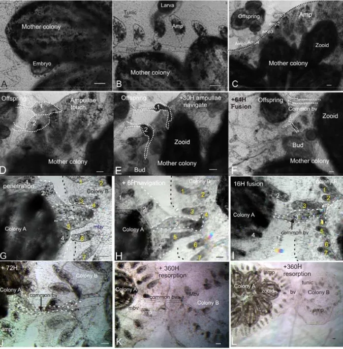
Fig. 1 Fusion process between kin. Using time laps microscopy (ImagExpress as described in Voskoboynik et al., 2008), we followed and documented the fusion process between 6 different pairs of compatible B. schlosseri
colonies. These observations revealed a more elaborate fusion process than the one described before.
1971). An extensive study of the Botryllid ascidians self-nonself recognition system in the last 25 years revealed that, while the colonial ascidian Fu/HC and the mammalian major histocompatibility complex (MHC) share phenomenological features, including polymorphism and specificity, their molecular structure is different. Similar to the MHC, the Fu/HC is highly polymorphic (Karakashian and Milkman 1967; Scofield et al., 1982; 1983; Grosberg and Quinn, 1986; Grosberg 1987; Rinkevich et al., 1995; Paz et al., 2003; De Tomaso et al., 2005; Ben-Shlomo et al., 2001, 2006, 2008). However, structural homologies were not found (Weissman et al., 1990; Rinkevich and Weissman, 1992b; Fagan and Weissman, 1997; Pancer et al., 1993, 1996a, b, c, 1997; Muller et al., 1994; Khalturin et al., 2003; De Tomaso et al., 2005; Nyholm et al., 2006). The
Botryllus Fu/HC is not homologous to any molecules of the vertebrate MHC-based histocompatibility system. Moreover, the whole Botryllus Fu/HC locus does not have a syntenic region in the Ciona genome or in the genomes of vertebrates (De Tomaso et al., 2005). Genes involved in adaptive immunity, which include the polymorphic MHC class I and II glycoproteins that present internal peptides to T cells, the clonally expressed T-cell receptors (TCRs), immunoglobulins (Igs) and the recombination activating genes (RAG1, RAG2), have not been identified in protochordates (Klein 1989, Laird et al., 2000; Dehal et al., 2002; Kaufman, 2002; Azumi et al., 2003; Khalturin et al., 2003; De Tomaso et al., 2005; Litman et al., 2005, 2007). In both mice and humans, the mediators of the adaptive immune system are thought to be responsible for the rejection of a transplant. The adaptive immune T cell system rejects grafts even if one of two alleles is shared, as T cells make an immune reaction against non-self allele gene products.
In contrast, allogeneic Botryllus colonies tolerate each other and create a chimera even if only one of the two Fu/HC alleles is shared. This is consistent with immune systems, like the NK system in vertebrates, wherein recognition of self prevents an immune reaction. Recently, NK cells have been recognized as active participants in the acute and chronic rejection of solid tissue grafts (reviewed in Kitchen et al., 2005). The importance of NK cell attributes has been first recognized in bone marrow transplantation, where NK cells are fully sufficient to reject hematopoietic cell transplants, even in the absence of T or B cell responses (lethally-irradiated or Lack MHC class I expression recipients; Hoglund
et al., 1991; Bix et al., 1991; Manilay and Sykes, 1998). Recent studies suggest that NK contribute to organ rejection indirectly, by activating or helping effector cells, such as cytotoxic and helper T cells (reviewed in Kitchen et al., 2005). Studies also point to a potential involvement of NK in the induction of tolerance to solid graft in mix chimerism, a tolerance regimen that employs the generation of mixed hematopoietic chimerism through donor stem cell engraftment (Zhao et al., 2003). Another aspect that might connect mammalian NK and the Botryllus Fu/HC was recently raised by Lighter et al. (2008). Studying uterine NK, Lighter et al., pointed to a unique phenotype that uterine NK and the Botryllus
Fu/HC might share. Uterine NK cells produce
angiogenic growth factors and are potential regulators of decidual angiogenesis in early pregnancy (Ashkar and Croy, 2001; Hanna et al., 2006; Manaster and Mandelboim, 2008). They suggested that, as Botryllus Fu/HC is probably involved in the generation of a common vascular system between two individuals, uterine NK may share the same evolutionary roots as the Botryllus
Fu/HC.
Cell parasitism induces development of a foreign entity within host colony
Inspired by the genetic control for allograft acceptance and creation of chimerism within kin in
Botryllus, Burnet (1971) hypothesized the emergence of intraspecific parasitism along the evolution. Indeed, his hypothesis was later confirmed in Botryllus chimeras, where the replacement of host germline by a donor genotype was demonstrated (Sabbadin and Zaniolo, 1979). In a remarkable set of experiments Sabbadin and Zaniolo (1979) demonstrated this kind of parasitism in B. schlosseri. Years later Pancer et al. (1995), Stoner and Weissman (1996) and Stoner et al.
(1999) confirmed these results and further showed that in a chimera, the blood, soma and germ cells, demonstrated the combine genotypes of both chimeric partners. Moreover, in many cases the circulating pluripotent cells of one partner parasitized either the soma or the germ line of the other partner and replaced the whole mass of gonads or the soma (bud/zooid) of several individuals in the host colony (termed gonads or somatic cell parasitism; G/SCP). In a few cases, a complete takeover of donor genotype occurred and the whole mass of gonads in the chimeric colony expressed solely the donor’s genotype (Sabbadin and Zaniolo 1979; Pancer et al., 1995; Stoner and Weissman 1996; Stoner et al., 1999). Under invariant environmental conditions, both germline and somatic cell parasitism followed repeatable hierarchies of “winner strains” and “loser strains” (Stoner et al., 1999). However, breeding experiments proved that only the hierarchical position of germ cell parasitism is sexually inherited (Stoner et al., 1999). The hierarchy of somatic parasitism in Botryllus chimeras is a plastic trait, as variations in the environmental conditions (such as seawater temperature) can be reversed; the winner – loser hierarchy at the somatic parasitism level (Rinkevich and Yankelevich, 2004).
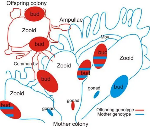
Botryllus colonies propagate a-sexually through budding, therefore somatic stem cell parasitism in host colonies can induce the development of partial allogeneic entities (buds) within the host colony. As a result, chimerism in protochordate could serve as a state that enables the development of a “virtual embryo” within the host colony (Voskoboynik et al., in press; Fig. 2).
Fig. 2 Virtual pregnancy: a development of a semi allogeneic entity in a host colony through asexual budding in a chimera of a mother colony and its offspring. Botryllus colonies propagate asexually through budding, therefore, somatic stem cell parasitism in host colonies can induce the development of a partial allogeneic entities (buds) within the host colony. In this illustration, an offspring colony (red) is fused with its mother colony (blue). Genetic analysis of the chimera’s buds and gonads can reveal several cell chimerism patterns. Either both genotypes are detected or only one genotype is detected. bv, blood vessel; Mbv, marginal blood vessel; red, offspring genotype; blue, mother genotype.
and reject another colony (Taneda, 1985; Sabbadin and Astorri, 1988). The presence, of a simultaneously fusion and rejection with a genotype that one of the chimera partner used to fuse with and the other partner used to reject, prove the persistence of both genotypes and suggest an uneven distribution of each genotype within the chimera. Different fusibility patterns that the chimeric entity presents on different time points suggests genomes fluctuation and competitive interaction of the different genomes within the chimera (Sabbadin and Astorri, 1988). Sabbadin and Astorri observed changes in the tolerance state of the chimeric colonies and linked it to changes in the dynamic of the chimeric cells within the chimera. Genetic analysis of the dynamic of chimeric cell revealed that chimeras exhibit either a sectorial pattern in which both genotypes are detected within some systems but not others, or a uniform pattern in which tissues throughout the entire chimera exhibit both genotypes (Stoner and Weissman, 1966). Colonies which showed rejection and fusion on the same time probably expressed sectorial pattern and the others expressed uniform pattern. The dynamic of chimeric cells within the host is changing with time, as different patterns are observed during different time points (Pancer et al., 1995; Stoner and Weissman, 1966; Stoner et al., 1999).
These studies show that the genetically controlled ability of Botryllus colonies to tolerate or reject other colonies can be altered by chimerism. The temporal and spatial dynamic of the chimeric cells, patterns of host/donor cells competition, niche occupation and immunoregulatory mechanisms for routing, timing and frequencies of chimeric cells have probably important role in the induction of tolerance or intolerance.
Stem cells mediated chimerism
we (Voskoboynik et al., 2008) have demonstrated that the anterior ventral region of the endostyle (termed endostyle niche) harbors adult stem cells and exports them to developing and regenerating organs, wherein they participate in tissue formation. As few as 5-20 engrafted cells transplanted from the donor endostyle niche sufficed to generate a somatic chimerism in compatible hosts; however, no germline chimerism was demonstrated. The induction of somatic chimerism demonstrates a remarkable stemness capacity of the cells in the endostyle niche (Voskoboynik et al., 2008). The endostyle produces thyroid hormones and serotonin and expresses several variety of factors that are involved in development and stem cell regulation like Wnt, Hox1, Pax 2/5/8, PL10, Cadherin, Raldh and PCNA (Canestro et al., 2008; Dunn, 1980; Hiruta et al., 2005; Nilsson et al., 1988; Pennati et al., 2001; Rosner et al., 2006; Rosner et al., 2007; Voskoboynik et al., 2008).
The endostyle niche is the first somatic stem cell niche ever described in a protochordate and is one of the most accessible stem cell niches for in vivo studies. The discovery of a major stem cell niche in an organism with Fu/HC controlled chimerism, pluripotent stem cell parasitism, and tolerance or intolerance induction via chimerism promotes the Botryllus as an evolutionary model for studying molecular regulations of tolerance induction by cellular chimerism in the absence of an adaptive immune system.
Acknowledgements
I thank A Voskoboynik for critical review, IL Weissman and B Rinkevich for discussion, P Brown, R Marinelli, R Pesich L Jerabek, KJ Ishizuka, and KJ Palmeri for support and helpful assistance.
References
Adams KM, Nelson JL. Microchimerism: An investigative frontier in autoimmunity and transplantation. JAMA 291: 1127-1131, 2004. Anderson D, Billingham RE, Lampkin GH,
Medawar PB. The use of skin grafting to distinguish between monozygotic and dizygotic twins in cattle. Heredity 5: 379-397, 1951.
Andrassy J, Kusaka S, Jankowska-Gan E, Torrealba JR, Haynes LD, Marthaler BR, et al. Tolerance to noninherited maternal MHC antigens in mice. J. Immunol. 171: 5554-5561, 2003.
Ashkar AA, Croy BA. Functions of uterine natural killer cells are mediated by interferon gamma production during murine pregnancy. Semin. Immunol. 13: 235-241, 2001.
Azumi K, Takahashi H, Miki Y, Fujie M, Usami T, Ishikawa H, et al. Construction of a cDNA microarray derived from the ascidian ciona intestinalis. Zool. Sci. 20: 1223-1229, 2003. Baker RR, Bellis MA. Human sperm competition:
copulation, masturbation and infidelity. Chapman & Hall, London, 1995.
Ballarin L, Cima F, Sabbadin A. Morula cells and histocompatibility in the colonial ascidian
Botryllus schlosseri. Zool. Sci. 12: 757-764, 1995.
Ballarin L, Cima F, Sabbadin A. Phenoloxidase and cytotoxicity in the compound ascidian Botryllus schlosseri. Dev. Comp. Immunol. 22: 479-492, 1998.
Ballarin L, Cima F, Floreani M, Sabbadin A. Oxidative stress induces cytotoxicity during rejection reaction in the compound ascidian
Botryllus schlosseri. Comp. Biochem. Physiol. 133C: 411-418, 2002.
Bancroft FW. Variation and fusion of colonies in compound ascidians. Proc. Calif. Acad. Sci. 3: 137-186, 1903.
Bauer M, Orescovic I, Schoell WM, Bianchi DW, Pertl B. Detection of maternal DNA in umbilical cord plasma by fluorescent PCR amplification of short tandem repeat sequences. Ann. N.Y. Acad. Sci. 945: 161-163, 2001.
Ben-Shlomo R, Douek J, Rinkevich B. Heterozygote deficiency and chimerism in remote populations of a colonial ascidian from New Zealand. Mar. Ecol. Prog. Ser. 209: 109-117, 2001.
Ben-Shlomo R, Paz G, Rinkevich B. Postglacial-period and recent invasions shape the population genetics of botryllid ascidians along European Atlantic coasts. Ecosystems 9: 1118-1127, 2006.
Ben-Shlomo R, Motro U, Paz G, Rinkevich B. Pattern of settlement and natural chimerism in the colonial urochordate Botryllus schlosseri. Genetica 132: 51-58, 2008.
Bianchi DW, Zickwolf GK, Weil GJ, Sylvester S, DeMaria MA. Male fetal progenitor cells persist in maternal blood for as long as 27 years postpartum. Proc. Natl. Acad. Sci. USA 93: 705-708, 1996.
Bianchi DW, Williams JM, Sullivan LM, Hanson FW, Klinger KW, Shuber AP. PCR quantitation of fetal cells in maternal blood in normal and aneuploid pregnancies. Am. J. Hum. Genet. 61: 822-829, 1997.
Bianchi DW, Simpson JL, Jackson LG, Elias S, Holzgreve W, Evans MI, et al. Fetal gender and aneuploidy detection using fetal cells in maternal blood: Analysis of NIFTY I data. national institute of child health and development fetal cell isolation study. Prenat. Diagn. 22: 609-615, 2002.
Bianchi DW. Fetomaternal cell trafficking: a story that begins with prenatal diagnosis and may end with stem cell therapy. J. Pediatr. Surg. 42:12-18, 2007.
Billingham RE, Lampkin GH, Medawar PB, Williams HL. Tolerance to homografts, twin diagnosis and the freemartin condition in cattle. Heredity 6: 200-212, 1952.
Billingham RE, Brent L, Medawar PB. Actively acquired tolerance of foreign cells. Nature 172: 603-606, 1953.
Billingham RE, Brent L. Acquired tolerance of foreign cells in newborn animals. Proc. R. Soc. Lond. B Biol. Sci. 146: 78-90, 1956.
Bix M, Liao NS, Zijlstra M, Loring J, Jaenisch R, Raulet D. Rejection of class I MHC-deficient haemopoietic cells by irradiated MHC-matched mice. Nature 349: 329-331, 1991.
receptors: new players in reproductive immunology. Immunol. Invest. 37: 483-497, 2008.
Boyd HC, Weissman IL, Saito Y. Morphologic and genetic verification that Monterey Botryllus and Woods Hole Botryllus are the same species. Biol. Bull. 178: 239-250, 1990.
Burighel P, Cloney RA. Urochordata: Ascidiacea. In: Harrison FW, Ruppert EE (eds), Microscopic anatomy of invertebrates, Wiley-Liss, Inc., NY, pp. 221-347, 1997.
Burnet FM. "Self-recognition" in colonial marine forms and flowering plants in relation to the evolution of immunity. Nature 232: 230-235, 1971.
Burnet FM. Multiple polymorphism in relation to histocompatibility antigens. Nature 245: 359-361, 1973.
Buss LW. Somatic cell parasitism and the evolution of somatic tissue compatibility. Proc. Natl. Acad. Sc.i USA 79: 5337-5341, 1982.
Buss LW, Grosberg RK. Morphogenetic basis for phenotypic differences in Hydroid competitive behavior. Nature 343: 63-66, 1990.
Canestro C, Bassham S, Postlethwait JH. Evolution of the thyroid: Anterior-posterior reorganization of the Oikopleura endostyle revealed by Otx, Pax 2/5/8, and Hox1 expression. Dev Dyn doi:10.1002/dvdy.21525, 2008
Cao YA, Wagers AJ, Beilhack A, Dusich J, Bachmann MH, Negrin RS, et al. Shifting foci of hematopoiesis during reconstitution from single stem cells. Proc. Natl. Acad. Sci. USA 101: 221-226, 2004.
Chadwick-Furman NE, Weissman IL. Life history plasticity in chimaeras of the colonial ascidian
Botryllus schlosseri. Philos. Trans. R. Soc. Lond.B Biol. Sci. 262: 157-162, 1995.
Cima F, Sabbadin A, Ballarin L. Cellular aspects of allorecognition in the compound ascidian
Botryllus schlosseri. Dev. Comp. Immunol. 28: 881-889, 2004.
Cima F, Sabbadin A, Zaniolo G, Ballarin L (2006) Colony specificity and chemotaxis in the compound ascidian Botryllus schlosseri. Comp. Biochem. Physiol. 145A: 376-382, 2006.
Claas FH, Gijbels Y, van der Velden-de Munck J, van Rood JJ. Induction of B cell unresponsiveness to noninherited maternal HLA antigens during fetal life. Science 241: 1815-1817, 1988.
Claas FH, Gijbels Y, von Veen A, de Waal LP, D'Amaro J, Persijn GG, et al. Selection of cross-match negative HLA-A and/or -B mismatched donors for highly sensitized patients. Transplant. Proc. 21: 665-666, 1989. Clark DA. Signaling at the fetomaternal interface.
Am. J. Repros. Immunol. 41: 169-173, 1999. De Tomaso AW, Nyholm SV, Palmeri KJ, Ishizuka
KJ, Ludington WB, Mitchel K, et al. Isolation and characterization of a protochordate histocompatibility locus. Nature 438: 454-459, 2005.
Dehal P, Satou Y, Campbell RK, Chapman J, Degnan B, De Tomaso A, et al. The draft genome of Ciona intestinalis: Insights into
chordate and vertebrate origins. Science 298: 2157-2167, 2002.
Delsuc F, Brinkmann H, Chourrout D, Philippe H. Tunicates and not cephalochordates are the closest living relatives of vertebrate. Nature 439: 965-968, 2006.
Dunn AD. Properties of an iodinating enzyme in the ascidian endostyle. Gen. Comp. Endocrinol. 40: 484-493, 1980.
Dunsford I, Bowley CC, Hutchison AM, Thompson JS, Sanger R, Race RR. A human blood-group chimera. Br. Med. J. 2: 80-81, 1953.
Emery D, McCullagh P. Immunological reactivity between chimeric cattle twins. I. homograft reaction. Transplantation 29: 4-9, 1980a. Emery D, McCullagh P. Immunological reactivity
between chimeric cattle twins. II. normal lymphocyte transfer. Transplantation 29: 10-16, 1980b.
Evans PC, Lambert N, Maloney S, Furst DE, Moore JM, Nelson JL. Long-term fetal microchimerism in peripheral blood mononuclear cell subsets in healthy women and women with scleroderma. Blood 93: 2033-2037, 1999.
Fagan MB, Weissman IL. HSP70 genes and historecognition in Botryllus schlosseri: Implications for MHC evolution. Hereditas 127: 25-35, 1997.
Gaillard MC, Ouvre E, Liegeois A, Lewin D. The concentration of fetal cells in maternal haematopoietic organs during pregnancy. an experimental study in mice (author's transl). J. Gynecol. Obstet. Biol. Reprod. (Paris) 7: 1043-1050, 1978.
Grosberg RK. Limited dispersal and proximity dependent mating success in Botryllus schlosseri. Evolution 41: 372-384, 1987. Grosberg RK. The evolution of allorecognition
specificity in colonial invertebrates. Quart. Rev. Biol. 63: 377-412, 1988.
Grosberg RK, Quinn JF. The genetic control and consequences of kin recognition by the larvae of a colonial marine invertebrate. Nature 322: 456-459, 1986.
Guleria I, Sayegh MH. Maternal acceptance of the fetus: true human tolerance. J. Immunol. 178: 3345-3351, 2007.
Hall JM, Lingenfelter P, Adams SL, Lasser D, Hansen JA, Bean MA. Detection of Maternal Cells in Human Umbilical Cord Blood using Fluorescence in Situ Hybridization. Blood 86: 2829-2832, 1995.
Hamada H, Arinami T, Kubo T, Hamaguchi H, Iwasaki H. Fetal nucleated cells in maternal peripheral blood: Frequency and relationship to gestational age. Hum. Genet. 91: 427-432, 1993.
Hanna J, Goldman-Wohl D, Hamani Y, Avraham I, Greenfield C, Natanson-Yaron S, et al.
Decidual NK cells regulate key developmental processes at human fetal-maternal interface. Nat. Med. 12: 1065-1074, 2006.
Hill JA. Immunological contribution to recurrent pregnancy loss. Baillieres Clin. Obster. Gynaecol. 6: 489-438, 1992.
Hirose E, Saito Y, Watanabe H. Anew type of the manifestation of the colony specificity in the compound ascidian, Botrylloides violaceus Oka. Biol. Bull. 175: 240-245, 1988.
Hirose E, Saito Y, Watanabe H. Allogeneic rejection induced by the cut surface contact in the compound ascidian, Botrylloides simodensis. Invertebr. Dev. Reprod. 17: 159-164, 1990. Hiruta J, Mazet F, Yasui K, Zhang P, Ogasawara M.
Comparative expression analysis of transcription factor genes in the endostyle of invertebrate chordates. Dev. Dyn. 233: 1031-1037, 2005. Höglund P, Ohlén C, Carbone E, Franksson L,
Ljunggren HG, Latour A, et al. Recognition of beta-2microglobulin-negative (beta 2m-) T-cell blasts by natural killer cells from normal but not from beta 2m-mice: nonresponsiveness controlled by beta 2m-bone marrow ib chimeric mice. Proc. Natl. Acad. Sci. USA 88: 10332-10336, 1991.
Hunt JS, Petroff MG, McIntire RH Ober C. HLA-G and immune tolerance in pregnancy. FASEB J. 19: 682-693, 2005.
Ishitani A, Sageshima N, Lee N, Dorofeeva N, Hatake K, Marquardt H, et al. Protein expression and peptide binding suggest unique and interacting functional roles for HLA-E, F, and G in maternal-placental immune recognition. J. Immunol. 171: 1376-1384, 2003.
Kammerer U, Kruse A, Barrientos G, Arck PC, Blois SM. Role of dendritic cells in the regulation of maternal immune responses to the fetus during mammalian gestation. Immunol. Invest. 37: 499-533, 2008.
Kaplan J, Land S. Influence of maternal-fetal histocompatibility and MHC zygosity on maternal microchimerism. J. Immunol. 174: 7123-7128, 2005.
Karakashian S, Milkman R. Colony fusion compatibility types in Botryllus schlosseri. Biol. Bull. 133: 473-?, 1967.
Kaufman J. The origins of the adaptive immune system: Whatever next? Nat. Immunol. 3: 1124-1125, 2002.
Khalturin K, Becker M, Rinkevich B, Bosch TC. Urochordates and the origin of natural killer cells: Identification of a CD94/NKR-P1-related receptor in blood cells of Botryllus. Proc. Natl. Acad. Sci. USA 100: 622-627, 2003.
Khosrotehrani K, Johnson KL, Guegan S, Stroh H, Bianchi DW. Natural history of fetal cell microchimerism during and following murine pregnancy. J. Reprod. Immunol. 66: 1-12, 2005. King A, Allan DS, Bowen M, Powis SJ, Joseph S,
Verma S, et al. HLA-E is expressed on trophoblast and interacts with CD94/NKG2 receptors on decidual NK cells. Eur. J. Immunol. 30: 1623-1631, 2000.
Kitchens WH, Uehara S, Chase CM, Colvin RB, Russell PS, Madsen JC. The changing role of natural killer cells in solid organ rejection and tolerance. Transplantation 81: 811-817, 2006.
Klein J. Are invertebrates capable of anticipatory immune responses?. Scand. J. Immunol. 29: 499-505, 1989.
Koopmans M, Kremer Hovinga IC, Baelde HJ, Fernandes RJ, de Heer E, Bruijn JA, et al. Chimerism in kidneys, livers and hearts of normal women: Implications for transplantation studies. Am. J. Transplant. 5: 1495-1502, 2005. Kurpisz M, Fernandez N, Fiszer D. The expression
of MHC genes in reproductive organs and their role in reproduction. In: Kurspiz M, Fernandez N (eds), Immunology of human reproduction, Bios Scientific Publishers, Oxford, pp163-184, 1995.
Laird DJ, De Tomaso AW, Cooper MD, Weissman IL. 50 million years of chordate evolution: Seeking the origins of adaptive immunity. Proc. Natl. Acad. Sci. USA 97: 6924-6926, 2000. Laird DJ, De Tomaso AW, Weissman IL. Stem cells
are units of natural selection in a colonial ascidian. Cell 123: 1351-1360, 2005.
Liegeois A. In: Edelman P, Sureau C (eds) Immunologie de le reproduction humanie, Edition Boz., Paris, pp 99-100, 1983.
Lightner A, Schust DJ, Chen YBA, Barrier BF. The fetal allograft revisited: Does the study of an ancient invertebrate species shed light on the role of natural killer cells at the maternal-fetal interface? Clin. Dev. Immunol. 2008;2008:631920.
Lillie FR. The theory of the free-martin. Science 43: 611-613, 1916.
Litman GW, Cannon JP, Dishaw LJ. Reconstructing immune phylogeny: New perspectives. Nat. Rev. Immunol. 5: 866-879, 2005.
Litman GW, Dishaw LJ, Cannon JP, Haire RN, Rast JP. Alternative mechanisms of immune receptor diversity. Curr. Opin. Immunol. 19: 526-534, 2007.
Lo ES, Lo YM, Hjelm NM, Thilaganathan B. Transfer of nucleated maternal cells into fetal circulation during the second trimester of pregnancy. Br. J. Haematol. 100: 605-606, 1998.
Lo YM, Patel P, Wainscoat JS, Sampietro M, Gillmer MD, Fleming KA. Prenatal sex determination by DNA amplification from maternal peripheral blood. Lancet 2: 1363-1365, 1989.
Lo YM, Lo ES, Watson N, Noakes L, Sargent IL, Thilaganathan B, et al. Two-way cell traffic between mother and fetus: Biologic and clinical implications. Blood 88: 4390-4395, 1996. Loubiere LS, Lambert NC, Flinn LJ, Erickson TD,
Yan Z, Guthrie KA, et al. Maternal microchimerism in healthy adults in lymphocytes, monocyte/macrophages and NK cells. Lab. Invest. 86: 1185-1192, 2006.
Maloney S, Smith A, Furst DE, Myerson D, Rupert K, Evans PC, et al. Microchimerism of maternal origin persists into adult life. J. Clin. Invest. 104: 41-47, 1999.
Manilay JO, Sykes M. Natural killer cells and their role in graft rejection Curr. Opin. Immunol. 10: 532-538, 1998.
Manni L, Burighel P. Common and divergent pathways in alternative developmental processes of ascians. BioEssays 28: 902-912, 2006.
Manni L, Zaniolo G, Cima F, Burighel P, Ballarin L.
Botryllus schlosseri: a model ascidian for the study of asexual reproduction. Dev. Dyn. 236: 335-352, 2007.
Marleau AM, Greenwood JD, Wei Q, Singh B, Croy BA. Chimerism of murine fetal bone marrow by maternal cells occurs in late gestation and persists into adulthood. Lab. Invest. 83: 673-681, 2003.
Mold JE, Michaelsson J, Burt TD, Muench MO, Beckerman KP, Busch MP, et al. Maternal alloantigens promote the development of tolerogenic fetal regulatory T cells in utero. Science 322: 1562-1565, 2008.
Mukai H. Experimental alteration of fusibility in compound ascidians. Sci. Rep. Tokyo Kyoiku Daigaku 13B: 51-73, 1987.
Mukai H, Watanabe H. Distribution of fusion incompatibility types in natural populations of the compound ascidian Botryllus primigenus. Proc. Jpn. Acad. 51: 44-47, 1975.
Muller WE, Pancer Z, Rinkevich B. Molecular cloning and localization of a novel serine protease from the colonial tunicate Botryllus schlosseri. Mol. Mar. Biol. Biotechnol. 3: 70-77, 1994.
Munn DH, Zhou M, Attwood JT, Bondarev I, Conway SJ, Marshall B, et al. Prevention of allogeneic fetal rejection by tryptophan catabolism. Science 281: 1191-1193, 1998. Nilsson O, Fredriksson G, Ofverholm T, Ericson LE.
Electron-microscopic immunocytochemistry of 5-hydroxytryptamine in the ascidian endostyle. Cell Tissue Res. 253: 137-143, 1988.
Nyholm SV, Passegue E, Ludington WB, Voskoboynik A, Mitchel K, Weissman IL, et al. Fester, A candidate allorecognition receptor from a primitive chordate. Immunity 25: 163-173, 2006.
O'Donoghue K, Chan J, de la Fuente J, Kennea N, Sandison A, Anderson JR, et al. Microchimerism in female bone marrow and bone decades after fetal mesenchymal stem-cell trafficking in pregnancy. Lancet 364: 179-182, 2004a.
O'Donoghue K, Fisk NM. Fetal stem cells. Best. Pract. Res. Clin. Obstet. Gynaecol. 18: 853-875, 2004b.
Oka H, Watanabe H. Colony specificity in compound ascidians as tested by fusion experiments (a preliminary report). Proc. Jpn. Acad. 33: 657-659, 1957.
Oka H, Watanabe H. problems of colony specificity in compound ascidians. Bull. Mar. Biol. Stat. Asamushi. 10: 153-155, 1960.
Oka H, Watanabe H. Problems of colony specificity, with special reference to the fusibility of ascidians. Kagaku 37: 307-313, 1967.
Oosterwijk JC, Mesker WE, Ouwerkerk-van Velzen MC, Knepfle CF, Wiesmeijer KC, Beverstock
GC, et al. Fetal cell detection in maternal blood: A study in 236 samples using erythroblast morphology, DAB and HbF staining, and FISH analysis. Cytometry 32: 178-185, 1998.
Owen RD. Immunogenetic consequences of vascular anastomoses between bovin twins. Science 102: 400-401, 1945.
Owen RD, Wood HR, Foord AG, Sturgeon P, Baldwin LG. Evidence for actively tolerance to Rh antigens. Proc. Natl. Acad. Sci. USA 40: 420-424, 1954.
Pancer Z, Gershon H, Rinkevich B. cDNA cloning of a putative protochordate FK506-binding protein. Biochem. Biophys. Res. Commun. 197: 973-977, 1993.
Pancer Z, Gershon H, Rinkevich B. coexistence and possible parasitism of somatic and germ cell lines in chimeras of the colonial urochordate
Botryllus schlosseri. Biol. Bull. 189: 106-112, 1995.
Pancer Z, Cooper EL, Muller WE. A tunicate (Botryllus schlosseri) cDNA reveals similarity to vertebrate antigen receptors. Immunogenetics 45: 69-72, 1996a.
Pancer Z, Leuck J, Rinkevich B, Steffen R, Muller I, Muller WE. Molecular cloning and sequence analysis of two cDNAs coding for putative anionic trypsinogens from the colonial urochordate Botryllus schlosseri (ascidiacea). Mol. Mar. Biol. Biotechnol. 5: 326-333, 1996b. Pancer Z, Scheffer U, Muller I, Muller WE. Cloning
of sponge (Geodia cydonium) and tunicate (Botryllus schlosseri) proteasome subunit epsilon (PRCE): Implications about the vertebrate MHC-encoded homologue LMP7 (PRCC). Biochem. Biophys. Res. Commun. 228: 406-410, 1996c.
Pancer Z, Diehl-Seifert B, Rinkevich B, Muller WE. A novel tunicate (Botryllus schlosseri) putative C-type lectin features an immunoglobulin domain. DNA Cell Biol. 16: 801-806, 1997. Paz G, Douek J, Mo CQ, Goren M, Rinkevich B.
Genetic structure of Botryllus schlosseri
(Tunicata) populations from the Mediterranean coast of Israel. Mar. Ecol. Prog. Ser. 250: 163-174, 2003.
Pennati R, Groppelli S, Sotgia C, Candiani S, Pestarino M, De Bernardi F. Serotonin localization in Phallusia mammillata larvae and effects of 5-HT antagonists during larval development. Develop. Growth Differ. 43: 647-656, 2001.
Petit T, Dommergues M, Socie G, Dumez Y, Gluckman E, Brison O. Detection of Maternal Cells in Human Fetal Blood during the Third Trimester of Pregnancy using Allele-Specific PCR Amplification. Br. J. Haematol. 98: 767-771, 1997.
Petit T, Gluckman E, Carosella E, Brossard Y, Brison O, Socie G. A highly sensitive polymerase chain reaction method reveals the ubiquitous presence of maternal cells in human umbilical cord blood. Exp. Hematol. 23: 1601-1605, 1995.
Rinkevich B. Immunological resorption in Botryllus schlosseri (Tunicata) chimeras is characterized by multilevel hierarchical organization of histocompatibility alleles. A speculative endeavor. Biol. Bull. 184: 342-345, 1993.
Rinkevich B. Primitive immune systems: are your ways my ways? Immunol. Rev. 198: 25-35, 2004a.
Rinkevich B. Will two walk together, except they have agreed? J. Evol. Biol. 17: 1167-1177, 2004b.
Rinkevich B. Rejection patterns in botryllid ascidian immunity: The first tier of allorecognition. Can. J. Zool. 83: 101-121, 2005.
Rinkevich B, Weissman IL. Chimeras in colonial invertebrates - a synergidtic symbiosis or somatic and germ cell parasitism. Symbiosis 4: 117-134, 1987.
Rinkevich B, Weissman IL. Allogeneic resorption in colonial protochordates: Consequences of nonself recognition. Dev. Comp. Immunol. 16: 275-286, 1992a.
Rinkevich B, Weissman IL. Incidents of rejection and indifference in Fu/HC incompatible protochordate colonies. J. Exp. Zool. 263: 105-111, 1992b.
Rinkevich B, Lilker-Levav T, Goren M. Allorecognition/Xenorecognition responses in
Botrylloides subpopulations from the Mediterranean coast of Israel. J. Exp. Zool. 270: 302-313, 1994.
Rinkevich B, Porat R, Goren M. Allorecognition elements on Urochordate histocompatibility locus indicate unprecedented extensive polymorphism. Proc. Biol. Sci. 259: 319-324, 1995.
Rinkevich B, Tartakover S, Gershon H. Contribution of morula cells to allogeneic responses in the colonial urochordate Botryllus schlosseri. Mar. Biol. 131: 227-236, 1998.
Rosner A, Paz G, Rinkevich B. Divergent roles of the DEAD-box protein BS-PL10, the urochordate homologue of human DDX3 and DDX3Y proteins, in colony astogeny and ontogeny. Dev. Dyn.235: 1508-1521, 2006. Rosner A, Rabinowitz C, Moiseeva E, Voskoboynik
A, Rinkevich B. BS-Cadherin in the colonial urochordate Botryllus schlosseri: One protein, many functions. Dev. Biol. 304: 687-700, 2007. Sabbadin A. Le basi geneticha della capacita di
fusion fra colonies in Botryllus schlosseri
(Ascidiacea). Rend. Accad. Naz. Lincei Ser. 32: 1031-1035, 1962.
Sabbadin A, Zaniolo G. Sexual differentiation and germ cell transfer in the colonial ascidian
Botryllus schlosseri. J. Exp. Zool. 207: 289-304, 1979.
Sabbadin A. Formal genetics of ascidians. Am. Zool. 22: 765-777, 1982.
Sabbadin A, Astorri C. Chimeras and histocompatibility in the colonial ascidian
Botryllus schlosseri. Dev. Comp. Immunol. 12: 737-747, 1988.
Sabbadin A, Zaniolo G, Ballarin L. Genetic and cytological aspects of histocompatibility in ascidians. Boll. Zool. 59: 167-173, 1992.
Saito Y, Watanabe H. Colony specificity in the compound ascidian Botryllus scalaris. Proc. Japn. Acad. 58B: 105-108, 1982.
Saito Y, Watanabe H. Partial biochemical characterization of humoral factors involved in the nonfusion reaction of a botryllid ascidian,
Botrylloides simodensis. Zool. Sci. 1: 229-235, 1984.
Saito Y, Hirose E, Watanabe H. Allorecognition in compound ascidians. Int. J. Dev. Biol. 38: 237-247, 1994.
Scofield VL, Schlumpberger JM, West LA, Weissman IL Protochordate allorecognition is controlled by a MHC-like gene system. Nature 295: 499-502, 1982.
Scofield VL, Schlumpberger JM, Nagashima LS. Morphology and genetics of rejection reactions between oozooids from the tunicate Botryllus schlosseri. Biol. Bull. 165: 733-744, 1983.
Sekizawa A, Kimura T, Sasaki M, Nakamura S, Kobayashi R, Sato T. Prenatal Diagnosis of Duchenne Muscular Dystrophy using a Single Fetal Nucleated Erythrocyte in Maternal Blood. Neurology 46: 1350-1353, 1996.
Socie G, Gluckman E, Carosella E, Brossard Y, Lafon C, Brison O. Search for maternal cells in human umbilical cord blood by polymerase chain reaction amplification of two minisatellite sequences. Blood 83: 340-344, 1994.
Stone WH, Cragle RG, Swanson EW, Brown DG. Skin grafts: Delayed rejection between pairs of cattle twins showing erythrocyte chimerism. Science 148: 1335-1336, 1965.
Stone WH, Cragle RG, Johnson DF, Bacon JA, Bendel S, Korda N. Long-term observations of skin grafts between chimeric cattle twins. Transplantation 12: 421-428, 1971.
Stoner DS, Weissman IL. Somatic and germ cell parasitism in a colonial ascidian: Possible role for a highly polymorphic allorecognition system. Proc. Natl. Acad. Sci. USA 93: 15254-15259, 1996.
Stoner DS, Rinkevich B, Weissman IL. Heritable germ and somatic cell lineage competitions in chimeric colonial protochordates. Proc. Natl. Acad. Sci. USA 96: 9148-9153, 1999.
Tanaka K, Watanabe H. Allogeneic inhibition in a compound ascidian, Botryllus primigenus oka. I. Processes and features of nonfusion reaction. Cell. Immunol. 7: 410-426, 1973.
Taneda Y. Simultaneous occurrence of fusion and nonfusion reaction in two colonies in contact of the compound ascidian Botryllus priminegus. Dev. Comp. Immunol. 9: 371-375, 1985.
Tippett P. Blood group chimeras. A review. Vox Sang. 44: 333-359, 1983.
van den Boogaardt DEM, van Rood JJ, Roelen DL, Claas FH. The influence of inherited and noninherited parental antigens on outcome after transplantation. Transpl. Int. 19: 360-371, 2006. van Dijk BA, Boomsma DI, de Man AJ. Blood group
chimerism in human multiple births is not rare. Am. J. Med. Genet. 61: 264-268, 1996.
of random PCR and a set of length polymorphisms. Hum. Genet. 99: 266-270, 1997.
Wachtel SS, Sammons D, Twitty G, Utermohlen J, Tolley E, Phillips O, et al. Charge Flow Separation: Quantification of Nucleated Red Blood Cells in Maternal Blood during Pregnancy. Prenat. Diagn. 18: 455-463, 1998. Voskoboynik A, Weissman IL, Rinkevich B. Stem
cells, chimerism, and tolerance: Lessons from mammals and ascidians. In: Rinkevich B, Matranga V (eds), Stem cells in marine organisms, Springer-Verlag [in press].
Weissman IL, Saito Y, Rinkevich B. Allorecognition histocompatibility in a protochordate species: Is the relationship to MHC somatic or structural?. Immunol. Rev. 113: 227-241, 1990.
Voskoboynik A, Soen Y, Rinkevich Y, Ueno H, Rosner A, Reshef R, et al. Identification of the endostyle as a stem cell niche in a colonial chordate. Cell Stem Cell 3: 456-464, 2008.