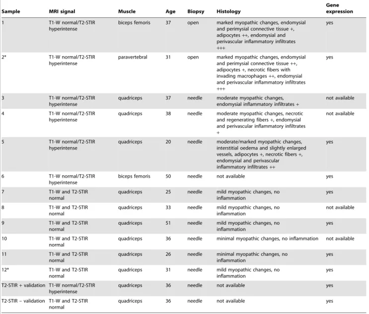
Different molecular signatures in magnetic resonance imaging-staged facioscapulohumeral muscular dystrophy muscles.
Texto
Imagem
Documentos relacionados
Magnetic resonance imaging and ultrasound measurements of extraocular muscles in thyroid-associated ophthalmopathy at different stages of the disease. Acta Ophthal-
Objective: To assess the importance of using conventional magnetic resonance imaging and T2 mapping to determine the pre-slip stage of the contralateral epiphysis in patients with
Objectives: To evaluate the use of magnetic resonance imaging in patients with β -thalassemia and to compare T2* magnetic resonance imaging results with
In this study, the feasibility of high resolution magic angle spinning (HR MAS) magnetic resonance spectroscopy (MRS) of small tissue biopsies to distinguish between tumor
We performed transcriptome analysis of pre-tumorous retina and retinal tumor tissue and found changes in gene expression signatures of radial glia and astrocytes ( slc1a3 ),
OBJECTIVE: To determine the frequency and localization of parenchymal abnormalities in cerebral venous thrombosis on magnetic resonance imaging and magnetic resonance angiography
The role of diffusion magnetic resonance imaging in Par - kinson’s disease and in the differential diagnosis with
The role of diffusion magnetic resonance imaging in Par- kinson’s disease and in the differential diagnosis with
