3 0 Arq Bras Oftalmol. 2014;77(1):30-3
Original Article
INTRODUCTION
Potential sightthreatening complications may occur if ocular in -flam mation is not diagnosed and treated early in the course of disea-se(1-5). However, initial evaluation of patients with uveitis is frequently
conducted in nonspecialized centers because intraocular inflamma-tion usually produces nonspecific symptoms such as pain, photopho-bia, redness, blurred vision, and floaters, which may easily be confused with other disorders(2,6). In Brazil, uveitis is one of the main diagnoses in
patients who attend institutions for visual rehabilitation(7) and accounts
for up to 7.4% visits to emergency eye care units.(8-11).
The etiology of uveitis can be broadly categorized into infectious and noninfectious, and it is frequently associated with systemic disea-se(12). Several studies have investigated the epidemiology of uveitis,
showing variations in etiology according to geographical region, gender, ethnicity, age, social aspects, and immunological factors. However, most of these studies have included patients from tertiary
uveitis centers and may have been influenced by selection bias(1,4,13-18).
Globally, anterior uveitis accounts for the majority of cases(1,4,17,18).
Ho-wever, posterior uveitis is the most common presentation in Brazilian patients, with toxoplasmic retinochoroiditis being the most frequent identifiable cause(14-16).
Identification of clinical and epidemiological patterns of uveitis is crucial to devise strategies for preventing late diagnosis and facili-tating prompt treatment. Therefore, this study aimed to analyze the clinical and epidemiological characteristics of patients with uveitis who visited an emergency eye care center.
METHODS
This prospective study included patients with a clinical diagnosis of active uveitis who were treated between May 2012 and July 2012 in the emergency eye care center of Fundação Altino Ventura, a reference eye hospital for patients from the public health system of
http://dx.doi.org/10.5935/0004-2749.20140009
Clinical and epidemiological characteristics of patients with uveitis in an emergency
eye care center in Brazil
Características clínicas e epidemiológicas das uveítes em um serviço de urgência otalmológica no Brasil
Eduardo NEry rossi Camilo1, GuilhErmE luCENa moura1, TiaGo EuGêNio FariaE araNTEs1,2
Submitted for publication: June 13, 2013 Accepted for publication: October 02, 2013 Study was carried out at Fundação Altino Ventura.
1 Fundação Altino Ventura, Recife, PE, Brazil. 2 Hospital de Olhos de Pernambuco, Recife, PE, Brazil.
Funding: No specific financial support was available for this study.
Disclosure of potential conflicts of interest: E.N.R. Camilo, None; G.L. Moura, None; T.E. Faria e Arantes, None.
Correspondence address: Eduardo Nery Rossi Camilo. Fundação Altino Ventura, 170 - Recife (PE) - 50070-040 - Brazil - E-mail: eduardo_nery@hotmail.com
The study was approved by Institutional Ethics Committee (Fundação Altino Ventura, no 053/2011). AbSTRACT
Purpose: To analyze the clinical and epidemiological characteristics of patients with uveitis in an emergency eye care center.
Methods: We conducted a prospective, observational study of patients with active uveitis admitted between May 2012 and July 2012 to an emergency eye care center.
Results: The majority of patients were male (63.2%), with a mean age of 43.2 years; 66.2% patients were of mixed ethnicity, 22.5% were Caucasian, and 11.3% were black. Anterior uveitis was observed in 70.1% patients, posterior uveitis in 26.5%, and panuveitis in 3.4%; no patient was diagnosed with intermediate uveitis. All patients had a sudden and acute presentation. The most frequent symptoms were ocular pain (76.9%), redness (59.8%), and visual blurring (46.2%). The majority of patients had unilateral disease (94.9%) with a mean symptom duration of 6.2 days. Diffuse and anterior uveitis were associated with ocular pain (p<0.001). Scotomata and floaters were more frequent in patients with posterior uveitis (p=0.003 and p=0.016, respectively). Patients with anterior uveitis presented with better visual acuity (p=0.025). Granulomatous keratotic precipitates were more frequent in patients with posterior uveitis (p=0.038). An etiological diagnosis based on the evaluation at the emergency center was made in 45 patients (38.5%).
Conclusions: Acute anterior uveitis was the most frequent form of uveitis. Initial patient evaluation provided sufficient information for deciding primary therapy and aided in arriving at an etiological diagnosis in a considerable number of patients.
Keywords: Uveitis/etiology; Uveitis/epidemiology; Uveitis/diagnosis; Uveitis/clas-sification; Emergencies
RESUMO
Objetivo: Analisar as características clínicas e epidemiológicas das uveítes em um serviço de atendimento oftalmológico de urgência.
Métodos: Estudo prospectivo, observacional de pacientes com uveíte ativa admitido entre maio e julho de 2012, em um serviço de atendimento oftalmológico de emergência. Resultados: A maioria dos pacientes eram do sexo masculino (63,2%) e a média de idade foi de 43,2 anos; 66,2% dos pacientes tinham etnia mista, 22,5% eram brancos e 11,3% negros. Uveíte anterior foi observada em 70,1% dos pacientes, uveíte posterior em 26,5%, e panuveíte em 3,4%, nenhum foi diagnosticado com uveíte intermediária. Todos os pacientes tiveram apresentação súbita e aguda. Os sintomas mais frequentes foram: dor ocular (76,9%), hiperemia conjuntival (59,8%) e baixa visual (46,2%). A maio-ria dos pacientes tinha doença unilateral (94,9%), com duração média dos sintomas de 6,2 dias. Uveítes anteriores e difusas foram associadas com dor ocular (p<0,001). Escotomas e a “floaters” foram mais frequentes na uveíte posterior (p=0,003 e p=0,016, respectivamente). Pacientes com uveíte anterior apresentaram melhor acuidade visual (p=0,025). Precipitados ceráticos granulomatosos foram mais frequentes em pacientes com uveíte posterior (p=0,038). Um diagnóstico etiológico com base na avaliação inicial no serviço de emergência foi possível em 45 pacientes (38,5%).
Conclusão: A uveíte anterior aguda foi a uveíte mais frequentemente encontrada no serviço de urgência oftalmológica. A avaliação inicial do paciente forneceu informações suficientes para a conduta terapêutica primária, e possibilitou diagnóstico etiológico em um número considerável de pacientes.
Camilo ENR, et al.
3 1
Arq Bras Oftalmol. 2014;77(1):30-3 the state of Pernambuco, Brazil, which admits self-referred and
pro-fessionally referred patients in all levels of care. Disease activity was defined by the presence of anterior chamber reaction, retinal or cho-roidal inflammation, and/or vitreous inflammation (if associated with macular edema or vasculitis). Patients with no signs of inflammatory activity of uveitis, those who had visited an outpatient uveitis clinic in the 3 months prior to consultation in the emergency department, those with a history of ocular trauma, and those with an uncertain diagnosis of uveitis were excluded.
The demographic and ophthalmological variables evaluated in -cluded age, gender, race, residence, symptoms, duration of symptoms, number of previous episodes, and clinical data from the ocular examination. Physical examination included presenting visual acuity (VA) measurement, external eye examination, slit-lamp biomicrosco-py, indirect ophthalmoscobiomicrosco-py, and applanation tonometry. Ancillary investigations were requested at the discretion of the examiners. Specific etiological diagnoses, when available, were based on the clinical data collected and tests requested at the initial consultation.
Anatomical and clinical classifications were determined accor-ding to established standard classification systems(12,19). Only one eye
of each patient was included in the analysis; in cases of bilateral invol-vement, the eye with more severe disease (higher grade of anterior chamber reaction or worse VA if the inflammation was symmetrical) was analyzed.
Statistical analysis was performed using SPSS 16.0 for Windows (SPSS Inc, Chicago, Illinois, USA). Continuous variables are expressed as means ± standard deviations, while categorical data are presen-ted as frequencies. Relationships between categorical variables were assessed using Fisher’s exact test. Analysis of variance (ANOVA) and Student’s t test were used for the analysis of continuous va -ria bles. A P-value of <0.05 was considered statistically significant. The study was approved by the Institutional Review Board of Funda-ção Altino Ventura (#053/2011). All patients signed a written informed consent form for this research.
RESULTS
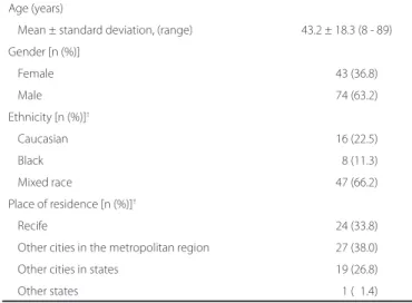
During the period from May 2012 to July 2012, 480 patients with uveitis were examined in the emergency eye care center of Fundação Altino Ventura. Among these, 117 who had active uveitis and fulfilled the study requirements were included in the analysis. The mean age of the evaluated patients was 43.2 ± 18.3 years, and 74 (63.2%) were male. Demographic data are shown in Table 1. There were no diffe-rences in the distribution of gender, race, and residence in relation to the anatomical classification of uveitis (p>0.05).
Anterior uveitis was observed in 82 patients (70.1%), posterior uveitis in 31 (26.5%), and diffuse uveitis in 4 (3.4%); none of the pa-tients were diagnosed with intermediate uveitis (Table 2). All papa-tients presented with sudden and acute symptoms (less than 3 months du-ration). Patients with posterior uveitis were younger than those with either anterior uveitis or diffuse uveitis (47.6 ± 17.0 years, 31.3 ± 16.1 years, and 46.0 ± 24.2 years, respectively, for anterior uveitis, posterior uveitis, and diffuse uveitis; p<0.001).
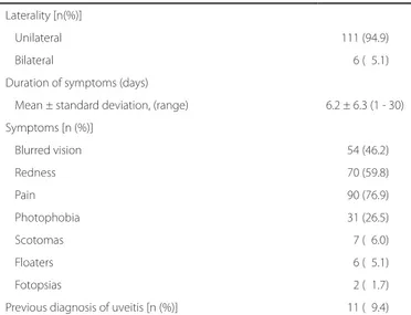
The most common symptoms observed were eye pain (n=90, 76.9%), redness (n=70, 59.8%), and visual blurring (n=54, 46.2%). The majority of patients had unilateral disease (n=111, 94.9%), with a mean symptom duration of 6.2 ± 6.3 days. Eleven patients (9.4%) had a previous diagnosis of uveitis and reported 1 to 5 previous epi-sodes. None of the patients was being treated for uveitis at the time of evaluation; however, one patient was being treated for iatrogenic conjunctivitis.
The clinical characteristics of the patients and the physical exami-nation findings are presented in Tables 3 and 4, respectively. Anterior and diffuse uveitis were associated with complaints of eye pain (86.6%, 48.4%, and 100.0%, respectively, for anterior uveitis, posterior uveitis, and diffuse uveitis; p<0.001). Scotomata were more frequent
in patients with posterior uveitis (1.2%, 19.4%, and 0.0%, respectively, for anterior uveitis, posterior uveitis, and diffuse uveitis; p=0.003). Com -plaints about floaters were associated with posterior uveitis (1.2%, 16.1%, and 0.0%, respectively, for anterior uveitis, posterior uveitis, and diffuse uveitis; p=0.016). Blurred vision was uncommon in patients with anterior uveitis when compared with inflammation at other sites (30.5%, 80.6%, 100.0%, respectively, for anterior uveitis, posterior uvei-tis, and diffuse uveitis; p<0.001). There was no significant association between the frequency of redness, photophobia, and photopsia with the anatomical classification of uveitis (p>0.05).
Patients with anterior uveitis showed better presenting VA (VA >20/63 in 57.0%, 38.7%, and 0.0%, respectively, for anterior uveitis, posterior uveitis, and diffuse uveitis; p=0.025). Conjunctival hype-remia was more common in patients with anterior uveitis (91.5%, 64.5%, and 75.0%, respectively, for anterior uveitis, posterior uveitis, and diffuse uveitis; p=0.003). Patients with granulomatous keratotic precipitates were most often diagnosed with posterior uveitis (3.7%, 19.4%, and 0.0%, respectively, for anterior uveitis, posterior uveitis, and diffuse uveitis; p=0.038). There was no association of the fre-quency of posterior synechiae, fine keratotic precipitates, iris nodules, and anterior chamber cells grade with the anatomical classification of uveitis (p>0.05). The mean intraocular pressure at presentation was 13.9 ± 7.2 mmHg (range, 2.0 to 40.0 mmHg), and there was no sta-tistical association between intraocular pressure and the anatomical classification of uveitis (p=0.598).
Table 1. Demographic characteristics of patients with active uveitis treated at the emergency eye care center of Fundação Altino Ventu-ra, Recife, brazil, between March and July 2012 (n=117)
Age (years)
Mean ± standard deviation, (range) 43.2 ± 18.3 (8 - 89)
Gender [n (%)]
Female 43 (36.8)
Male 74 (63.2)
Ethnicity [n (%)]†
Caucasian 16 (22.5)
Black 08 (11.3)
Mixed race 47 (66.2)
Place of residence [n (%)]†
Recife 24 (33.8)
Other cities in the metropolitan region 27 (38.0)
Other cities in states 19 (26.8)
Other states 01 (01.4)
†=data on ethnicity and residence were not collected for 46 patients (n =71).
Table 2. Anatomical classiication and frequency of determination of a cause or clinical syndrome on the basis of initial evaluation of 117 patients with active uveitis treated at the emergency eye care center of Fundação Altino Ventura, Recife, brazil between March and July 2012 [n (%)]
Anatomical classiication
Total patients n (%)
Patients with speciic diagnoses n (%)
Clinical and epidemiological characteristics of patients with uveitis in an emergency eye care center in Brazil
3 2 Arq Bras Oftalmol. 2014;77(1):30-3
Etiological diagnosis was established in 45 patients (38.5%) on the basis of the clinical evaluation and ancillary laboratory tests re quested at the initial visit. Among the 82 patients with anterior uveitis, 68 (82.9%) had an unknown etiology (of these, 63 had their first episode of anterior uveitis and were not investigated), 8 (9.8%) had uveitis associated with rheumatological disease, and 6 (7.3%) had a herpetic etiology. Among the 31 patients with posterior uveitis, 26 (87.1%) had an etiological diagnosis established during the initial visit, 25 (83.9% of posterior uveitis) had toxoplasmic retinochoroidits, and 1 (3.2%) patient had herpetic retinitis (acute retinal necrosis). Among the 4 patients with diffuse uveitis, 2 (50%) were diagnosed with Vogt-Koyanagi-Harada disease, 1 (25%) with fungal endophthal-mitis, and 1 (25%) with hypersensitivity uveitis caused by a corneal bee sting (Table 2).
DISCUSSION
This study prospectively evaluated patients from an emergency eye care center, in contrast to most uveitis epidemiological studies that have been conducted retrospectively in tertiary specialized cen-ters(1,4). In previous studies from referring uveitis centers in Brazil,
in-cluding our institution, posterior uveitis accounted for the majority of cases, particularly toxoplasmosis(14-16). In contrast, anterior uveitis
was responsible for 70.1% patients in our study. This can be explained by the fact that our sample mostly included individuals with first-epi sode anterior uveitis; these patients are typically not referred to specialized centers for investigation. The high frequency of anterior uveitis was in accordance with that reported in studies conducted in specialized uveitis centers in other countries(1,4,17) and studies
conduc-ted in community-based eye care centers(18).
The mean age of patients in this study (42.6 years) was higher than that reported in a previous study conducted at our institution betwe-en 1998 and 1999 (32.1 years)(14) and in studies conducted in re ferring
centers in Colombia and Tunisia (31.7 and 34.0 years, respectively)
(13,17). Nevertheless, the mean age of patients in the present study was
similar to that in studies from North America (45 years)(18) and
Sou-theastern Brazil (41 years)(16). Most patients in our study were working
adults, similar to the patients in the previous studies. The incidence of uveitic entities has been associated with ethnicity(1,4); however, we
could not find such an association, possibly because of the mixed race background of the Brazilian population.
The most frequently reported symptoms were pain, redness, blur-red vision, and photophobia. Such symptoms are nonspecific and can be easily misdiagnosed as other conditions, including conjuncti vitis and keratitis(6). Anterior uveitis was associated with a higher fre
quen-cy of redness and pain, while posterior uveitis was greatly associated with blurred vision and scotomata. Granulomatous keratotic precipi-tates were most common in patients with the primary site of inflam-mation in the posterior segment. Therefore, an accurate medical his tory and physical examination are imperative for establishing a diag nosis in patients with uveitis.
Determination of a cause or clinical syndrome on the basis of cli-nical presentation and ancillary examination requested at the initial visit was possible in 38.5% patients, a diagnostic rate lower than that observed in community-based, comprehensive ophthalmological units and uveitis referral centers (46% to 79.4%)(1,4,13,17,18). In this study,
first-episode acute anterior uveitis accounted for the majority of cases. It should be noted that further investigations are usually not per-formed in patients presenting with the first episode of uncomplica-ted anterior uveitis(20), which explains the high frequency of uveitis of
unknown etiology.
This research mostly included patients who were visiting the hos-pital for the first time, and the onset and course of disease were based on symptomatology, which can lead to misclassification. For example, a patient with an exacerbation of undiagnosed chronic uveitis could have been incorrectly diagnosed with sudden-onset acute uveitis(18).
Another limitation of this study was the small sample size, which may have restricted the inclusion of less common uveitic entities.
Table 3. Clinical characteristics of patients with active uveitis treated at the emergency eye care center of Fundação Altino Ventura, Recife, brazil between March and July 2012 (n=117)
Laterality [n(%)]
Unilateral 111 (94.9)
Bilateral 006 (05.1)
Duration of symptoms (days)
Mean ± standard deviation, (range) 6.2 ± 6.3 (1 - 30)
Symptoms [n (%)]
Blurred vision 054 (46.2)
Redness 070 (59.8)
Pain 090 (76.9)
Photophobia 031 (26.5)
Scotomas 007 (06.0)
Floaters 006 (05.1)
Fotopsias 002 (01.7)
Previous diagnosis of uveitis [n (%)] 011 (09.4)
Table 4. Ocular examination indings of the patients with active uveitis treated at the emergency eye care center of Fundação Altino Ventura, Recife, brazil between March and July 2012 (n=117)
Visual acuity [n (%)]†
>20/63 57 (50.0)
20/63 to 20/200 29 (25.4)
<20/200 28 (24.6)
Anterior segment [n (%)]
Conjunctival hyperemia 98 (83.8)
Fine keratic precipitates 48 (41.0)
Granulomatous keratic precipitates 09 (07.7)
Corneal edema 19 (16.2)
Keratitis 10 (08.5)
Posterior synechiae 24 (20.5)
<180o 15 (12.8)
>180o 09 (07.7)
Iris nodules 01 (00.9)
Anterior chamber cell reaction‡
0+ cells 10 (08.8)
0.5 to 2+ cells 63 (55.3)
>2+ cells 41 (36.0)
Hypopyon 03 (02.6)
Posterior segment [n (%)]§
Vitreous opacity 24 (21.6)
Retinochoroiditis 24 (21.6)
Retinitis 01 (00.9)
Exudative retinal detachment 02 (01.8) Intraocular pressure(mmHg)¶
Mean ± standard deviation, (range) 13.9 ± 7.2 (2.0 - 40.0)
†= visual acuity measurement was not possible in 3 patients (n=114); ‡= evaluation of
anterior chamber reactions was not possible in 3 patients (n=114); §= evaluation of
pos-terior segment was not possible in 6 patients (n=111); ¶= applanation tonometry was
Camilo ENR, et al.
3 3
Arq Bras Oftalmol. 2014;77(1):30-3 In conclusion, this study shows that anterior uveitis is observed
more frequently in primary health care centers than in tertiary referral centers. Initial evaluation of the patient in the emergency room pro-vided sufficient information for deciding primary therapy and aided in arriving at an etiological diagnosis in a considerable number of patients. These findings are important for prioritization of education and training for general ophthalmologists.
REFERENCES
1. Chang JH, Wakefield D. Uveitis: a global perspective. Ocul Immunol Inflamm. 2002; 10(4):263-79. Review.
2. Gutteridge IF, Hall AJ. Acute anterior uveitis in primary care. Clin Exp Optom. 2007; 90(5):390; author reply 390.
3. Prieto-del-Cura M, González-Guijarro J. [Complications of uveitis: prevalence and risk factors in a series of 398 cases]. Arch Soc Esp Oftalmol. 2009;84(10):523-8. Spanish. 4. Rathinam SR, Namperumalsamy P. Global variation and pattern changes in
epidemio-logy of uveitis. Indian J Ophthalmol. 2007;55(3):173-83. Review.
5. Durrani OM, Meads CA, Murray PI. Uveitis: a potentially blinding disease. Ophthalmo-logica. 2004;218(4):223-36. Review.
6. Mahmood AR, Narang AT. Diagnosis and management of the acute red eye. Emerg Med Clin North Am. 2008;26(1):35-55, vi. Review.
7. Kara-José N, Carvalho KM, Pereira VL, Venturini NH, Gasparetto ME, Gushiken MT. [Retrospective study of first 140 cases attended the Clinica de Visäo Sub-Nor mal of the Hospital de Clínicas da Unicamp]. Arq Bras Oftalmol. 1988;51(2):65-9. Portuguese. 8. Carvalho Rde S, José NK. Ophthalmology emergency room at the University of São
Paulo General Hospital: a tertiary hospital providing primary and secondary level care. Clinics (Sao Paulo). 2007;62(3):301-8.
9. Campos Júnior JC. [Profile of ophthamological attendance of emergency]. Rev Bras Oftalmol. 2004;63(2):89-91. Portuguese.
10. Kumar NL, Black D, McClellan K. Daytime presentations to a metropolitan ophthalmic emergency department. Clin Experiment Ophthalmol. 2005;33(6):586-92. 11. Pierre Filho PT, Gomes PR, Pierre ET, Pinheiro Neto FB. [Profile of ocular emergencies
in a tertiary hospital from Northeast of Brazil]. Rev Bras Oftalmol. 2010;69(1):12-7. Por-tuguese.
12. Deschenes J, Murray PI, Rao NA, Nussenblatt RB; International Uveitis Study Group. International Uveitis Study Group (IUSG): clinical classification of uveitis. Ocul Immu-nol Inflamm. 2008;16(1):1-2.
13. de-la-Torre A, López-Castillo CA, Rueda JC, Mantilla RD, Gómez-Marín JE, Anaya JM. Clinical patterns of uveitis in two ophthalmology centres in Bogota, Colombia. Clin Experiment Ophthalmol. 2009;37(5):458-66.
14. Diniz JR, Toscano JL, Campelo DE, Delgado AC, Leal SD. [Occurency of uveitis in Per -nambuco state, Brazil]. Rev Bras Ciênc Saúde. 2001;5(1):59-64. Portuguese. 15. Gehlen ML, Dabul VM, Obara SS, Grebos SP, Moreira CA. [Incidence and etiology of
uveitis in Curitiba]. Arq Bras Oftalmol. 1999;62(5):622-6. Portuguese.
16. Gouveia EB, Yamamoto JH, Abdalla M, Hirata CE, Kubo P, Olivalves E. [Causes of uveitis in a tertiary center in São Paulo city, Brazil]. Arq Bras Oftalmol. 2004;67(1):139-45. Portuguese. 17. Khairallah M, Yahia SB, Ladjimi A, Messaoud R, Zaouali S, Attia S, et al. Pattern of uveitis
in a referral centre in Tunisia, North Africa. Eye (Lond). 2007;21(1):33-9.
18. McCannel CA, Holland GN, Helm CJ, Cornell PJ, Winston JV, Rimmer TG. Causes of uveitis in the general practice of ophthalmology. UCLA Community-Based Uveitis Study Group. Am J Ophthalmol. 1996;121(1):35-46.
19. Jabs DA, Nussenblatt RB, Rosenbaum JT; Standardization of Uveitis Nomenclature (SUN) Working Group. Standardization of uveitis nomenclature for reporting clinical data. Re-sults of the First International Workshop. Am J Ophthalmol. 2005;140(3):509-16. Review. 20. Forooghian F, Gupta R, Wong DT, Derzko-Dzulynsky L. Anterior uveitis investigation
by Canadian ophthalmologists: insights from the Canadian National Uveitis Survey. Can J Ophthalmol. 2006;41(5):576-83.

