Arq. Bras. Cardiol. vol.96 número1 en v96n1a19
Texto
Imagem
Documentos relacionados
If the G894T variant of the eNOS gene could reduce the enzymatic activity, individuals with the mutant allele (T) would show a smaller increase in muscle blood flow in
The stimulation of the apex of the right ventricle promotes an inversion of the natural sequence of cardiac electrical activation, generates an artificial left bundle
Manuscript received April 22, 2010; revised manuscript received June 28, 2010; accepted July 06,
matter deserves further consideration, since there is some dissociation between the methods and the conclusion: 1) The authors concluded that the morbidity-mortality of
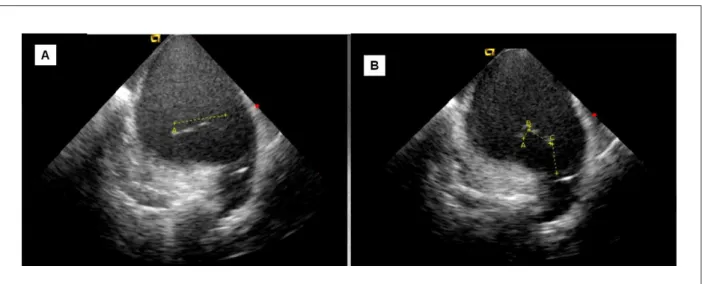
Figure 2 - Echocardiogram shows a sharp increase of the right cavities in 4-chamber apical section, in A, the septal tricuspid valve was not very mobile and “attached”
In fact, previous investigations clearly demonstrated that exercise training dramatically reduced muscle sympathetic nerve activity and significantly improved muscle
The impairment of the right ventricle, and particularly that of the interventricular septum, associated with a large and indefinite mass compressing the right cavities,
En la ablación de fibrilación atrial demuestra gran utilidad por proveer datos anatómicos del atrio izquierdo y venas pulmonares, auxiliar en las
