C
ASER
EPORT| R
ELATO DEC
ASO217
Reversal of uremic tumoral calcinosis by optimization of
clinical treatment of bone and mineral metabolism disorder
Reversão da calcinose tumoral urêmica pela otimização do
tratamento clínico da desordem do metabolismo ósseo e mineral
Authors
Mariana Espiga Maioli 1,2
Vinicius Daher Alvares Delfino 1,2
Amanda Carolina Damasceno Zanuto Guerra 1,2
Luiz Fernando Kunii 1,2
Raquel Ferreira Nassar Frange 2
1 Universidade Estadual de Londrina (UEL).
2 Instituto do Rim de Londrina.
Submitted on: 7/1/2016. Approved on: 8/30/2016.
Correspondence to: Mariana Espiga Maioli. Instituto do Rim de Londrina. Rua Engenheiro Omar Rupp, nº 100, Jardim Ipiranga, Londrina, PR, Brazil. CEP: 86015-360 E-mail: mariana. [email protected]
DOI: 10.5935/0101-2800.20170037
A calcinose tumoral é um tipo raro de calcificação extraóssea caracterizada por grandes massas císticas e elásticas con-tendo depósitos de fosfato de cálcio. A condição é mais prevalente no tecido periarticular e preserva estruturas os-teoarticulares. A elevação do produtos cálcio-fósforo e o hiperparatireoidismo secundário grave estão presentes na maio-ria dos pacientes com calcinose tumoral urêmica (UTC). O relato de caso em questão refere-se a um homem de 22 anos, branco, obeso, com doença renal crônica secundária à glomerulonefrite crônica, em diálise peritoneal ambulatorial contínua (CAPD), que apresentou aparecimento de tumores indolores na face medial do quin-to quirodáctilio e braço esquerdo. A calci-nose tumoral foi confirmada por biópsia do bíceps esquerdo. O paciente apresen-tava baixa adesão à CAPD. Foi transferido para a modalidade de diálise peritoneal e depois iniciou tratamento por hemodiálise. Apesar da persistência do hiperparatire-oidismo grave, houve redução progressiva da UTC, com resolução próxima do seu desaparecimento completo. Há 1 ano o paciente foi submetido a transplante renal, doador falecido, e apresentou melhora do hiperparatiroidismo secundário. A UTC deve ser incluída na elucidação de calcifi-cação periarticular de pacientes em diálise. Os achados laboratoriais relevantes, tais como hiperparatiroidismo secundário e elevação dos produtos cálcio-fósforo na presença de calcificação periarticular, de-vem chamar a atenção para o diagnóstico da UTC.
R
ESUMOPalavras-chave: distúrbios do
metabolis-mo do cálcio; distúrbios do metabolismetabolis-mo do fósforo; nefrologia.
Tumoral calcinosis is an uncommon type of extraosseous calcification char-acterized by large rubbery or cystic masses containing calcium-phosphate deposits. The condition prevails in the periarticular tissue with preservation of osteoarticular structures. Elevated calcium-phosphorus products and se-vere secondary hyperparathyroidism are present in most patients with ure-mic tumoral calcionosis (UTC). Case report of an obese secondary to chronic glomerulonephritis, undergoing con-tinuous ambulatory peritoneal dialysis (CAPD) reported the appearance of painless tumors in the medial surface of fifth finger and left arm. Tumoral calcinosis was confirmed by left biceps biopsy. Poor adherence to CAPD. The patient was transferred to the "tidal" modality of peritoneal dialysis and after was treated by hemodialysis, de-spite the persistence of severe hyper-parathyroidism progressive reduction of UTC until near to its complete dis-appearance. Nowadays, one year after patient received deceased-donor kidney transplantation, he presents with an improvement in secondary hyperpara-thyroidism. UTC should be included in the elucidation of periarticular cal-cification of every patient on dialysis. Relevant laboratory findings such as secondary hyperparathyroidism and elevated calcium- phosphorus products in the presence of periarticular calcifi-cation should draw attention to the di-agnosis of UTC.
A
BSTRACTJ Bras Nefrol 2017;39(2):217-219
Calcinose tumoral urêmica
218
I
NTRODUCTIONTumoral calcinosis is an uncommon type of extraosseous calcification characterized by large rubbery or cystic masses containing calcium-phosphate deposits.1 The condition prevails in the periarticular
tissue with preservation of osteoarticular structures. Genetic causes and metabolic disorders are associated with their appearance.2 The tumoral calcinosis that
occurs in chronic kidney disease (CKD) patients is known for uremic tumoral calcinosis (UTC).
In this condition, calcifications can also contain hydroxyapatite (Ca5 (PO4) 3OH).1,3 Although,
most often described in hemodialysis (HD) patients (0.5- 1.2% prevalence),2 UTC may also occur in
patients undergoing peritoneal dialysis. Elevated calcium-phosphorus products and severe secondary hyperparathyroidism are present in most patients with UTC.4
Common sites of involvement are the blood vessels, periarticular region, heart, lungs, kidneys, gastric mucosa, central nervous system, breast and eyes. Clinical treatment for UTC includes dietary phosphorus restriction, non-calcium phosphate binders, calcimimetics, optimal control of hyperparathyroidism, and intensive hemodialysis with low calcium dialysate. Surgical excision of tumoral calcinosis, parathyroidectomy, and kidney transplantation are recommended for persistent or refractory UTC.1,5
C
ASEREPORTAn obese (Body Mass Index 38.5) young white man of 22 years with CKD secondary to chronic glomerulonephritis, undergoing continuous ambulatory peritoneal dialysis (CAPD) for uremia management, reported, after 6 months of treatment, the appearance of painless tumors in the medial surface of fifth finger and left arm (biceps region).
Laboratory results: serum calcium 8.5 mg/dL (reference: 8.4 to 10.2 mg/dL), serum albumin 3.6 mg/ dL (reference: 3.4 to 5.2 mg/dL), serum phosphorus 11.1 mg/dL (reference: 2.5 to 4.5 mg/dL), and intact PTH (iPTH) 1867 pg/mL (reference: 15 to 65 pg/ml), serum uric acid 8.7 mg/dL (reference: 3.6 to 7.7 mg/dL).
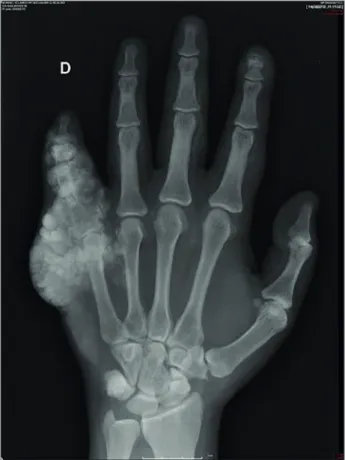
Computed tomography of the left arm and radiography of the right hand (Figure. 1) showed periarticular, irregular and multilobular calcifications. Tumoral calcinosis was confirmed by left biceps biopsy:
lesion characterized by the presence of abundant giant cells scattered throughout the proliferation of fibroblasts, with associated areas of hemorrhage and extensive calcifications. Ultrasonography of anterior cervical region showed no nodules in the four parathyroid glands.
Figure 1. Right hand X-ray: Multiple calcifications around the phalanges and the phalangeal-metacarpal transition of 5th right hand finger.
Questioned, the patient admitted poor adherence to CAPD (with 3.5 mEq/L calcium concentration in dialysate), sometimes doing only one or two exchanges a day. The patient was transferred to the “tidal” modality of peritoneal dialysis in order to increase the efficiency of uremia treatment, while waiting for AVF maturation for starting HD.
J Bras Nefrol 2017;39(2):217-219
Calcinose tumoral urêmica
219
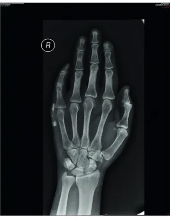
Figure 2. Right hand X-ray after 4 months of the transference of the patient to hemodialysis showing markedly reduction of UTC.
After 3 months on hemodialysis, it was observed, despite the persistence of severe hyperparathyroidism (calcium 8.9 mg/dL, P 7.6 mg/dL and PTH 1840 pg/ mL), progressive reduction of UTC until near to its complete disappearance (Figure. 2). In November 2014, the patient received deceased-donor kidney transplantation (immunosuppression: tacrolimus, mycophenolate mofetil, and prednisone).
Nowadays, three months after surgery, he presents with a serum creatinine of 0.9 mg/dL and calcium, phosphorus and iPTH values of 9.4 mg/ dL; 1.7 mg/dL and 368.8 pg/mL, respectively. Since there was improvement in hyperparathyroidism by normalization of renal function, no hypercalcemia, moderate hypophosphatemia, thus was vitamin D nutritional supplemented by low serum.
Since no abnormal parathyroid glands was identified at their usual position at ultrasound, it was planned to observe for a few months more the possibility of reversal the secondary hyperparathyroidism, as suggested in the Literature,6
instead considering parathyroidectomy as a first approach of treatment.6
D
ISCUSSIONUTC should be included in the elucidation of periarticular calcification of every patient on dialysis. The diagnosis is often not done due to its rarity and the difficulty of clinical recognition. In patients on peritoneal dialysis, there is a report of adherence to dialysis, as exemplified in this case.5
Relevant laboratory findings such as secondary hyperparathyroidism and elevated calcium- phosphorus products in the presence of periarticular calcification should draw attention to the diagnosis of UTC. Treatment involves dialysis improvement, and appropriate control of calcium and phosphorus. Parathyroidectomy and kidney transplantation may be required for its management.
R
EFERENCES1. Chu HY, Chu P, Lin YF, Chou HK, Lin SH. Uremic tumoral calcinosis in patients on peritoneal dialysis: clinical, radiologic, and laboratory features. Perit Dial Int 2011;31:430-9. DOI: http://dx.doi.org/10.3747/pdi.2009.00250
2. Olsen KM, Chew FS. Tumoral calcinosis: pearls, polemics, and alternative possibilities. Radiographics 2006;26:871-85. PMID: 16702460 DOI: http://dx.doi.org/10.1148/rg.263055099 3. Floege J. When man turns to stone: extraosseous calcification
in uremic patients. Kidney Int 2004;65:2447-62. DOI: http:// dx.doi.org/10.1111/j.1523-1755.2004.00664.x
4. Carvalho M, de Menezes IA, Riella MC. Massive, painful tu-moral calcinosis in a long-term hemodialysis patient. Hemodial Int 2011;15:577-80. DOI: http://dx.doi.org/10.1111/j.1542-4758.2011.00581.x
5. Raju DL, Podymow T, Barre P. Tumoral calcinosis in a peri-toneal dialysis patient. Kidney Int 2006;70:1887. DOI: http:// dx.doi.org/10.1038/sj.ki.5001752
6. Triponez F, Clark OH, Vanrenthergem Y, Evenepoel