ORIGINAL
ARTICLE
ENGLISH VERSION
Intra and interdays reliability of the 4-second exercise test
Claudio Gil Soares de Araújo
1, Djalma Rabelo Ricardo
2and Marcos Bezerra de Almeida
2CVI reliability assessed by T4s, and confirmed the need for
two consecutive trials, as prescribed in its protocol.
Key words: 4-second exercise test. Physical exercise. Heart rate.
Cardiac vagal tone. Reliability.
INTRODUCTION
The behavior of heart rate (HR) in exercises transient has
been investigated in a number of studies over the past few
years1-4, which shows the importance of its clinical and
physiological investigations. HR modulation is mediated
by the sympathetic and parasympathetic branches of the
autonomous nervous system (ANS), and its integrity is
asso-ciated to a decrease in mortality risk from cardiac events5,6.
In a study carried out by Nolan et al.7, it was seen that
patients with heart failure (moderate and severe), with au-tonomic dysfunction, had a mortality rate 10-fold higher than their peers whose parasympathetic activity was closer to normal ranges. These results confirm the impression that cardiac vagal activity is a powerful and independent
prog-nostic marker5.
A number of physiological and clinical procedures have
been proposed to assess autonomic condition8-11. Among
them, one can mention the 4-second exercise test (T4s),
orig-inally proposed by Araújo et al.12. The T4s is intended to
indirectly assess cardiac vagal tone through the initial heart rate transient of a dynamic short term exercise performed
in apnea13. This test was pharmacologically validated and
has been applied in clinical trials and sports medicine,
prov-ing to be quite useful both as a diagnostic tool14,15, and
lon-gitudinal follow-up of cardiac parasympathetic activity16.
Thus, in order to consolidate this procedure, it is
impor-tant to determine T4s consistency and reliability in
assess-ing cardiac vagal autonomic activity of a heterogeneous sample. Therefore, the purposes of this study were to
de-termine intra and interdays T4s reliability in measuring
car-diac vagal tone, and the need for two measurements, as described in the original protocol.
MATERIALS AND METHODS
To meet the intended purposes, the results from two dif-ferent labs, presented separately, were assessed, looking
1. Programa de Pós-Graduação em Educação Física da Universidade Gama Filho – Rio de Janeiro, RJ, CLINIMEX – Clínica de Medicina do Exercí-cio – Rio de Janeiro, RJ.
2. Programa de Pós-Graduação em Educação Física da Universidade Gama Filho – Rio de Janeiro, RJ.
Received in 12/7/03 Approved in 22/9/03
Correspondence to:
Dr. Claudio Gil S. Araújo
Clínica de Medicina do Exercício – CLINIMEX (www.clinimex.com.br) Rua Siqueira Campos, 93/101
22031-070 – Rio de Janeiro, RJ E-mail: [email protected] ABSTRACT
The 4-second exercise test (T4s) is pharmacologically
validated to assess cardiac vagal tone, and consists in ped-aling, as fast as possible, a cycle-ergometer unloaded, from
the 4th to the 8th second of a 12-second maximum
inspira-tory apnea. An dimensionless cardiac vagal index (CVI) is
calculated from the ratio of the duration of the cardiac
cy-cles (RR intervals at the electrocardiogram) from
immedi-ately before the exercise and the shorter of the exercise.
Our objective was to determine T4s intra and interdays
reli-ability, and the actual need for two trials, as described on the original protocol. In study 1, the interday reliability of the results was assessed prospectively from 15
asymptom-atic subjects (28 ± 6 years) submitted to T4s for five
con-secutive days, being two trials carried out at each day. To
determine CVI intraday reliability, in one of the five days,
randomly selected, nine T4s consecutive trials were made.
In study 2, the CVI intraday reliability was calculated from
1699 subjects (47 ± 17 years) in two trials. CVI presented
high intraday and interday reliability (ri = 0.92; 95%CI =
0.84 to 0.97 and ri = 0.77; 95%CI = 0.49 to 0.92,
respective-ly) for study 1 and for study 2 (ri = 0.89; 95%CI = 0.88 to
0.90). In spite of high reliability, there were some minor
differences between the means (mean ± SEM = 1.32 ± 0.01
for better understanding of the experimental procedures used. Furthermore, all tests and their measurements and interpretations were performed by experienced evaluators
in the T4s protocol, and strictly followed its standards. The
subjects submitted to the T4s read and signed an informed
consent form before the procedures.
The 4-second Exercise Test – T4s
The purpose of the T4s is to assess exclusively the
integ-rity of the parasympathetic branch of the autonomic
ner-vous system through the initial HR transient at
rest-exer-cise transition. The T4s consists in pedaling as quickly as
possible a unloaded cycleergometer, from the 4th to the 8th
second of a maximum inspiratory apnea of 12 seconds. Four consecutive commands are given at each four seconds: (a) a maximum and swift inhale through the mouth and then sustain apnea; (b) to pedal as fast as possible; (c) suddenly stop pedaling, and (d) normally exhale.
To minimize anticipatory responses to the commands, the subject should not see the chronometer nor the electro-cardiograph, while a continuous trace is recorded from only
one ECG lead (typically CC5or CM5) for 35 seconds at a
speed of 25 mm/s, started five seconds before the com-mand for maximum inhale.
To determine the magnitude of the cardiac vagal tone,
one must identify the immediately before or the first RR
interval of the exercise (which is the longest) (RRB), and
the shortest one during the exercise, usually the last (RRC).
The quotient or ratio between these two intervals shows
the cardiac vagal index (CVI), a T4s dimensionless index.
Two consecutive trials are made with a one-to-two-mi-nute interval between them, which is typically enough for heart rate to return to resting level, being selected the
high-est of the two CVI values as representative of the subject’s
cardiac vagal tone.
Prior methodological studies with T4s showed that CVI
value independs on the presence or absence of resistance
opposing pedal movement17,if the exercise is performed
actively or passively18, or if performed by lower or upper
limbs19.
Study 1
To assess T4s reliability, we made a prospective analysis
of the results from 15 subjects (eight women), and 13 sub-jects (six women), for intra and interdays, respectively. The subjects were considered asymptomatic, aged 28 ± 6 (21 to 42) years, who volunteered for the study carried out in a lab.
Data were collected from a 380B electrocardiograph
(Si-emens, Germany), from a single lead (CC5), at a speed of
25 mm/s, and T4s was performed in a mechanic-breaking
cycle-ergometer Monark. Measurement of the RR intervals
on the recordings was made by a single evaluator, experi-enced in the test, with a 10 ms resolution.
The subjects were asked to rest in a room for about five
minutes prior to the beginning of the test protocol. T4s was
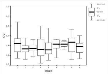
repeated for five days, always at the same time of the day, in order to measure interdays reliability. Intraday reliabili-ty was investigated in nine consecutive trials carried out in one of those five days, randomly selected for each subject.
Study 2
In a second assessment, intraday reliability was retro-spectively determined from the results of 1,699 subjects (613 women), aged 47 ± 17 (8 to 85) years, including subjects of different clinical and fitness conditions (even athletes), who spontaneously went for a detailed medical-functional assessment in one of the labs between 1994 and 2003.
For this analysis, a TEC 7100 electrocardiograph
(Nihon-Kohden, Japan) was used in the tests carried out until 2001, and a digital electrocardiograph with a specific software (ErgoPC Elite version 3.2.1.5. Micromed, Brazil), in the
tests from 2001 on, both in single lead (CC5 or CM5),
re-corded at a speed of 25 mm/s, and an electro-magnetic-break cycle-ergometer Cateye model Ergociser EC 1600 (CatEye, Japan). The measurement of the duration of the
RR intervals on the electrocardiograph recordings were
made by experienced evaluators, using a 10 ms resolution.
In order to assess T4s reliability in subjects with different
magnitudes of vagal tone, we sorted the values as a
func-tion of the CVI from the second trial, and then used cut-off
points arbitrarily defined in our database, of less than 1.20, between 1.20 and 1.70, and higher than 1.70 for vagal hy-potonic, normal, and hypertonic (vagotonic), respectively (unpublished data).
Statistical analysis
Intraclass correlation coefficient was used in the studies to measure the degree of association between the trials.
Furthermore, to compare the means, ANOVA for repeated
measures, and paired t-test (for study 1 and study 2, re-spectively) were employed. Significance level of 5% and 95% confidence interval was used for all results.
RESULTS
Study 1 – The CVI obtained in the T4s showed high intra
and interdays reliability, as one can see from the intraclass
correlation coefficients (ri = 0.92; 95%CI = 0.84 to 0.97 and
ri = 0.77; 95%CI = 0.49 to 0.92, respectively). These results
were confirmed by ANOVA, and no differences between the
9 8 7 6 5 4 3 2 1 2, 2 2, 0 1, 8 1, 6 1, 4 1, 2 1, 0 Máximo P75 Mediana Mínimo P25
Study 2 – In a larger and more heterogeneous sample,
the CVI between the two T4s trials was considerably
associ-ated (ri = 0.89; 95%CI = 0.88 to 0.90), attesting once more
its reliability (figure 3). However, the t-test showed
differ-ences between the means (mean ± SEM = 1.32 ± 0.01 vs
1.37 ± 0.01; p < 0.001), and in only 15% this difference was higher than 0.20 (figure 4). We also noticed that in
65% of the observations, the second trial had higher CVI
results.
Likewise, by splitting the sample in three groups accord-ing to reference values from the second trial (selected for
including most of CVI best results), we observed
differenc-es between the two trials for all groups (table 1). However,
the number of cases in which the difference between trials was higher than 0.20 was lower than 1% for hypotonic, 9% for normals, and 5% for hypertonic subjects, being worth to mention that intraclass correlation coefficient had a significant association between the trials, in each group (p < 0.001). Moreover, 27% of the subjects would have been mistakenly classified if only one trial was to be made.
DISCUSSION
HR behavior is an important marker of the cardiac vagal
activity, which, if decreased, is strongly associated to
mor-tality risk2,3,6,20-22, showing an increase of cardiac
vulnera-bility due to a potential and lethal risk of ventricular
ar-rythmia5,23,24.
The clinical contributions of autonomic assessment in stratifying mortality risk from cardiovascular events and from all causes have been broadly used by the scientific
community25-28, particularly because vagal activity is the
main autonomic dysfunction marker29-32. Thus, it is
inter-Fig. 1 – Study 1 intraday reliability Fig. 2 – Study 1 interdays reliability
IVC da tentativa 2
2,40 2,20 2,00 1,80 1,60 1,40 1,20 1,00 n i 2,40 2,20 2,00 1,80 1,60 1,40 1,20 1,00
Y = 0,68.X + 0,39 EPM = 0,13 r2= 0,32
p < 0,001
CVI of trial 2
2,40 2,20 2,00 1,80 1,60 1,40 1,20 1,00 2,40 2,20 2,00 1,80 1,60 1,40 1,20 1,00
Y = 0,68.X + 0,39 EPM = 0,13 r2= 0,32
p < 0,001
CVI of trial 1
Fig. 3 – Association between two consecutive T4s trials for values of
subjects classified as normal – 1.20 to 1.70 – in the second trial (study 2) 0 100 200 300 400 500 600 700 < -0 ,6 0 -0 ,6 0 a -0 ,5 1 -0 ,5 0 a -0 ,4 1 -0 ,4 0 a -0 ,3 1 -0 ,3 0 a -0 ,2 1 -0 ,2 0 a -0 ,1 1 -0 ,1 0 a -0 ,0 1 0 a 0 ,10 0, 1 1 a 0, 2 0 0, 2 1 a 0, 3 0 0, 3 1 a 0, 4 0 0, 4 1 a 0, 5 0 0, 5 1 a 0, 6 0 > 0 ,60
CVI difference between T4s first and second trial
N
umbe
r of o
bs e rv a ti ons
Fig. 4 – Differences of CVI between trials (study 2)
2, 2 2, 0 1, 8 1, 6 1, 4 1, 2 1, 0 Máximo P75 Mediana Mínimo P25
Day 1 Day 2 Day 3 Day 4 Day 5
C
VI
C
VI
Trials
M axim um
M edian
M inim um
M axim um
M edian
esting to stress the importance of a valid and reliable test to assess cardiac vagal tone.
As we could see from study 1, the measure of the vagal
cardiac tone by T4s showed high intra and interdays
reli-ability, evidencing T4s consistency to assess vagal cardiac
function, expressed by CVI. It is also important to mention
that ANOVA confirmed these results, with no differences
found among the sample means.
Study 2 was based on a very large sample, and again CVI
obtained in T4s was highly reliable, in spite of the diverse
clinical conditions and populations (children, adolescents, adults, elders, athletes, even of Olympic levels, and asymp-tomatic non-athletes).
By confronting our results with those of other reliability studies on cardiovascular autonomic tests, we observed
some important aspects that favored our T4s studies, such
as: the size of the sample, as studies that investigate such topic have a significantly smaller number of subjects in
their sample33,34; the characteristics of the sample (age group
and clinical conditions)35,36; and the magnitude of
intrac-lass correlation coefficients, similar to some and higher than
others37,38.
One should also add that, regarding study 2, we observed differences between the first and second trial (t-test), for the whole sample and for the subgroups, even though these results do not indicate clinical or physiological relevance. This fact may be observed from the number of cases in which discrepancy between trials was higher than 0.20, par-ticularly among subjects referred as vagal hypotonic, in whom a possible association with cardiovascular condi-tions and complex arrhythmias due to a lower vagal
cardi-ac protection draws attention28,39-41.
In practice, the physician, when supervises the proce-dure, frequently advises the subject to correct minor dis-tortions or errors from the first trial, so that better results from the second trial of study 2 are to be expected. In fact,
about 2/3 of the subjects achieve higher CVI values in the
second trial, probably because they are more familiar with the procedure, and thus perform it more appropriately. Fur-thermore, the second trial prevented more then 1/4 of the
subjects to have their CVI wrongly classified or
interpret-ed. These results prove the need for two trials, as described in the original protocol.
T4s seems to comply with scientific authenticity criteria
(reliability and validity), considering the clinical conditions and the significant diversity of the sample, as we could see
in this and in prior studies13,15. Moreover, the simplicity
and applicability of this test should be stressed, in addition to its low operational cost.
Thus, incorporating T4s in the routine of pre-exercise test
– either conventional or with exhaled gases measurements42
–, has the potential ability to wide open the clinically rele-vant information to be obtained with the use of physical exercise in health or unhealthy subjects.
In summary, this study evidenced T4s reliability in
as-sessing cardiac vagal tone, expressed by the CVI, and
con-firmed the need for two consecutive trials, as recommend-ed in its protocol.
ACKNOWLEDGMENTS
The authors thank Prof. Flavia Dias de Oliveira for her role in the data collection of study 1.
All the authors declared there is not any potential conflict of interests regarding this article.
REFERENCES
1. Pierpont GL, Stolpman DR, Gornick CC. Heart rate recovery as an in-dex of parasympathetic activity. J Auton Nerv Syst 2000;80:169-74. 2. Lauer MS, Francis GS, Okin PM, Pashkow FJ, Snader CE, Marwick
TH. Impaired chronotropic response to exercise stress testing as a pre-dictor of mortality. JAMA 1999;281:524-9.
TABLE 1
Intraclass reliability for each group, according to cardiac vagal index (CVI) of the second trial
Groups n Trial 1 Trial 2 Intraclass t-test CVI difference
M ean ± SEM M ean ± SEM correlation p higher than
(min-max) (min-max) ri (CI 95%) 0.20
(CI 95%)
Hypotonic 0.502 1.14 ± 0.004 1.12 ± 0.002 0.51 0.001 1
(1.00-1.82) (1.00-1.19) (0.41 a 0.58) (0.02 a 0.03)
Norm al 1,004 1.34 ± 0.01 1.39 ± 0.004 0.72 0.001 9
(1.00-2.21) (1.20-1.69) (0.68 a 0.75) (0.06 a 0.04)
Hypertonic 0.193 1.71 ± 0.02 1.89 ± 0.01 0.67 0.001 5
3. Cole CR, Blackstone EH, Pashkow FJ, Snader CE, Lauer MS. Heart rate recovery immediately after exercise as a predictor of mortality. N Engl J Med 1999;341:1351-7.
4. Morshedi-Meibodi A, Larson MG, Levy D, O’Donnell CJ, Vasan RS. Heart rate recovery after treadmill exercise testing and risk of cardio-vascular disease events (The Framingham Heart Study). Am J Cardiol 2002;90:848-52.
5. Buch AN, Coote JH, Townend JN. Mortality, cardiac vagal control and physical training – What’s the link? Exp Physiol 2002;87:423-35. 6. Tapanainen JM, Thomsen PE, Kober L, Torp-Pedersen C, Makikallio
TH, Still AM, et al. Fractal analysis of heart rate variability and mortal-ity after an acute myocardial infarction. Am J Cardiol 2002;90:347-52. 7. Nolan J, Batin PD, Andrews R, Lindsay SJ, Brooksby P, Mullen M, et al. Prospective study of heart rate variability and mortality in chronic heart failure: results of the United Kingdom heart failure evaluation and assessment of risk trial (UK-Heart). Circulation 1998;98:1510-6. 8. Marfella R, Guigliano D, di Maro G, Acampora R, Giunta R, D’Onofrio
F. The squatting test. A useful tool to assess both parasympathetic and sympathetic involvement of the cardiovascular autonomic neuropathy in diabetes. Diabetes 1994;43:607-12.
9. Castro CLB, Nóbrega ACL, Araújo CGS. Testes autonômicos cardio-vasculares. Uma revisão crítica. Parte I. Arq Bras Cardiol 1992;59:75-85.
10. Castro CLB, Nóbrega ACL, Araújo CGS. Testes autonômicos cardiovas-culares. Uma revisão crítica. Parte II. Arq Bras Cardiol 1992;59:151-8. 11. European Society of Cardiology. Heart rate variability: standards of
measurement, physiological interpretation, and clinical use. Task Force of the European Society of Cardiology and the North American Society of Pacing Electrophysiology. Circulation 1996;93:1043-65.
12. Araújo CGS, Nóbrega ACL, Castro CLB. Vagal activity: effect of age, sex and physical pattern. Braz J Med Biol Res 1989;22:909-11. 13. Araújo CGS, Nóbrega ACL, Castro CLB. Heart rate responses to deep
breathing and 4-seconds of exercise before and after pharmacological blockade with atropine and propranolol. Clin Auton Res 1992;2:35-40. 14. Lazzoli JK, Castro CLB, Nóbrega ACL, Araújo CGS. Acurácia de crité-rios para vagotonia no eletrocardiograma de repouso de 12 derivações: uma análise com curvas ROC. Rev Bras Med Esporte 2002;8:50-8. 15. Lazzoli JK, da Silva Soares PP, da Nóbrega AC, de Araújo CG.
Electro-cardiographic criteria for vagotonia-validation with pharmacological para-sympathetic blockade in healthy subjects. Int J Cardiol 2003;87:231-6. 16. Castro CLB, Nóbrega ACL, Araújo CGS. Cardiac vagal activity is still
depressed two years after acute myocardial infarction. Med Sci Sports Exerc 1993;25:S106.
17. Araújo CGS. Fast “on” and “off” heart rate transients at different bicy-cle exercise levels. Int J Sports Med 1985;6:68-73.
18. Nóbrega ACL, Araújo CGS. Heart rate transient at the onset of active and passive dynamic exercise. Med Sci Sports Exerc 1993;25:37-41. 19. Araújo CGS, Nóbrega ACL, Castro CLB. Similarities between fast
ini-tial heart rate response to arm and leg cycling exercise. J Cardiopulm Rehabil 1993;13:348.
20. Cole CR, Foody JM, Blackstone EH, Lauer MS. Heart rate recovery after submaximal exercise testing as a predictor of mortality in a cardio-vascularly healthy cohort. Ann Intern Med 2000;132:552-5.
21. Lauer MS, Okin PM, Larson MG, Evans JC, Levy D. Impaired heart rate response to graded exercise. Prognostic implications of chronotro-pic incompetence in the Framingham Heart Study. Circulation 1996;93: 1520-6.
22. Shelter K, Marcus R, Froelicher VF, Vora S, Kalisetti D, Prakash M, et al. Heart rate recovery: validation and methodological issues. J Am Coll Cardiol 2001;38:1980-7.
23. Nissinen SI, Makikallio TH, Seppanen T Tapanainen JM, Salo M, Tulp-po MP, et al. Heart rate recovery after exercise as a predictor of
mortal-ity among survivors of acute myocardial infarction. Am J Cardiol 2003; 91:711-4.
24. Prakash M, Myers J, Froelicher VF, Marcus R, Do D, Kalisetti D, et al. Clinical and exercise test predictors of all-cause mortality. Results from > 6,000 consecutive referred male patients. Chest 2001;120:1003-13. 25. Makikallio T, Høiber S, Køber L, Torp-Pedersen C, Peng C, Goldberger
AL, et al. Fractal analysis of heart rate dynamics as a predictor of mor-tality in patients with depressed left ventricular function after acute my-ocardial infarction. Am J Cardiol 1999;83:836-9.
26. Curtis B, O’Keefe JR J. Autonomic tone as a cardiovascular risk factor: the dangers of chronic fight or flight. Mayo Clin Proc 2002;77:45-54. 27. Sosnowski M, MacFarlane P, Czyz Z, Skrzypek-Wanha J,
Boczkowska-Gaik E, Tendera M. Age-adjustment of HRV measures and its prognos-tic value for risk assessment in patients late after myocardial infarction. Int J Cardiol 2002;86:249-58.
28. Lombardi F, Makikallio T, Myerburg RJ, Huikuri H. Sudden cardiac death: role of heart rate variability to identify patients at risk. Cardio-vasc Res 2001;50:210-7.
29. Robinson TG, Dawson SL, Eames PJ, Panerai RB, Potter JF. Cardiac baroreceptor sensitivity predicts long-term outcome after acute ischem-ic stroke. Stroke 2003;34:705-12.
30. Sevre K, Lefrandt JD, Nordby G, Os I, Mulder M, Gans RO, et al. Auto-nomic function in hypertensive and normotensive subjects: the impor-tance of gender. Hypertension 2001;37:1351-6.
31. La Rovere MT, Bersano C, Gnemmi M, Specchia G, Schwartz PJ. Exer-cise-induced increase in baroreflex sensitivity predicts improved prog-nosis after myocardial infarction. Circulation 2002;106:945-9. 32. Braith RW, Edwards DG. Neurohormonal abnormalities in heart failure:
impact of exercise training. Congest Heart Fail 2003;9:70-6.
33. Vardas P, Kochiadakis G, Orfanakis A, Kalaitzakis M, Manios E. In-traindividual reproducibility of heart rate variability before and during postural tilt in patients with syncope of unknown origin. Pacing Clin Electrophysiol 1994;17:2207-10.
34. Hartwig MS, Cardoso SS, Hathaway DK, Gaber AO. Reliability and validity of cardiovascular and vasomotor autonomic function tests. Dia-betes Care 1994;17:1433-40.
35. Kim SY, Euler DE. Baroreflex sensitivity assessed by complex demod-ulation of cardiovascular variability. Hypertension 1997;29:1119-25. 36. Ziegler D, Laux G, Dannehl K, Spuler M, Muhlen H, Mayer P, et al.
Assessment of cardiovascular autonomic function: age-related normal ranges and reproducibility of spectral analysis, vector analysis, and stan-dard tests of heart rate variation and blood pressure responses. Diabet Med 1992;9:166-75.
37. Gerritsen J, TenVoorde BJ, Dekker JM, Kingma R, Kostense PJ, Bouter LM, et al. Measures of cardiovascular autonomic nervous function: agree-ment, reproducibility, and reference values in middle age and elderly subjects. Diabetologia 2003;46:330-8.
38. Amara CE, Wolfe LA. Reliability of noninvasive methods to measure cardiac autonomic function. Can J Appl Physiol 1998;23:396-408. 39. Bikkina M, Alpert M, Mukerji R, Mulekar M, Cheng B, Mukerji V.
Di-minished short-term heart rate variability predicts inducible ventricular tachycardia. Chest 1998;113:312-6.
40. Partington S, Myers J, Cho S, Froelicher V, Chun S. Prevalence and prognostic value of exercise-induced ventricular arrhythmias. Am Heart J 2003;145:139-46.
41. Frolkis J, Pothier CE, Blackstone EH, Lauer MS. Frequent ventricular ectopy after exercise as a predictor of death. N Engl J Med 2003;348: 781-90.