(1) Universidade do Vale do Itajaí – UNIVALI, Itajaí, SC, Brasil. (2) Universidade do Vale do Itajaí- UNIVALI, Itajaí, SC, Brasil. (3) Universidade do Vale do Itajaí – UNIVALI, Itajaí, SC, Brasil. Conlict of interest: non-existent
suck the mother’s breast, interfering even in weight gain4,5.
It is worth mentioning that there are in the speech therapy literature different terms used to classify the lingual frenulum as altered: frenulum with anterior insertion, deined as one that has insertion anywhere on the underside of the tongue, half forward, or even near the apex impeding or preventing the elevation of the anterior third of this; short frenulum of the tongue, though inserted in the correct place, is small in size, preventing or hindering isolated movements of the tongue and / or the performance of orofacial functions, and there is another alteration of the frenulum of the tongue called short frenulum with anterior insertion, which is smaller compared to normal frenulum and with insertion into the forward half of the lower surface of the tongue 2,6-8.
The lingual frenulum when altered, interferes with the functions of the stomatognathic system, and the major interferences may be related to speech 1-4,8-16,swallowing 4,11, chewing 5,16, the patient may present dificulties to make moves with tongue 1,5, alterations in the development of the structures of the bones of the face17, dificulty of playing wind instruments and in adulthood may occur instability
INTRODUCTION
The lingual frenulum is a structure that connects the tongue to the loor of the mouth, allowing the handling of this. This is not muscle1 tissue, but a median fold of mucous membrane passing from the gums to the posteroinferior surface of the tongue
and covers the lingual aspect of the anterior alveolar ridge1-3.
Among the alteration the lingual frenulum are ankyloglossia or lisp, which can be characterized with the fold of the tongue tip down to protrude out of the mouth, forming a heart at its apex, the diastema between the mandibular central incisors, abrasion or cut on the underside of the tongue and swallowing disorders and their consequences on feeding, especially during breastfeeding4, because the baby who has alteration in the frenulum of the tongue may have problems in the handle, unable to
CLINICAL ANALYSIS OF PROPOSED CLASSIFICATION
OF THE LINGUAL FRENULUM BY INDEX AND PERCENTAGE
Análise clínica das propostas de classiicação
do frênulo da língua por índice e porcentagem
Ligia Patron Witwytzkyj(1), Mariane Cristina Cordeiro(2), Tânia Terezinha Tozi Coelho(3)
ABSTRACT
Purpose: to determine the clinical applicability of the proposed classiication of the lingual frenulum
by index and percentage. Methods: the sample of this study consisted of 287 subjects of both sexes
aged between 23 and 28 years. The lingual frenulums were classiied based on two measures deined with aid of a compass and a digital caliper and it were analyzed in the formulas and proposed pattern from a research of corpses which resulted in formulas for index and percentage calculations. Results:
it was observed that the pattern established in the classiication of lingual frenulum by the proposal is reliable by percentage. At the index classiication a new pattern was found in the studied population.
Conclusion: after clinical application, it is suggested that the new pattern proposed by the index be
adopted. In percentage proposal, the initial pattern should be maintained, providing effectiveness to both ways of classiication of the lingual frenulum in speech-language practice.
of both sexes and of different ages, who agreed to participate. It were excluded all the participants who did not it the criteria as: be performed surgeries on the tongue (lingual frenectomy), students who wore piercing in the oral cavity and those which had limitations in mouth opening.
The population studied was composed of 574 students, of which 287 was sampled. This number was divided proportionately between the three courses, so that everyone could have the oppor
-tunity to participate. Thus, the division resulted in: 96 students of the course in Speech Therapy and Audiology, 96 of the course of Psychology and 95 students of the course of Physical Education. The sample size was calculated using the software Epi Info 3.4 with the following calculation parameters: population of 574 students; expectation of change and deletion of the 5% sample, sampling error of 1% and a conidence level of 99.99%. This calculation resulted in a minimum value of 252, but the inal sample established was 287 participants.
The data collection started from brief expla
-nation to students of the purpose of the research and methodology, as well as information regarding ethical and professional secrecy of the data collected by the researchers who carried out the approach in the classroom, in groups or individually in hallways of the Institution. Students who agreed to participate were asked to leave their classroom after consent of the professors, two by two, for an average of 5 (ive) minutes, and were taken to an empty classroom provided by the coordinator of each course. There, the students received more details about the survey and clariied other questions. The participants who agreed, signed the Form of Free Consent and Informed in which allow the use of your data for purposes of study.
After the acceptance and signatures, the data collection consisted of two measures, through the use of digital caliper and a compass. It were used for the measurement, examination gloves, disposable masks, alcohol, gauze and wooden spatula to accord the biosecurity rules. The compass used underwent adaptation of the researchers who withdrew from the irst rod the pointed metal tip and it was inserted unlike, getting a cylindrical surface. On the contrary stem, just was removed the grafiti. The two modiications aimed at preventing damage to the mucosa of the participants of the research.
The irst measure (M1) consisted of the distance between the origin and insertion of the frenulum and the second measure (M2) in measuring the distance between the insertion of the frenulum and the apex of the tongue. These two measure
-ments were performed three times each, and the measure adopted was averaged between the of dental prosthesis and also, prejudice in some
social activities18.
Once the frenulum of the tongue is altered, shortened or their anteriorized origin, the speaker may have a loss on the accuracy of isolated movements of the tongue, or the articulatory movements necessary for the production of certain speech sounds, such as: elevation of the tip of the tongue characteristic of the production of alveolar consonants12.
The literature points to the existence of controversies between health professionals in the assessment and classiication of the lingual frenulum. These controversies also extend as to the action to be taken, making evident the dificulty in diagnosis, noting the uncertainty of patients and their families when they receive conlicting opinions on the same issue from different professionals 2-4.
Considering the differences between the profes
-sionals who evaluate and treat the frenulum of the tongue, as well as the insecurity of patients before diverging opinions on the same clinical picture, this research came from two proposed classiication of the frenulum of the tongue raised in a study of 40 cadavers, which the authors suggest to verify the clinical application and eficacy results in clinical practice19.
Thus, the purpose of this research was to verify the applicability of the classiications by index and percentage, proposed in the study by Oliveira, Lindoso, Coelho (2010)19, in order to establish a quantitative and standardized method, generating a greater reliability on diagnostic, for both the evaluator as to the evaluated in the classiication of normal and abnormal frenulum.
METHODS
This study was approved by the Ethics Committee of the home institution by opinion nº 492/10ª.
This is a quantitative descriptive research conducted in the home institution that aimed to apply the proposed classiication of the lingual frenulum de Oliveira, Lindoso and Coelho (2010)19 in students of the Center for Health Sciences of the home institution. The courses involved were Psychology, Physical Education and Speech Therapy and Audiology, all them of the nighttime. This study was authorized by the director of the Centre for Health Sciences (CHS) of the home institution, as well as by the coordinators of these courses.
values . Measurements were performed by a single researcher who had less variation between the results of three equal measures, so that the result did not suffer alteration. The annotation of the results and the achievement of the objective questions were conducted by another researcher.
The measurements were taken with the assessed student sitting, and the researcher positioned right next to the participant. The order for the participant was to open the mouth as wide as he could and put the apex of the tongue on the lingual papillae. It is noteworthy, that the method used (tongue supported by the incisive papilla) was based on a study done earlier, where measurement with the language in the incisive papilla showed up as the best form of measurement2.
The explanation was followed by the statement made by the researcher who conducted the measurements and, when appropriate, the apex of the tongue and palate papillae were sensitized with wooden spatula. If the lingual frenulum was not visible, the student was asked to perform the suction of the tongue against the palate, to make it visible. In the research with cadavers, opened the compass until their tips coincides with the origin and insertion of the lingual frenulum.
After the bar set this angle, the digital caliper that was closed, was open and their tips positioned along the edges of the bar, thus obtaining the measurement. After the compass set this angle, the digital caliper that was closed, was open and their tips were positioned along the edges of the compass, thus obtaining the measurement.
The second measure consisted in obtaining the length between the insert of the frenulum and the apex of the tongue. The participant received the same order, to open the mouth as wide as he could with the apex of the tongue supported on the papillae and the researcher performed the measurement of the length of insertion of the frenulum and the apex of the tongue.
Remember that the apex of the tongue was supported in the incisive papilla, so it was necessary to introduce the measure behind the incisor teeth to get to the correct spot. At this time, to prove that the measurement was being held in the proper place, besides visual inspection, the researcher made use of proprioception of the patient and asked if they were feeling the tip of the compass at the apex of his tongue. These measurements were performed three times, and between one and another measure, the compass and calipers were completely closed to not interfere in the following measure.
The same procedure was used to measure the distance between the insertion of the frenulum and the apex of the tongue.
Figure 1 - Length of the measure M1 and M2
Each measurement was performed three times, and after this procedure, the values was written in a table. Finished the exam, it was calculated the average of the three values and the two measures were stored in an spreadsheet.
The formula proposed by index refers to the value found between the total length of the frenulum (M1) less the length of completion of the frenulum to the apex of the tongue (M2), being divided by the length value of the frenulum (M1) plus the length of the end of the frenulum to the apex of the tongue (M2) which result in a value that ranks as normal, short or anterior lingual frenulum (Equation 1).
In the proposed by percentage, the formula is developed in order to classify the frenulum propor
-tional to the size of the tongue, which is the value obtained in the measurement of the extent of frenulum (M1) multiplied by one hundred and divided by the total length of the tongue (M1 + M2) obtaining the percentage of the tongue that is occupied by the frenulum (Equation 2).
At the end of the measurements, the students were asked to ill the form containing two objective questions involving the perception and possible discomfort of the exam.
Completed data collection, the researchers performed the classiication of the frenulum of the tongue proposed by index and percentage according to the formulas and standards suggested in research with cadavers.
to the standard and the formula established by Oliveira, Lindoso and Coelho (2010)19 proposed by the index and percentage.
After the classiication was observed that in the proposed of porcentage, 91% of the frenulum (261) were classiied as normal and 9% (26) were classiied as short.
were analyzed using the Pearson Test of assessing the degree of association between variables.
RESULTS
The igure 2 shows the results obtained after the classiication of the frenulum of the tongue according
Figure 2 - Distribution of the lingual frenulum as the classiication by index and percentage according
to the standard of research in cadavers
In proposed by index, 166 of 287 participants (58%) showed alterations in lingual frenulum and this was classiied as short. The remaining 121, representing 42%, were classiied as having normal frenulum.
In both proposals were not found frenulum classiied as frenulum with anterior insertion.
Regarding the average age of the participants and the average of the results for the index and porcentage classiication according to the gender, the data are presented in Tables 1 and 2.
Table 1 - Distribution of ages average according to the gender
Gender N Mean Median Standard
deviation Standard error
Female 219 23,45 21,00 7,43 0,50
Male 68 27,82 25,00 10,34 1,25 Total 287 24,48 21,00 8,40 0,50
were statistically eficient. (Index - (p): 0.0234599; Percentage - (p): 0.0265663).
One aspect also observed, was the degree of association between classiications according to the measurements obtained in the present study. Figure 3 shows the statistical results of the distribution of the degree of association between variables index and percentage according to the linear correlation coeficient of Pearson, where there was the greatest concentration of measures in the distribution line. In other words, the coeficient is between 0.90 and 1.00 indicating a very strong association between the classiications.
Table 1 shows the signiicance (p) <0.05: 0.001685867 in the difference in the average age by gender, with age average 23 years to female and 28 years in males, which can be justiied by an academic population.
Table 2 shows the average of results obtained in the classiication index and percentage according to gender. The mean values in the ratio as the percentage was lower in males, where the index variable, the average was -0.05 and 47.55 respectively.
It was veriied that both classiications the average of results were lower in males. There was signiicance in the results, and the values obtained
Table 2 - Distribution of average results of classiication according to gender
Variable Gender Number Mean Median Standard deviation
Standard error
Index Female
219 -0,01 -0,02 0,14 0,01 Male 68 -0,05 -0,07 0,13 0,02 Total 287 -0,02 -0,03 0,14 0,01
Percentage
Female 219 49,30 48,80 6,45 0,44
Male 68 47,55 46,75 6,59 0,80 Total 287 48,88 48,50 6,51 0,38
evaluates the formula according to the observation of the visible insert. Also, a factor associated with not feasibility of the values is the pattern established by the proposed by index in relation to a normal and short frenulum of tongue.
Considering the increased reliability for studies on living, considering that the visible observation occurs upon muscle mobility, it is suggested that the standard be adapted for the classiication of short frenulum, for smaller or equal to -0.21 and not less than 0 (zero), as proposed in the research with cadavers.
From the values found in research with students, it was noted was that the degree of association between the variables, index and the percentage, where it was observed that the highest concen
-tration of measurements was found in the margin of 50% (percentage) and 0.00 (index), indicating the normality of the lingual frenulum. Measures that showed lower values of 40% (percentage) and -0.20 (index) indicated short frenulum. These data are considered statistically eficient, where he obtained a very strong degree of association between variables.
The data analyzed show that the pattern estab
-lished in corpses in the proposed classiication of lingual frenulum by percentage shown to be reliable in the population of living human beings, classifying the structure in proportion to the size of the tongue (the ixed point on the loor of the mouth to the apex of language).
In the proposal by index, differences were observed in the pattern between cadavers and live humans, since, as evidenced in the above results, it was found an increased amount of altered frenulum, according to the established pattern in the study of cadavers.
It is noteworthy that the facts discussed so far show that in the proposed of percentage is possible the application of standard measures suggested in the study with cadavers 19. As regards the proposed by index, the pattern found in the cadavers must be replaced by new standard of measures of living humans demonstrated by this work.
The threshold values are part of a range of existing variation, considering that it refers to a measuring of soft structure, being the professional appraiser to reach a borderline result, supplement your evaluation with qualitative methods and clinical history of the patient to conirm the results obtained on classiication.
Data classiication of lingual frenulum in cadavers, proposed by Oliveira, Lindoso, Coelho
(2010)19 are shown in Table 3. Table 4 presents the values suggested in the present study on the classi
-ication of the lingual frenulum in living humans.
DISCUSSION
The classiications of the frenulum of the tongue are used to assess and characterize the structure in normal and altered, but depend on the criteria used by professional appraiser.
There are few studies that propose methods for classiication of frenulum of the tongue and among these; the minority seeks to assess the frenulum by quantitative means.
Current literature suggests the importance of establishing a quantitative method to classify frenulum of the tongue which, added to the quali
-tative data and medical history of the patient, will allow to create a reliable parameter to differentiate normal and altered frenulum 2,3,8.
A survey of the insertion of the lingual frenulum in 40 cadavers in the Anatomy Laboratory of the University of Vale do Itajaí (UNIVALI) showed that, with the data collected would be possible to propose two new ways of classiication the lingual frenulum, by index and percentage, since there is still no consensual objective way to classify the frenulum clinically 19.
Whereas the classiication standard of proposals was created from measurements found in cadavers, where the question: mass and muscle mobility should be emphasized, it brought need for implementing these proposals and veriies the effectiveness of the standard for living humans. The relevance of the research is given by considering the possibility of using one of the formulas in clinical practice aiming to reduce the assessment time, providing greater reliability of the results and less divergence between
evaluators.
According to scientiic study, it was shown that the best way to measure the lingual frenulum was taking the measure with wide open mouth with tongue in papilla 2. This method was also used in this study, noting the ease in measurements when the apex of the tongue is supported by the incisive papilla.
Through the analysis of the results obtained with humans alive, after the measurements and classii
-cation of frenulum in accordance with the formulas and standards set in cadavers, it was veriied that the values found in the proposal of classiication by percentage are feasible, inding the same pattern established in the proposal made in cadavers 19, so, so, it were obtained larger amount of normal frenulum that altered frenulum.
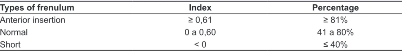
Table 3 - Distribution of patterns of classiication of the lingual frenulum in research with cadavers
Types of frenulum Index Percentage
Anterior insertion ≥ 0,61 ≥ 81%
Normal 0 a 0,60 41 a 80%
Short < 0 ≤ 40%
Table 4 - Distribution of patterns of classiication of the frenulum of the tongue suggested in
research on living human beings
Types of frenulum Index Percentage
Anterior insertion ≥ 0,41 ≥ 71%
Normal -0,20 a 0,40 40 a 70%
Short ≤ -0,21 ≤ 39%
According to the standard set in the proposal by index and percentage in study done in cadavers, did not were found frenulum with anterior insertion, where the value would be, according to the proposal by index, greater than or equal to 0.61, and in the proposal by percentage greater or equal to 81%. Thus, the pattern described in Table 4 refers to the borderline value found in the normalcy.
The standard established by Oliveira, Lindoso; Coelho (2010) 19 refers to altered frenulum (short), with lower values than the standard normalcy, in other words, altered frenulum in the index should present itself less than 0 (zero) and in the percentage classiication, equal to or less than 40%.
The average of the results obtained (Table 2) is in the classiication of the frenulum as normal, but as in males the results were lower, it is noted that they are closer to altered frenulum than in females. It is not possible to afirm that these data extend to all subjects by the signiicant difference in the number of participants, being 219 females and 68 males.
Found in the literature, studies report a higher incidence of abnormal frenulum in males14,20. Thus, even without signiicant evidence and the average frenulum, in both variables do not classify as altered in accordance with the model proposed by Oliveira, Lindoso and Coelho (2010) 19. Results show a trend towards the existence of difference of measures of altered frenulum according to sex.
It is noteworthy that to make measurements of the lingual frenulum presented here, is required the contribution of the patient, since him should be able to understand and execute a command.
Regarding the measurements made in this study, only eight of the 287 participants reported discomfort when performing the examination, cited as tongue position, discomfort in the temporomandibular joint (TMJ) and dificulty in opening the mouth.
In the item about perception of the test, 249 participants reported tranquility, 04 participants reported being uncomfortable, 6 participants reported tiring, and 43 considered the test as rapid. It is noteworthy that some participants chose more than one option as it could be multiple alternatives, justifying the value of the sum of the items evaluated be larger than the number of respondents. It is important to mention that the perception of limited mouth opening did not exist by the patient before the exam, which justiies its inclusion in the study and the previously reported perception.
Considering the responses obtained, it is observed that the proposed method is feasible from the perspective of the evaluator and evaluated.
CONCLUSION
In the proposal by percentage is possible to apply the standard measures suggested in the study with cadavers. As regards the proposal by index, the pattern found in the cadavers must be replaced by new standard measures of living humans observed in this work.
So we suggest the use of both classiications in clinical practice, thus beneiting the patient, as it will be assessed by a standardized and safe method, ensuring greater diagnostic accuracy for both the
evaluator and evaluated.
Because the measurements in soft structure, it is up to the evaluator professional complement their evaluation with qualitative methods and clinical history of the patient.
REFERENCES
1. Melo NSFO, Lima AAS, Fernandes A, Silva RPGVC. Anquiloglossia: Relato de caso. Rev
Sul-Bras Odontol. 2011;8(1):102-7.
2. Marchesan IQ. Frênulo lingual: proposta de avaliação quantitativa. Rev CEFAC.
2004;6(3):288-93.
3. Marchesan IQ, Teixeira AN, Cattoni DM. Correlações entre diferentes frênulos linguais e alterações na fala. Rev Disturb Comun.
2010;22(3):195-200.
4. Vargas BC, Monnerat, LHP, Favilla EE, Pinto LAPF, Gandelmann IHA, Cavalcante MAA. Anquiloglossia: quando indicar a frenectomia lingual? Rev Uningá.
2008;(18):195-204.
5. Silva MC, Costa MLVCM, Nemr K, Marchesan IQ. Frênulo de língua alterado e interferência na mastigação. Rev CEFAC. 2009;11(3):363-9.
6. Brito SF, Marchesan IQ, Bosco CM, Carrilho ACA, Rehder MI. Frênulo lingual: classiicação e conduta segundo ótica fonoaudiológica, odontológica e otorrinolaringológica. Rev CEFAC.
2008;10(3):343-51.
7. Comitê de Motricidade Orofacial da Sociedade Brasileira de Fonoaudiologia. Documento oicial 04/2007. São Paulo: Sociedade Brasileira de Fonoaudiologia; 2007.
8. Marchesan IQ. Frênulo de língua: classiicação e interferência na fala. Rev CEFAC. 2003;5(4):341-5. 9. Braga LAS, Silva J, Pantuzzo CL, Motta AR. Prevalência de alteração no frênulo lingual e suas implicações na fala de escolares. Rev CEFAC.
2009;11(3):378-90.
10. Corrêa MSNP, Alvarez JA, Corrêa FNP, Bonini GAVC, Alves FBT. Anquiloglosia y amamantamiento: Revisión y reporte de caso. Rev Estomatol Herediana. 2008;18(2):123-7.
11. Advíncula CEE, Paz MP. Frenillo Lingual: cuándo es un problema? Rev Odontol Pediatr.
2010;9(1):71-7.
12. Gonçalves CS, Ferreiro MC. Estudo da relação entre presença de frênulo lingual curto e/ ou anteriorizado e a dorsalização do fone [r] na articulação da fala. Rev CEFAC. 2006;8(1):56-60. 13. Marchesan IQ. Alterações de fala de origem musculoesquelética. In: Ferreira LP, Bei- Lopes DM, Limongi SCO. Tratado de fonoaudiologia. São Paulo: Roca; 2004. p. 292-303.
14. Marchesan IQ, Rehder MIBC, Oliveira LR, Araújo RLT, Costa MLVCM, Martinelli RLC. Incidência de alterações de frênulo da língua em uma população de crianças de 1ª a 3ª série do ensino fundamental. 16º Congresso Brasileiro de Fonoaudiologia, SBFa; 24-27 de setembro; Campos do Jordão. São Paulo;
2008.
15. Marchesan IQ. Protocolo de avaliação do frênulo da língua. Rev CEFAC. 2010;12(6):977-89.
16. Silva HA, Bordon AKCB, Junqueira JLC, Miyamura ZY. Frenectomia Lingual: cirurgia em odontopediatria. Rev Ortodontia. 2002;157-60. 17. Lee SK, Kim YS, Lim CY. A pathological consideration of ankyloglossia and lingual myoplasty. Taehan Chikkwa Uisa hyophoe Chi.
1989;27(3):287-308.
18. Kotlow LA. Ankyloglossia (tongue-tie): a diagnostic and treatment quandary. Quintessence
Int. 1999;30(4):259-62.
RESUMO
Objetivo: veriicar a aplicabilidade clínica das propostas de classiicação do frênulo da língua por
índice e porcentagem. Métodos: a casuística desta pesquisa foi composta por 287 sujeitos de ambos
os sexos com idade variando entre 23 e 28 anos. Os frênulos da língua foram classiicados a partir de duas medidas deinidas com o auxilio de compasso e de paquímetro digital, e analisadas nas fórmu
-las e padrões propostos a partir de uma pesquisa realizada com cadáveres que resultou em fórmu-las para mensurar o frênulo da língua por índice e porcentagem. Resultados: observou-se que o padrão
estabelecido na classiicação do frênulo da língua pela proposta por porcentagem é idedigno. Na proposta por índice um novo padrão foi encontrado na população pesquisada. Conclusão: após apli
-cação clínica, sugere-se que um novo padrão na classii-cação por índice seja adotado. Na proposta por porcentagem o padrão inicial deve ser mantido, proporcionando a efetividade de ambas as formas de classiicação do frênulo da língua na prática fonoaudiológica.
20. Vieira EMM, Salineiro FS, Hespanhol D, Musis CR, Jardim Junior EG. Frequência de anquiloglossia em uma comunidade indígena brasileira. Rev Gaucha Odontol. 2010;58(2):215-8.
19. Oliveira IC, Lindoso RL, Coelho TTT. Comprimento Anatômico e Funcional do Frênulo da Língua. [Monograia]. Itajaí (SC): UNIVALI; 2010.
Received on: September 17, 2012 Accepted on: January 24, 2013
Mailing address: Tânia T. Tozi Coelho
Rua Uruguai, 458 – Bloco F5
Itajaí – SC
CEP: 88302-202