Pulmonary embolism: multicenter registry in
tertiary hospitals
Embolia pulmonar: registro multicêntrico da prática clínica em
hospitais terciários
INTRODUCTION
Pulmonary embolism (PE) is a challenge to clinical practice. Presence of risk factors for venous thromboembolism associated with signs and symptoms of PE is the initial condition to raise clinical suspicion. he nonspeciicity of clinical manifestations and limited availability of complementary methods, the main obstacles to diagnosis, distort epidemiological assessments, generat-ing approximate and unreliable results. Complementary tests are essential for diagnosis of patients with clinical suspicion and to evaluate prognosis of those with an already diagnosed EP.
André Volschan1, Denílson Campos
de Albuquerque2, Bernardo Rangel
Tura3, Marcos de Freitas Knibel4,
Paulo César Pereira da Silva e Souza5, Maria Luiza Toscano6 e
Investigadores do EMEP – Estudo Multicêntrico de Embolia Pulmonar
1. Master, Physician at the Emergency Unit of Hospital ProCardíaco, Rio de Janeiro (RJ), Brazil.
2. PhD, Adjunct Professor of
Cardiology, Universidade do Estado do Rio de Janeiro UERJ - Rio de Janeiro (RJ), Brazil.
3. PhD, Physician at the Instituto Nacional de Cardiologia - Rio de Janeiro (RJ), Brazil.
4. Master, Physician at the Intensive Care Center, Hospital São Lucas, 5. Physician at the Intensive Care Center, Hospital de Clínicas de Niterói, Niterói (RJ), Brazil.
6. Physician at the Hospital Municipal Miguel Couto - Rio de Janeiro (RJ), Brazil.
ABSTRACT
Introduction: he clinical proile as well as the therapeutic and diagnostic strategies for patients with pulmonary embolism, describes clinical practice in the approach of the disease. Such in-formation, scarce in national studies, enables a better understanding of pul-monary embolism.
Methods: A multicenter trial in-cluded 727 patients with pulmonary embolism who were admitted in emer-gency or intensive care unit. Diagnostic criteria for inclusion were: 1. Visibility of thrombus in the pulmonary artery at pulmonary arteriography, helical com-puter tomography, magnetic resonance or echocardiogram. 2. High probability at pulmonary scintigraphy. 3. Venous duplex-scan with thrombus and clini-cal manifestations of pulmonary em-bolism. Clinical and complementary exams were analyzed.
Results: Mean age was 68 years, 42% were male. Most prevalent risk factors were: age>40 years, bed rest and neoplasm. More frequent signs
and symptoms were: dyspnea, tachyp-nea, sinus tachycardia, and chest pain. Changes were observed at electro-cardiogram in 30%, at chest X-ray in 45%, at venous duplex-scan in 67%, at transthoracic echocardiogram in 37%. . D-dimer, troponin I and CKMB were positive in, respectively, 93, 9 and 8%. Most frequently used methods to con-irm diagnosis were helical computer tomography and non-fractioned hepa-rin was the treatment most used. In-hospital mortality was 19.5%.
Conclusions: It was observed that age>40 years, prolonged rest and neo-plasms were the most prevalent risk fac-tors and dyspnea and tachypnea were the more frequent clinical manifesta-tions. Helical computer tomography was the most often used method to conirm diagnosis and non-fractioned heparin was the main form of treat-ment.
Keywords: Pulmonary embolism/ diagnosis; Pulmonary embolism/drug therapy; Heparin/therapeutic use; To-mography, X-ray computed/methods
Received fromEmergency Unit of Hospital ProCardíaco, Rio de Janeiro (RJ), Brazil.
Submitted on July 29, 2009 Accepted September 23, 2009
Author for Correspondence
André Volschan
Rua General Polidoro, 192 - Botafogo CEP: 22471-270 - Rio de Janeiro (RJ), Brazil.
he standard treatment with unfractionated heparin has as alternative low molecular weight heparin (LMWH) and thrombolytic therapy, the irst being restricted to stable patients and the second given to patients with he-modynamic instability.(1) Although the therapeutic op-tion following general rules, it may present variaop-tions for certain subgroups of patients.
he scarcity of records representing clinical practice was a stimulus for this study. here are few Brazilian publications on the subject and those with the largest casuistry are relating to necropsy studies or those carried out by medical chart review.(2,3)
he observational study that aims to assess the clini-cal proiles of the diagnosis and treatment strategies is the one that best portrays the reality of clinical practice. With this type of evaluation results will not be subject to interferences generated in controlled studies, with close surveillance of the patients included.
Based upon this premise, a record was designed to analyze the clinical proile and the diagnostic s and treat-ment approach for patients with pulmonary embolism.
METHODS
Multicenter prospective cohort study conducted in 20 research centers from tertiary hospitals in Brazil (Chart 1), from January 1998 to May 2003 (EMEP-
Chart 1- Research centers
Investigating center City State
Hospital Procardíaco* Rio de Janeiro Rio de Janeiro
Hospital Samaritano Rio de Janeiro Rio de Janeiro
Hospital RioMar Rio de Janeiro Rio de Janeiro
Clínica São Vicente Rio de Janeiro Rio de Janeiro
Casa de Saúde São José (CU) Rio de Janeiro Rio de Janeiro
Casa de Saúde São José (ICU) Rio de Janeiro Rio de Janeiro
Hospital São Lucas (CU) Rio de Janeiro Rio de Janeiro
Hospital São Lucas (ICU) Rio de Janeiro Rio de Janeiro
Hospital Cardiotrauma Rio de Janeiro Rio de Janeiro
Hospital Prontocor Rio de Janeiro Rio de Janeiro
Hospital do Câncer Rio de Janeiro Rio de Janeiro
Hospital Barra D’Or Rio de Janeiro Rio de Janeiro
Hospital Copa D’Or Rio de Janeiro Rio de Janeiro
Hospital São Vicente de Paulo Rio de Janeiro Rio de Janeiro
Hospital de Clínicas de Niterói Niterói Rio de Janeiro
Hospital Mario Lioni Duque de Caxias Rio de Janeiro
Hospital Português Salvador Bahia
Hospital Prontocor Belo Horizonte Minas Gerais
Hospital São Lucas Porto Alegre Rio Grande do Sul
Santa Casa da Misericórdia Santos São Paulo
*Coordinating center CU – Coronary unit; ICU - Intensive care unit
Multicenter Study of Pulmonary Embolism).
Clinical suspicion was deined by the physician as-sessing the patient, based upon risk factors, signs and symptoms of PE. In addition to the clinical suspicion documentation on EP by one or more of the following complementary methods was necessary:
Angiography with visualization of the thrombus in pulmonary artery.
Helical computed angiotomography with visualiza-tion of the thrombus in the pulmonary artery.
Magnetic resonance angiography with visualization of the thrombus in the pulmonary artery.
Echocardiogram with visualization of the thrombus in the pulmonary artery.
Pulmonary ventilation / perfusion with high prob-ability of pulmonary embolism.
Duplex-scan with visualization of the thrombus and reduced compressibility in the deep venous system.
In-hospital mortality was deined as that occurring in the same hospitalization that motivated admission by EP, regardless of the cause of death.
, smoking, cardiac failure, stroke, chest pain, tachycar-dia (heart rate> 100 bpm), syncope, dyspnea, tachypnea (respiratory rate > 20 bpm), fever (axillary temperature> 37°C), coughing, cyanosis and hemoptysis and/or he-mopics.
In the multicenter registry, data on the electrocardio-gram, chest X-ray, d-dimer, troponin I, the creatinine kinase MB, the duplex of the lower limbs and echocar-diography were collected and analyzed about frequen-cies of completion and percentage of positivity. Were considered as changes in the standard electrocardiogram S1Q3T3 (presence of S wave in V1 derivation and pres-ence of Q wave and inverted T wave in lead D3), known as McGuinn - White standard, T-wave inversion in leads V1 to V4, right bundle branch block or QRS axis de-viation to the right, changes related to right ventricular overload.(4)
At chest X-ray, presence of pleural efusion, iniltrates or pulmonary atelectasia were the changes observed.(5) In relation to troponin I and the d-dimer, the cut- of point was accepted for positivity of each surveyed center and in this case, three diferent methods (ELISA, or latex or immunoturbidimetry were used depending on the avail-ability at each center. Visualization of the thrombus or reduction of venous compressibility was the factor that deined the change in venous duplex scan,(6) and the presence of dilation or right ventricular dysfunction cor-related with echocardiography.(7)
Use of unfractionated heparin, intravenous unfrac-tionated subcutaneous heparin, low molecular weight subcutaneous heparin, , thrombolytic agents (rt-PA or streptokinase), coumarin, vena cava ilter and surgical embolectomy. he mortality rate was analyzed in the irst 24 hours of diagnosis and during hospital stay.
Data was statistically analyzed using: tables contain-ing frequency distributions, percentages, averages, me-dians, standard deviations, minimum and maximum values. Continuous variables were reported as mean ± standard deviation.
he study was approved by the Ethics Research Com-mittee of the Pro-Cardiac Hospital, according to Resolu-tion 196/96 and 251/97 of the NaResolu-tional Health Coun-cil. he original database (EMEP Study - Multicenter Study of Pulmonary Embolism) was partially funded by the Sanoi - Aventis Laboratory.
RESULTS
We evaluated 727 patients with a mean age of 68.9 ± 15.8 years, and 421 (57.9%) were female.
More prevalent risk factors were age over 40 years (93.4%) and bed rest> 72 h (38.5%), in 24.3% of pa-tients. there was presence of neoplasm. Prevalence of risk factors was documented in all patients included, as de-scribed in table 1.
Table 1 – Prevalence of risk factors
Risk factor N %
Age > 40 years 679 93.4
Bed rest > 72h 280 38.5
Neoplasm 177 24.3
Previous history of DVP / PE 119 16.4
Smoking 115 15.8
CCF 112 15.4
Abdominal or pelvic surgery 70 10.7
Hip/lower limb fracture < 90 dias 52 7.2
Stroke 46 6.3
Chronic cor pulmonale 44 6.1
Estrogen use 33 7.8*
Pregnancy and postpartum 3 0.7*
CCF - Congestive cardiac failure; DVT/PE Deep venous thrombosis or pulmonary thrombolisis; * percentage relating to female gender pa-tients.
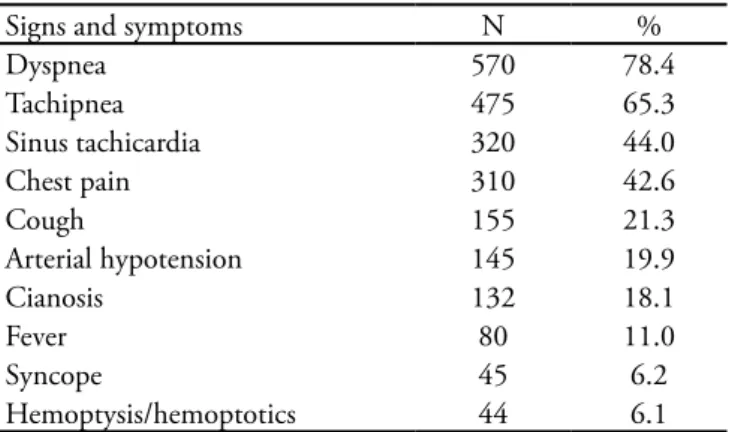
Dyspnea (78.4%) and tachypnea (65.3%) - were the most frequent clinical respiratory manifestations while 44% of patients had tachycardia and 42.6%. Chest pain Indicators of hemodynamic instability - such as hypotension and syncope were observed re-spectively in 19.9% and 6.2% of the sample, as shown in table 2.
Table 2 – Prevalence of signs and symptoms
Signs and symptoms N %
Dyspnea 570 78.4
Tachipnea 475 65.3
Sinus tachicardia 320 44.0
Chest pain 310 42.6
Cough 155 21.3
Arterial hypotension 145 19.9
Cianosis 132 18.1
Fever 80 11.0
Syncope 45 6.2
Hemoptysis/hemoptotics 44 6.1
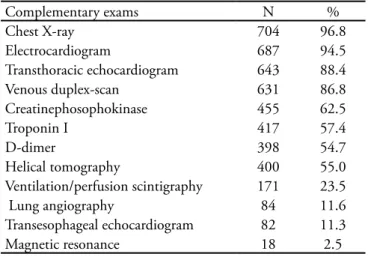
78 (35.1%) standard S1Q3T3, 78 (35.1%) wave “T” inversion in leads V1 to V4 and 108 (48.6%) right bundle branch block. Chest X-ray showed specific changes in 333 (45.8%) of which, 128 (38.4%) had pleural effusion, 69 (20.7%) atelectasis, 31 (9.3%) area of hypoperfusion and 201 (60.3%) pulmonary infiltrates. Determination of d-dimer was positive in 93.2%, CK-MB in 8.39% and troponine in 9.63% of the samples. The venous duplex scan with specific changes, visualization of thrombus or reduction of ve-nous compressibility, was found in 426 (67.5%) and echocardiogram showed right ventricular dysfunction in 182 (36.5%) and TEE in 78, 1% of the exams. Methods considered to confirm diagnosis are listed in table 4.
Table 3 – Frequency of complementary exams
Complementary exams N %
Chest X-ray 704 96.8
Electrocardiogram 687 94.5
Transthoracic echocardiogram 643 88.4
Venous duplex-scan 631 86.8
Creatinephosophokinase 455 62.5
Troponin I 417 57.4
D-dimer 398 54.7
Helical tomography 400 55.0
Ventilation/perfusion scintigraphy 171 23.5
Lung angiography 84 11.6
Transesophageal echocardiogram 82 11.3
Magnetic resonance 18 2.5
Table 4- Determinant criteria for inclusion
Complementary exams %
Helical computed tomography 47.2
Lung scintigraphy 14.0
Vernous duplex-scan 14.0
Echocardiogram 12.7
Lung angiography 9.1
Magnetic resonance 1.7
Of the 727 patients analyzed, 99.2% underwent some form of treatment. Untreated died between con-irmation of diagnosis and onset of therapy. Heparins (unfractionated heparin and low molecular weight) were the most often used form of treatment, with 7 patients treated with placement of a vena cava ilter. In these patients the procedure was established because of their previous use of coumarin. One patient treat-ed only with coumarin, which was already using the drug, the assisting physician did not elect to place the
vena cava ilter. Table 5 shows frequency of each type of treatment use. Mortality rate within 24 hours of ad-mission and during hospital stay was respectively 3.4% and 19.5%.
Table 5 – Type of treatment used
Treatment %
Nonfractionated heparin IV 50.2
Low m olecular weight heparin 29.2
hrombolytic 11.7
Vena cava ilter 3.3
Nonfractionated heparin SC 2.6
Cumarin 1.0
Embolectomy 0.1
Others 0.5
Not treated 0.8
IV- intravenous; SC - sub-cutaneous
DISCUSSION
Findings of this study demonstrate the clinical prac-tice in the approach to PE in which risk factors, clini-cal manifestations, diagnosis and therapy were assessed. he study was limited to document data, without any interference in medical conducts employees, thus depict-ing the care given to patients with EP. he technological availability of research centers in the study was the basis for laboratory tests, vital for investigation of EP. Inclu-sion criteria, which required anatomic or functional sub-stantiation of pulmonary embolism, although limiting the number of patients, warranted more accurate diag-nosis.
Patients included were admitted to coronary emer-gency rooms, or coronary intensive care in tertiary cen-ters which may have led to select the more severe cas-es. he mean age observed in our study was 68.9 years therefore higher than those observed in the ICOPER(8) (62.3 years), MAPPET(9) (63.5 years) and JASPER (60 years)(10) studies. Most studies show a tendency towards even distribution by gender. In our study we observed a higher prevalence of the n female gender (57.9%), which was also reported in ICOPER (55%)(8) and JASPER (60.5%),(10) but not in the MAPPET study (41%).(9)
he emergence of endothelial degenerative changes - associated with presence of diseases that predispose to thrombus formation, more frequent in patients over 40 years of age - explains the high prevalence of this risk fac-tor in the EP. With the increasing life expectancy of the population, age should become an increasingly prevalent risk factor.
he importance of the muscle pump action of to maintain adequate venous returncan be assessed by the high prevalence of restraint to prolonged bed rest in pa-tients with EP.
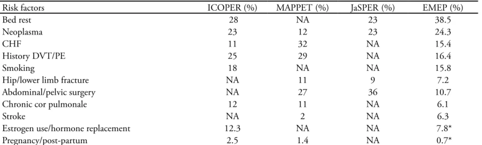
he tumor able to promote activation of the coagula-tion system(13) was detected in almost one quarter of cases, corroborating data of the studies PIOPED (18.3%),(14) ICOPER (22.5%)(8) and JaSPER (23%),(10) as shown in table 6. his high prevalence promotes discussion on the need of routine investigation of cancer in patients with EP, as well as strategies of a comprehensive primary pre-vention of thrombosis.
he endothelial changes caused by a irst episode of deep vein thrombosis (DVT) predispose to disease re-currence. Previous occurrence of DVT or PE had been reported in 14% of our cases, 25% in ICOPER(8) and 29% in MAPPET.(9) hese observations indicate possible beneit of continued use of elastic stockings for such pa-tients.
A hip or lower limb fracture was documented in 9% of the sample, similar to JaSPER (9%)(10) and MAPPET (11%).(9) Such similarities are not repeated in the history of abdominal or pelvic surgery, with prevalence of 8% in our sample and 36% and 27% respectively, in the other two reports. his diference can be explained by the
char-Table 6- Comparison between prevalences of risk factors
Risk factors ICOPER (%) MAPPET (%) JaSPER (%) EMEP (%)
Bed rest 28 NA 23 38.5
Neoplasma 23 12 23 24.3
CHF 11 32 NA 15.4
History DVT/PE 25 29 NA 16.4
Smoking 18 NA NA 15.8
Hip/lower limb fracture NA 11 9 7.2
Abdominal/pelvic surgery NA 27 36 10.7
Chronic cor pulmonale 12 11 NA 6.1
Stroke NA 2 NA 6.3
Estrogen use/hormone replacement 12.3 NA NA 7.8*
Pregnancy/post-partum 2.5 1.4 NA 0.7*
CHF- Congestive heart failure; DVT/PE - Deep venous thrombosis /pumonary thrombolisis; * percentage related to female gender patients; NA - not available; ICOPER - International Cooperative Pulmonary Embolism Registry; MAPPET - Management Strategy and Prognosis of Pulmonary Embolism Registry; JaSPER - Japanese Society of Pulmonary Embolism Research; EMEP - Estudo Multicêntrico de Embolia Pulmonar.
acteristics of hospitals involved in each of the studies. Congestive heart failure (CHF) and chronic pulmo-nale was present respectively in 15.4% and 6.1% of our patients and in 11% and 12% in the ICOPER study.(8) he situation in favor of thrombosis involving vein sta-sis, activation of procoagulant substances(15) and present endothelial changes suggest the possible underestimation of PE diagnosis in this population based surveys.
Prevalence of 0.7% of expectant mothers is some-what lower than those observed in ICOPER(8) (2.5%) and MAPPET (1.4%),(9) while the use of estrogen was higher in ICOPER (12.3%)(8) than in the women exam-ined in this study (7.8%). Diference in use of estrogen suggests greater use of hormone replacement therapy in the United States and Europe, where the ICOPER(8) was carried out.
he small number of patients with a history of stroke (6.3%) was also reported in MAPPET(9) (2%). he low prevalence of history of stroke, a subgroup often with multiple risk factors, may have been inluenced by the chronic use of aspirin by these patients by promoting prevention of DVT.
Smoking was reported by 15.8% of patients and quit-ting should be encouraged as it is one of the few risk factors considered avoidable.
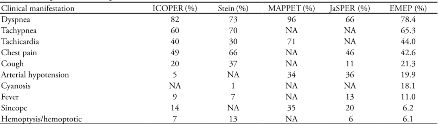
In the analysis of signs and symptoms, respiratory symptoms and chest pain, were as prevalent as in other studies.(16.17) In the JaSPER(10) dyspnea was present in 66% and chest pain in 49%, the most frequent symp-toms reported.
inlamma-tory lung diseases isolated or in sets are the irst diferen-tial diagnoses. he sudden onset of dyspnea is the feature that alerts the physician on diagnosis of PE, although the symptoms may appear gradually or aggravating chronic cardiopulmonary conditions. In 727 patients evaluated, dyspnea was present in 78.4%, similar to the result of Stein et al.(16) that demonstrated presence of dyspnea in 78% and Hoellerich et al. 74%.(17)
Pleuiritic chest pain is more correlated with the EP, but e angina pain may occur in patients with ischemia of the right ventricle (RV). Pleuritic chest pain was re-ported in 85% of patients in the Bell et al.,(18) study and 66% in the PIOPED study.(14) In this study a prevalence of 42.6% of chest pain was found, similar to the indings of ICOPER(8) (49%) and JaSPER(10) (46%).
Clinical signs of hemodynamic instability were pres-ent in one ifth of patipres-ents evaluated with hypotension observed in 19.9%, regardless of volume replacement. In the main studies, prevalence of forms of presentation diverge greatly as shown in table 7. In ICOPER(8), only 5% had arterial hypotension, while MAPPET(9) and JaS-PER(10) showed higher prevalence that our observations. Notwithstanding these diferences there is agreement that this is a greater severity subgroup, for which throm-bolytics or embolectomy may be indicated.(1)
Cough was present in approximately 22% of our pa-tients, with lesser prevalence than in the two studies of Stein et al. that documented it in 37% and 55% of pa-tients.(16,19) Hemoptysis was observed in 6.1%, while in 7% of patients in ICOPER.(8) he low occurrence of he-moptysis suggests a lower number of cases of pulmonary infarction in the sample.
Fever was observed in 11%, similar to the 14% re-ported in all the sub-studied by PIOPED,(14)
demon-strating the importance of diferential diagnosis of PE in inlammatory lung diseases.(20)
Overall, prevalence of signs and symptoms of PE is similar in most studies as exempliied in table 7.
he electrocardiogram, chest x-ray, transthoracic echocardiogram and venous duplex scan of lower limbs were carried out n more than 90% of patients studied. Laboratory evaluation of d-dimer, creatinine kinase, tro-ponin I, blood gases, in more than half of the cases, al-lowed analysis of large volumes of data. Compared to the ICOPER,(8) study, less scintigraphy and angiography were made as seen in, table 8, a condition explained by the widespread use of helical computed tomography for diagnosis in our sample.
Table 8 – Percentage of exams performed
Exam ICOPER (%) MAPPET (%) EMEP (%)
Chest X-ray 95 NA 96.5
Electrocardiogram 90 98 98.5
Cintilograia 84* 56 21.2
Echocardiogram 47 74 95.5
D-dímer (Latex) 21 NA 48.5
D-dimer (ELISA) 18 NA 21.2
Lung angiography 19 17.5 11.3
*only perfusion scintigraphy. NA- not available; ICOPER - Internatio-nal Cooperative Pulmonary Embolism Registry; MAPPET- Manage-ment Strategy and Prognosis of Pulmonary Embolism Registry; EMEP - Estudo Multicêntrico de Embolia Pulmonar.
he electrocardiogram was positive in one third of patients and only four types of changes were considered, representing RV overload. Stein(21) showed electrocardio-graphic abnormalities in 94% of cases of massive PE and 77% of submassive however the patterns of right ven-tricular hypertrophy were present in only 26%.
Table 7 – Comparison between prevalence of clinical manifestations
Clinical manifestation ICOPER(%) Stein(%) MAPPET(%) JaSPER (%) EMEP (%)
Dyspnea 82 73 96 66 78.4
Tachypnea 60 70 NA NA 65.3
Tachicardia 40 30 71 NA 44.0
Chest pain 49 66 NA 46 42.6
Cough 20 37 NA 11 21.3
Arterial hypotension 5 NA 34 36 19.9
Cyanosis NA 1 NA NA 18.1
Fever 9 7 NA 13 11.0
Síncope 14 NA 35 20 6.2
Hemoptysis/hemoptotic 7 13 NA 6 6.1
ICOPER - International Cooperative Pulmonary Embolism Registry; Stein(18); MAPPET - Management Strategy and Prognosis of Pulmonary
While radiographic changes were observed in 47.2%, the ICOPER analyzed chest X-rays of 2322 patients and found abnormal in 78%, although it as-sessed more variables. he ICOPER showed that pleu-ral efusion was present in 23%, atelectasis in 18% and areas of pulmonary iniltrates in 17%.(22) Our indings identiied pleural efusion in 17%, 10% in atelectasis and pulmonary iniltrates in 27%. Given the possibil-ity of interobserver variabilpossibil-ity and subjective analysis of changes, we consider this diference between the obser-vations acceptable.
he realization of d-dimer in suspected PE, has greater application for exclusion of diagnosis due to its high negative predictive value, as shown by Gingsberg et al. in 1998.(23) In our analysis, the technique of latex was positive in 87.7% and the ELISA in 93%, conirm-ing the higher sensitivity of the latter and corroboratconirm-ing the data of Kline et al.(24) Probably association of low clinical probability of the negative d-dimer enables ex-clusion of PE diagnosis with large safety margin.(25)
he elevation of markers of myocardial necrosis in 19.7% of samples taken was lower than expected in a group in which 37.8% had RV dysfunction. When sep-arately analyzed, creatininekinase was high n 16.9%, while troponin I in 11.2%. hese data contrast with the work of Meyer et al., who identiied 39% of patients with PE, troponin I value above the normal range.(26)
Studies using objective criteria for diagnosis of RV dysfunction on transthoracic two-dimensional difer on the methodology employed. Goldhaber et al. diagnosed the RV dysfunction by assessing the end-diastolic area measured by planimetry(27) while the study of Hamel et al. used the relationship between end-diastolic vol-ume of right and left ventricles> 0.6.(28) In our study, the subjective analysis of RV dysfunction through tow-dimensional echocardiogram may have caused bias in the results. While we recognize this methodological limitation, we believe that the information regarding the RV is in most cases, results of this subjective analy-sis. herefore, we believe we are portraying the actual practice, as proposed in this work. he percentage of patients with RV dysfunction on echocardiography was 37.8%, similar to results found in ICOPER(25) and low-er than those of Ribeiro et al.(29) Choice of the method was directly inluenced by the availability and the clini-cal practice of each investigator’s center. Considered a standard, the pulmonary arteriography substantiated diagnosis in 11.3% while scintigraphy in 21.2% and helical tomography (CT) in 44.5% of the patients. Pa-tient inclusion by the venous duplex scan criterion with
thrombus and the presence of signs or symptoms of PE may generate questions about t absence of evidence of disease in the lung. he incorporation of this criterion was deined by the embolic potential of DVT and the high probability of PE, when associated with clinical manifestations.
Although available in most centers participating in the study, the AGP is still little used. If in the past, technical limitations and risk of complications of pul-monary arteriography are not often requested, the main reason is, today, the possibility of diagnosis through less complex exams.
A scintigraphic study of ventilation and perfusion was the diagnostic method for co-21.2%, and similarly to the AGP, the low utilization can be explained by the expand use of spiral CT.
Spiral CT has a sensitivity between 66 and 93% and speciicity of 89 to 97%,(30) furthermore, enable the eval-uation of diferential diagnosis of PE, such as acute dis-eases of the aorta, pulmonary inlammations and pneu-mothorax. hese characteristics have made the spiral CT exam I mandatory in latest diagnostic algorithms.
MRI remains underused, mainly due to lower avail-ability of the-helical CT. he diagnostic accuracy simi-lar to CT and the use of contrast without risk of neph-rotoxicity or anaphylaxis should encourage greater use of MRI for diagnosis of PE in the near future.
he HNF is the most widely used treatment in our patients (55.1%). he proven eicacy of UFH and low risk of bleeding make this therapeutic strategy most of-ten employed for EP.
LMWH has proved as efective as UFH in the treat-ment of EP, as well as its advantages, such as subcuta-neous administration and no need for laboratory con-trol.(31) hese characteristics are increasing in use of this therapeutic strategy in the EP, and this study, LMWH was used in 23.7% of patients. LMWH should be only be used for clinically stable patients without RV dys-function and treated in hospital setting.
hrombolytic treatment was used in 15.2% of pa-tients in this study. he inding that 20.8% had hy-potension suggests that we may be underdoing the thrombolytic agent in our patients. In the JaSPER(10) the thrombolytic was administered to 50% of patients in the sample, in which 36% had cardiogenic shock. Goldhaber,(32) in an editorial, reviews results of the main publications and reinforces the need for more evidence to stresses thrombolytic therapy for clinically stable pa-tients and RV dysfunction, such as submassive PE.
throm-bolytic therapy approved by the FDA for treatment of PE may vary, depending on clinical presentation. he only study comparing heparin with thrombolytic thera-py, which showed a reduction in mortality, used dose of streptokinase 1.500.000U,(33) diferent from the scheme approved by the FDA.
he intravenous route is the most used for the ad-ministration of thrombolytic therapy in patients with EP. he patients undergoing pulmonary arteriography as the diagnostic method may use thrombolytic agents directly into the pulmonary artery. his strategy re-quires a lower dose of the drug and may be indicated for hemodynamically unstable patients with high risk of bleeding.
Probably, just as today we stratify patients for use of heparin or thrombolytic therapy in the future we may deine doses and administration routes depending on the patient and on the presentation of disease.
Treatment of PE should not only be deined by clinical instability or dysfunction of the RV, but also by variables such as age, presence of proximal DVT and previous cardio respiratory condition. A possible subtilization of thrombolytics in this study is suggested when comparing percentage of patients with hypoten-sion or syncope (25.8%) with use of thrombolytic ther-apy (15.2%).
In-hospital mortality of 22.7% is higher than the ICOPER study(8) formerly reported 17.5% of deaths in three months, and in the group with clinical instability, the mortality rate was 58.3%. Kasper et al. showed a steady increase in mortality according to worsening of the hemodynamic status.(9)
he assessed sample was of patients admitted to emergency rooms and intensive care and advanced age which may relect a selection bias, regarding severity of the population studied. Among the deaths observed, 54.3% occurred in the irst seven days of stay. his sug-gests that almost half of the patients had complications of the EP’s, which led to longer hospital stays, these complications may have contributed to death.
Finally, this study was limited to descriptive infor-mation, and the intention to establish a correlation be-tween the variables studied was never intended. With the continuation of the project certainly these reviews will be carried out.
his study has some limitations. he research cen-ters that participated in the study have the structure for diagnosis and treatment above the average of the Brazilian hospitals. herefore, this study demonstrates the clinical practice of qualiied centers. Data from
ad-ditional tests were included according to diagnosis of the researcher; a center for independent validation of them was not available. he patients were admitted in emergency and intensive care units, which may have led to selection of a population of greater severity.
CONCLUSIONS
Presence of multiple risk factors was common in pa-tients with PE, where age over 40 years and bed rest were the most prevailing, while dyspnea and tachyp-nea were the most common clinical manifestations ob-served. Among the complementary methods, classical research strategies in percentages in some cases, higher than the average described in literature. he most wide-ly used diagnostic criteria for inclusion by researchers was helical CT angiography also reproducing a tenden-cy of diagnostic studies and heparin the most often pre-scribed treatment following the guidelines of therapeu-tic consensus. Perhaps, in-hospital mortality of 19.5%, slightly higher than in most studies published, is due to characteristics of the greater severe in the population admitted to intensive care units.
ACKNOWLEDGMENTS
We appreciate the participation of researchers from the EMEP (Multicentric Study of Pulmonary Embo-lism)) for collecting study data
Jose Pericles Esteves and Carolina Barbosa (Portuguese Hospital, Salvador, BA), Francisco Silveira (Prontocor Hospital, Belo Horizonte, MG), Alex Macedo (Hos-pital Santa Casa, Santos, SP), Luis Carlos Bodanese (Hospital Sao Lucas, Porto Alegre, RS).
RESUMO
Introdução: O peril clínico e as estratégias diagnósticas e terapêuticas nos pacientes com embolia pulmonar demons-tram a prática clínica na abordagem da doença. Essas infor-mações, escassas nos estudos nacionais, possibilitam melhor conhecimento da embolia pulmonar.
Métodos: Estudo multicêntrico de 727 pacientes admiti-dos em unidades de emergência ou terapia intensiva, com o diagnóstico de embolia pulmonar conirmado por um ou mais dos seguintes exames: arteriograia pulmonar angiotomograia computadorizada helicoidal angioressonância magnética, eco-dopplercardiograma, cintilograia pulmonar ou duplex-scan venoso. Dados demográicos, comorbidades, manifestações clínicas e métodos complementares foram analisados.
Resultados: A média de idade foi 68 anos, sendo 42%
homens. Os fatores de risco mais prevalentes foram: idade > 40 anos, repouso no leito e neoplasia. A dispnéia, taquipnéia, taquicardia, dor torácica, foram as manifestações clínicas mais frequentes. O eletrocardiograma apresentou alterações em 30%, a radiograia de tórax em 45%, o duplex-scan venoso em 69% e o ecodopplercardiograma em 37%. O D-dímero a troponina e a CKMB foram positivos em respectivamente 93, 9 e 8%. Os métodos mais utilizados para o diagnóstico foram: tomograia computadorizada: 47%, duplex-scan ve-noso: 14% e cintilograia pulmonar: 14%. As formas mais freqüentes de tratamento foram: heparina não fracionada 50%, heparina de baixo peso molecular 30% e trombolítico 12%. A mortalidade intra-hospitalar foi de 19,5%.
Conclusões: Observou-se que a idade > 40 anos, imobi-lização prolongada e neoplasia foram os fatores de risco de maior prevalência e a dispnéia a apresentação clínica mais freqüente. A angiotomograia computadorizada helicoidal foi o método mais utilizado para o diagnóstico e a heparina não fracionada a principal forma de tratamento.
Descritores: Embolia pulmonar/diagnóstico; Embolia pulmonar/quimioterapia; Heparina/uso terapêutico; Tomo-graia computadorizada por raios-x/métodos
REFERENCES
1. Torbicki A, Perrier A, Konstantinides S, Agnelli G, Galiè
N, Pruszczyk P , Bengel F, Brady AJ, Ferreira D, Janssens U, Klepetko W, Mayer E, Remy-Jardin M, Bassand JP, Vahanian A, Camm J, De Caterina R, Dean V, Dickstein K, Filippatos G, Funck-Brentano C, Hellemans I, Kristen-sen SD, McGregor K, Sechtem U, Silber S, Tendera M, Widimsky P, Zamorano JL, Zamorano JL, Andreotti F, As-cherman M, Athanassopoulos G, De Sutter J, Fitzmaurice D, Forster T, Heras M, Jondeau G, Kjeldsen K, Knuuti J, Lang I, Lenzen M, Lopez-Sendon J, Nihoyannopoulos P, Perez Isla L, Schwehr U, Torraca L, Vachiery JL; Task Force for the Diagnosis and Management of Acute Pulmonary Embolism of the European Society of Cardiology. Guideli-nes on the diagnosis and management of acute pulmonary embolism: the Task Force for the Diagnosis and Mana-gement of Acute Pulmonary Embolism of the European Society of Cardiology (ESC). Eur Heart J. 2008; 29(18): 2276-315.
2. Menna-Barreto S, Cerski MR, Gazzana MB, Stefani SD,
Rossi R. Tromboembolia pulmonar em necropsias no Hos-pital de Clínicas de Porto Alegre, 1985-1995. J Pneumol. 1997;23(3):131-6.
3. Mesquita CT, Morandi Júnior JLB, Perrone FT, Olivei-ra CS, BarreiOlivei-ra LJ, Nascimento SSCA, et al. Diagnóstico clínico versus conirmaçäo patológica de embolia pulmo-nar fatal em pacientes hospitalizados. Arq Bras Cardiol. 1999;73(3): 251-8.
4. Abecasis J, Monge J, Alberca D, Grenho MF, Arroja I, Aleixo AM. Electrocardiographic presentation of massive and submassive pulmonary embolism. Rev Port Cardiol. 2008;27(5):591-610.
5. Greenspan RH, Ravim CE, Polansky SM, and McLoud T.
Accuracy of chest radiography in diagnosis of pulmonary embolism. Invest Radiol. 1982; 17(6): 539-43.
6. Lensing AW, Prandoni P, Brandjes D, Huisman PM, Vigo
M, Tomasella G, et al. Detection of deep-vein thrombo-sis by real-time B-mode ultrasonography. N Engl J Med. 1989;320(6):342-5.
7. Torbicki A, Pruszczyk P. he role of
echocardiogra-phy in suspected and established PE. Semin Vasc Med. 2001;1(2):165-74.
8. Goldhaber SZ, Visani L, De Rosa M. Acute pulmonary embolism: clinical outcome s in the International Coope-rative Pulmonary Embolism Registry (ICOPER). Lancet. 1999;353(9162):1386-9.
9. Kasper W, Konstatinides S, Geibel A, Olschewski M,
Heinrich F, Grosser KD, et al. Management strategies and determinants of outcome in acute major pulmonary em-bolism: results of a multicenter registry. J Am Coll Cardiol. 1997;30(5):1165-71.
Kline JA. Frequency of thrombophilia-related genetic variations in patients with idiopathic pulmonary em-bolism in an urban emergency department. Clin Chem. 2006;52(6):1026-32.
12. Anderson FA Jr, Wheller HB, Goldberg RJ, Hosmer DW, Patwardhan NA, Jovanovic B, et al. A population-based perspective of the hospital incidence and case-fatality rates of deep vein thrombosis and pulmonary embolism. he Worcester DVT Study. Arch Intern Med. 1991;151(5): 933-8.
13. Luzzatto G, Schafer Al. he prethrombotic state in cancer. Semin Oncol. 1990;17(2):147-59. Review.
14. Value of the ventilation/perfusion scan in acute pulmona-ry embolism. Results of the prospective investigation of pulmonary embolism diagnosis (PIOPED). he PIOPED Investigators. JAMA. 1990;263(20):2753-9.
15. Yamamoto K, Ikeda U, Furuhashi K, Irikawa M,Nakayama T, Shimada K. he coagulation system is activated in idiopathic cardiomyopathy. J Am Coll Car-diol. 1995;25(7):1634-40.
16. Stein PD, Willis PW 3rd, DeMets DL. History and physi-cal examination in acute pulmonary embolism in patients without preexisting cardiac or pulmonary disease. Am J Cardiol. 1981;47(2):218-23.
17. Hoellerich VL, Wigton RS. Diagnosing pulmona-ry embolism using clinical indings. Arch Intern Med. 1986;146(9):1699-704.
18. Bell WR, Simon TL, DeMets DL. he clinical features of submassive and massive pulmonary emboli. Am J Med. 1977; 62(3):355-60.
19. Stein PD, Terrin ML, Hales CA, Palevsky HI, Saltzman HA, hompson BT, Weg JG. Clinical, laboratory, roent-genographic, and electrocardiographic indings in patients with acute pulmonary embolism and no pre-existing car-diac or pulmonary disease. Chest. 1991;100(3):598-603. 20. Stein PD, Afzal A, Henry JW, Villareal CG. Fever in acute
pulmonary embolism. Chest. 2000;117(1): 39-42. 21. Stein PD, Dalen JE, McIntyre KM, Sasahara AA, Wenger
NK, Willis PW 3rd. he electrocardiogram in acute pulmo-nary embolism. Prog Cardiovasc Dis. 1975;17(4):247-57. 22. Elliott CG, Goldhaber SZ, Visani L, DeRosa M. Chest
radiographs in the acute pulmonary embolism. Results from the International Cooperative Pulmonary Embolism Registry. Chest. 2000;118(1): 33-8.
23. Ginsberg JS, Wells PS, Kearon C, Anderson D, Crowther M, Weitz JI, et al. Sensitivity and speciicity of a rapid
whole-blood assay for D-dimer in the diagnosis of pulmo-nary embolism. Ann Intern Med. 1998;129(12):1006-11. 24. Kline JA, Johns KL, Colucciello SA, Israel EG. New diag-nostic test for pulmonary embolism. Ann Emerg Med. 2000;35(2):168-80.
25. Carrier M, Righini M, Djurabi RK, Huisman MV, Perrier A, Wells PS, et al. VIDAS D-dimer in combination with clinical pre-test probability to rule out pulmonary embo-lism. A systematic review of management outcome studies. hromb Haemost. 2009;101(5):886-92.
26. Meyer T, Binder L, Hruska N, Luthe H, Buchwald AB. Cardiac troponin I elevation in acute pulmonary embo-lism is associated with right ventricular dysfunction. J Am Coll Cardiol. 2000;36(5):1632-6.
27. Goldhaber SZ, Haire WD, Feldstein ML, Miller M, Tolt-zis R, Smith JL, et al.. Alteplase versus heparin in acute pulmonary embolism: randomised trial assessing right-ventricular function and pulmonary perfusion. Lancet. 1993;341(8844): 507-11.
28. Hamel E, Pacouret G, Vincentelli D, Forissier JF, Peycher P, Pottier JM, Charbonnier B. hrombolysis or heparin therapy in massive pulmonary embolism with right ven-tricular dilatation: results from a 128-patient monocenter registry. Chest. 2001;120(1):120-5.
29. Ribeiro A, Lindmarker P, Juhlin-Dannfelt A, Johnsson H, Jorfeldt L. Echocardiography Doppler in pulmonary embolism: right ventricular dysfunction as a predictor of mortality rate. Am Heart J. 1997;134(3):479-87.
30. Hogg K, Brown G, Dunning J, Wright J, Carley S, Foex B, Mackway-Jones K. Diagnosis of pulmonary embolism with CT pulmonary angiography: a systematic review. Emerg Med J. 2006;23(3):172-8.
31. Simonneau G, Sors H, Charbonnier B, Page Y, Laaban JP, Azarian R, et al. A comparison of low-molecular-weight heparin with unfractionated heparin for acute pulmona-ry embolism. he THESEE Study Group. Tinzaparine ou Heparine Standard: Evaluations dans l’Embolie Pulmonai-re. N Engl J Med. 1997;337(10):663-9.
32. Goldhaber SZ. hrombolysis in pulmonary embo-lism: a large-scale clinical trial is overdue. Circulation. 2001;104(24):2876-8.