i
José Miguel de Oliveira Dias Prudente dos Santos
Licenciado em Ciências de Engenharia do Ambiente
Comparative effects of sediments contaminated by
carcinogenic and non-carcinogenic PAHs in
Dicentrarchus
labrax
: a semi-quantitative histopathological approach
Dissertação para obtenção do Grau de Mestre em Engenharia do
Ambiente
Orientador: Prof. Doutora Maria Helena Ferrão Ribeiro da
Costa, Professora Associada com Agregação, Faculdade de
Ciência e Tecnologia da Universidade Nova de Lisboa
Co-orientador: Engª Marta Susana Silvestre Gouveia Martins,
Investigadora do IMAR- Instituto do Mar, Faculdade de
Ciências e Tecnologia da Universidade Nova de Lisboa
Faculdade de Ciências e Tecnologia
Universidade Nova de Lisboa
iii
Comparative effects of sediments contaminated by carcinogenic and non-carcinogenic PAHs in Dicentrarchus labrax: a semi-quantitative histopathological approach
José Miguel de Oliveira Dias Prudente dos Santos
Statement of Copyright
v Agradecimentos
À professora Maria Helena Costa por me ter dado a oportunidade de fazer algo novo e me ter despertado (ainda mais) o interesse pela biologia.
À Marta Martins, por toda a paciência dispendida a organizar a minha cabeça, ajudar a pôr ideias em texto e pelos momentos de descontracção.
Ao Pedro Costa, também pela paciência, para os meus “porquês” e por me transmitir conhecimentos e o entusiasmo pela ciência/biologia.
Aos colegas todos de laboratório, pelo apoio que me deram e pelas conversas de grande cariz intelectual à hora de almoço, sempre inovadoras.
Aos migos, por ordem alfabética, Cátia, Francisco, Joana, Rita, Sílvia, Sofia e Vanessa, pelas visitas ao laboratório e me aturarem mesmo quando não mereço.
À malta de Leiria, demasiados para numerar, e pelas incessantes perguntas sobre o estado da tese e conselhos, pelas noitadas e jogatanas para aliviar o stress.
A todos os amigos que fiz ao longo deste curso, cujos contactos e amizades não quero perder. À avó Zé e ao avô Tomás, sem os quais a minha vida universitária seria mais difícil e por todo o apoio e carinho.
Aos meus irmãos, João e Manuel, pelo enorme companheirismo e amizade, pelo apoio mútuo neste ano de enorme pressão para todos.
vii Abstract
Polycyclic Aromatic Hydrocarbons (PAHs) are considered priority pollutants due to their high risk to environmental and human health. Due to their hydrophobic character, in aquatic environments, these substances tend to adsorb to the particulate fraction and accumulate in the sediments. Despite their division into carcinogenic, potentially carcinogenic and non-carcinogenic to humans, little is known about the differences between modes of action of carcinogenic and non-carcinogenic PAHs in aquatic organisms.
In order to understand the toxicity mechanisms of these two classes, laboratory assays were performed with juvenile basses (Dicentrarchus labrax) exposed to contaminated artificial
sediments for 28 days. Sediment were spiked with environmentally-relevant concentrations of benzo[b]fluoranthene (a carcinogenic PAH) and phenanthrene (non-carcinogenic), either isolated or in mixture. Exposure effects were analysed through an indice-based semi-quantitative histopathological approach in hepatic tissue, due to the role of liver in the accumulation and detoxification of xenobiotics.
Overall, significant alterations in the hepatic tissue were detected relatively to control tests, either for isolated or mixture assays, despite the low levels of exposure. Individuals exposed to benzo[b]fluoranthene presented higher severity and number of hepatic lesions compared to phenanthrene. Furthermore different toxicants caused different patterns of histopathological lesions and alterations. The results also show that histopathological condition indices of mixture-exposed individuals do not match the expected additive effects, suggesting a possible synergistic interaction effect between the contaminants. This work allows the conclusion that, albeit considered low, environmentally-relevant concentrations of PAHs in sediments may cause adverse effects in organisms, in this case, a demersal fish. On the other hand, results also suggest that a non-carcinogenic PAH may be responsible for considerable toxic effects, even in moderate concentrations. Altogether, requalifying risk assessment for these substances becomes of the upmost importance since PAHs (as other pollutants) are usually present in the environment in complex mixtures.
ix Resumo
Os hidrocarbonetos aromáticos policíclicos (PAHs) são poluentes considerados prioritários devido ao seu elevado risco para a saúde ambiental e humana. Devido ao seu carácter hidrofóbico, estas substâncias tendem a adsorver à fracção particulada e a acumular-se no ambiente sedimentar. Apesar da divisão dos PAHs em cancerígenos, potencialmente cancerígenos e não-cancerígenos para humanos, pouco se sabe sobre a diferença entre o modo de acção destas substâncias em organismos aquáticos.
Com o intuito de compreender as diferenças entre os mecanismos de toxicidade entre duas classes de PAHs, realizaram-se ensaios laboratoriais com robalos juvenis (Dicentrarchus labrax) expostos
durante 28 dias a sedimentos artificiais contaminados com diferentes concentrações ambientalmente relevantes de benzo[b]fluoranteno (PAH cancerígeno) e fenantreno (PAH não cancerígeno), isolados ou em mistura. Considerando o fígado como órgão-alvo, devido ao seu papel na acumulação e desintoxicação de xenobióticos, os efeitos da exposição aos PAHs foram analisados através de uma abordagem de índices histopatológicos semi-quantitativos.
No geral, foram detectadas alterações significativas no tecido hepático causadas por ambas as substâncias, isoladas ou em mistura, comparativamente ao teste controlo. Indivíduos expostos a benzo[b]fluoranteno, classificado como cancerígeno, apresentaram mais lesões hepáticas, em termos de severidade e disseminação, comparativamente à exposição a fenantreno. Para além disso, foram identificados padrões diferentes de lesões histopatológicas consoante o contaminante. Os resultados mostraram também que o índice de condição histopatológica em indivíduos expostos à mistura não corresponde aos efeitos cumulativos esperados, o que sugere uma possível interacção sinergística entre os contaminantes. Este trabalho permite concluir que concentrações de PAHs nos sedimentos, mesmo que consideradas baixas, embora ambientalmente relevantes, podem causar efeitos adversos nos organismos, neste caso um peixe de carácter demersal. Por outro lado, os resultados sugerem também que um PAH não cancerígeno pode ser responsável por efeitos tóxicos consideráveis, mesmo se presente em concentrações moderadas, possivelmente exponenciados pela co-exposição a PAHs cancerígenos. Desta forma torna-se premente requalificar os níveis de risco destas substâncias uma vez que os PAHs (tal como outros poluentes) estão normalmente presentes no meio ambiente incluídos em misturas complexas de substâncias.
xi Index
Figure Index ... xiii
Table Index ... xv
Abbreviation list ... xvii
1. Introduction ... 1
2. Objectives ... 5
3. Material and Methods... 7
3.1. Sediment spiking ... 7
3.2. Bioassays ... 8
3.3. Sample preparation for histological analyses ... 8
3.4. Histopathological condition indices ... 9
3.5. Statistical analyses... 10
4. Results ... 11
4.1. Liver histopathology ... 11
4.2. Hepatic histopathological condition indices... 14
5. Discussion ... 21
6. Conclusions ... 27
xiii Figure Index
Fig. 4.1. Histopathological alterations in the livers of D. Labrax……….……13
Fig. 4.2. Histopathological alterations in the livers of D. Labrax……….14
Fig. 4.3. Average global histopathological indice (Ih)………..…….15
Fig. 4.4. Average reaction pattern histopathological indice ……….….….…..16
xv Table Index
Table 3.1.Nominal PAH concentrations used ...……...……….8
Table 3.2.Histopathological alterations observed ...………...………...10
Table 4.1. Results from discriminant analysis for isolated assays ………..………...17
Table 4.2. Results from discriminant analysis for mixture assays………….………..18
xvii Abbreviation list
AHR – Aryl Hydrocarbon Receptor
ARNT – Aryl Hydrocarbon Nuclear Translocator B[a]P – Benzo[a]pyrene
B[b]F – Benzo[b]fluoranthene B[k]F - B[k]fluoranthene CAT - Catalase
CYP – Cytochrome P450 DMSO – Dimethyl sulfoxide DNA - Deoxyribonucleic acid
EPA – Environmental Protection Agency EQS – Environmental Quality Standards EROD - ethoxyresorufin-O-deethylase
EU – European Union
FAO – Food and Agriculture Organization FF – Sediment Fine Fraction
GPx – Glutathione peroxidase GSH – Reduced glutathione GST – Glutathione S-Transferase
IARC – International Agency for Research on Cancer Ih– Histopathological condition indice
MFO – Mixed Function Oxygenase
MSFD – Marine Strategy Framework Directive PAH – Polycyclic Aromatic Hydrocarbon PCD – Programmed Cell Death
PEL – Probable Effects Level Phe – Phenanthrene
RNA – Ribonucleic Acid
1 1. Introduction
The rising worldwide concern for water pollution and its effects confirms that this may be one of the biggest environmental issues in today’s world. In an attempt to set water protection policies, the European Union (EU) adopted a legislative tool entitled Water Framework Directive (WFD, updated through the Directive 2008/105/EC), later followed by the Marine Strategy Framework Directive (MSFD, Directive 2008/56/EC), both pointing objectives for water protection in the future by setting quality standards and suggesting local policies to determine the degree of impacts and ensuring that clean waters remain unpolluted. The WFD states that EU Member States should not only apply Environmental Quality Standards (EQSs) for superficial waters, but also, if possible, for the biota or sediments in order to achieve good ecological and chemical status. The WFD lists various priority substances for which ESQs are set, one major group of those contaminants being Polycyclic Aromatic Hydrocarbons (PAHs). Also, the International Agency for Research on Cancer (IARC) classified PAHs as non-carcinogenic, potentially carcinogenic and carcinogenic to humans.
Polycyclic aromatic hydrocarbons have two or more fused benzene rings, often containing alkyl side groups, and they are present in the marine environment as a result of forest fires, volcanism and petroleum sweeps, combustion processes and petroleum–based products (reviewed by Meador et al., 1995). Low molecular-weight PAHs are usually defined as those possessing two or three aromatic rings while high-molecular PAHs possess four or more rings. PAHs are usually associated with sediments due to their high hydrophobicity and low solubility in water, whereas hydrophobicity increases with their molecular weight (Meador et al., 1995). In the aquatic environment, those contaminants are present as complex mixtures of aromatic and aliphatic compounds (see for instance Douben, 2003 for a review).
Owing to PAH partitioning between the different environmental compartments and due to different routes of uptake for organisms, differential accumulation is expected for different species (Meador et al., 1995). High hydrophobicity ensures that, in an organism with weak metabolic ability for these compounds, PAHs are accumulated in fatty tissues, which is hardly reversible (Eisler, 1987). Various aquatic organisms like mussels,clams, crabs, polychaetes and others, with slow rates of PAH metabolization and excretion, have shown adverse effects when exposed to PAHs (eg: Pisoni et al., 2004; Martins et al., 2013).
2
more excretable, metabolites. PAHs are easily metabolized by the phase I enzymes of the mixed function oxygenase system (MFO), to more hydrophilic products like phenols, dihydrodiols, quinones and epoxides, although most PAHs are only excreted after conjugation (adding a large polar group) by phase II enzymes such as GST-glutathione through conjugation with GSH-reduced glutathione (refer to van der Oost et al., 2003, for a review).
However, the biotransformation of metabolizable PAHs (termed bioactivation) may yield a metabolite that is more toxic than the parent compound, while producing reactive oxygen species (ROS) as by-products (reviewd by Altenburger et al., 2003). The different size and structure of various PAH compounds also influence the type and magnitude of toxicological effects such as narcotic effects, carcinogenicity, mutagenicity, and genotoxicity (Logan, 2007). In general, toxicity exerted by metabolites increases as molecular weight from parent PAH increases. In fact, metabolites from higher weight PAHs are known to be highly genotoxic and carcinogenic, since some of which (e.g. PAH epoxides) bind covalently to DNA or RNA, forming bulky adducts that are not, if at all, easily repaired (as reviewed by Douben, 2003).
3
distinct histological changes may not share the same impact (biological significance) to the animal. In this case, the empirical value of a given histopathological alteration is based on two factors: the extension of a pathological change and the pathological importance of the alteration or “weight” (Bernet et al., 1999; Costa et al., 2011).
Fish are often chosen as models in PAH bioassay-based ecotoxicology studies for several reasons, such as: being particularly vulnerable receptors to PAH contamination; their habitats may be close to human settlements and thus close to potential sources of contaminations (which is of particular relevance for coastal ecosystems); and also ecological importance as well as recreational and commercial value (Logan, 2007). Studies dealing with PAH exposure in fish vary widely in their approach, mainly because there are different possible exposure routes and apical organs of entry, such as gills for their role in gas exchange and salt uptake and excretion; gut from food, sediment or detritus ingestion, and even direct absorption through the integument (see, for instance: Fragoso et al., 2006; Gonçalves et al., 2008; Kopecka-Pilarczyk and Correia, 2009; Sanchez et al., 2009). Also, bioassays may be performed in laboratory or in situ (field). While the latter may yield more ecologically-relevant outcomes, the former holds the considerable advantage of eliminating much effect from noise variables on biomarker responses, including histopathological traits, even when complex toxicant matrices are involved, such as contaminated sediments (Costa et al., 2012).
The European sea bass (Dicentrarchus labrax Linnaeus, 1758, Perciformes: Moronidae) is an
5 2. Objectives
In order to compare the effects and responses of a sediment-bound carcinogenic and non-carcinogenic PAHs in a benthic fish, animals were exposed to phenanthrene (Phe), a low molecular PAH classified as a non-carcinogenic to humans and benzo[b]fluoranthene (B[b]F), a high molecular PAH, considered as possibly carcinogenic to humans, but estimated as carcinogenic for fish and other wildlife (IARC, 2013) and included the list of priority substances (WFD).
Phenanthrene is commonly used as a model substrate for studies involving the metabolism of low molecular PAHs, since it is composed of three benzene rings. Although neither considered mutagenic nor carcinogenic, its toxicity has been demonstrated in aquatic organisms (USEPA, 1990). On the other hand, benzo[b]fluoranthene is a five-ring PAH and knowledge about its toxicological effects is currently scarce. Nevertheless, a similar PAH, benzo[a]pyrene (also 5-ring), is probably the best known model PAH, and its toxicity, as well as carcinogenicity, is widely acknowledged in ecotoxicological research (Varanasi et al., 1986; Akcha et al., 2000; Gravato and Guilhermino, 2009).
Specifically, the main objectives of this thesis may be summarized as follows:
To identify histological lesions and alterations in the liver of Dicentrarchus labrax exposed
to ecologically-relevant concentrations of sediment-bound phenanthrene and benzo[b] fluoranthene;
Estimate individual weighted indices through a semi-quantitative histopathological approach;
Distinguish the effects in the hepatic parenchyma caused by exposure to a carcinogenic and a non-carcinogenic PAH, whether isolated or in mixture.
7 3. Material and Methods
3.1. Sediment spiking
The artificial sediment was prepared by mixing sandy and muddy estuarine sediments (1:3) in order to obtain a final sediment holding 5-10% total organic matter (TOM) and 50% fine size particles, i.e.< 67 µm (FF), which stands for the average composition of estuarine sediments from potentially polluted areas in Portugal (see for instance Carreira, 2013). For the purpose, sediment samples were collected from the Mira estuary, an area considered devoid of direct input of hazardous substances and one of the least impacted coastal areas in Portugal (Vasconcelos et al., 2007). Final sediment FF was determined by hydraulic sieving after digestion with H2O2 and disaggregation
with pyrophosphate, yielding the value of 46.2%, relative to sediment dry weight. Total organic matter was inferred from carbon loss-on-ignition by combustion at 450 ± 50 ºC. The final sediment TOM was recorded to be 6%.
Sediments were spiked with two different concentrations of phenanthrene (Phe) and benzo[b]fluoranthene (B[b]F), hereforth termed “low” C1) and “high” C2). In order to achieve ecological relevance, the choice of the concentrations was based on the thresholds determined by (MacDonald et al., 1996) as numerical sediment quality guidelines (SQGs). Accordingly, the toxicant SQGs may attain the concentrations below which adverse effects only rarely occur (TEL - Threshold Effects Level) or surpass the concentration above which adverse effects frequently occur (PEL - Probable Effects Level). In accordance, the concentrations referred to as “low” were targeted between TEL and PEL, whereas “high” as directly above PEL. ue to the lack of a guideline available for benzo[b]fluoranthene, the guideline used referred to benzo[a]pyrene, considering the chemical similarity between the two compounds. The TEL and PEL values are, respectively, 86.7 ng g-1 and 544 ng g-1 for phenanthrene and 88.8 ng g-1 and 763 ng g-1 for
benzo[a]pyrene (McDonald et al., 1996).
Sediments were spiked according to the method described by Hickey and Roper (1992) and Martins et al (2013). The contaminants (dissolved in Dimethyl sulfoxide - DMSO) were directly added to the artificial sediment expected nominal concentrations of 250 ng g-1 (C1) and 600 ng g-1
(C2) for phenanthrene and 250 ng g-1 (C1) and 800 ng g-1 (C2) for benzo[b]fluorenthene (Table
8
equilibrate for 48h at 4ºC after 15 min of mechanical mixing. The control sediments were prepared in a similar way and spiked only with DMSO.
3.2. Bioassays
The laboratory assay was prepared according to Costa et al. (2009) and Martins et al (2013). Briefly: 2 L of freshly-collected sediments were placed in 15 L-capacity white polyvinyl tanks with blunt edges to which 10 L of clean, 0.45 µm-filtered water was added. Sediments were then allowed to settle for 48h before the beginning of the assay. The tanks were supplied with continuous aerations. A weekly 25% water change was done to maintain constancy of parameters with minimal removal of suspended particles and contaminants. Photoperiod was set at 16:8 h light:dark. Water parameters were monitored weekly and were observed to be: pH = 7.9 ± 0.2, salinity = 32 ± 1g L-1, temperature = 18 ± 1 °C, dissolved O
2 ranged between 92 and 95% and total
ammonia was maintained within 2-4 mg L-1. Fish were fed daily with M2 grade commercial fish
pellets (Aquasoja, Portugal). The full experimental procedure was divided into nine experimental treatments: Control, Phe-C1, Phe-C2, B[b]F-C1, B[b]F-C2, plus the combination assays, termed M1, M2, M3 and M4 (see Table 3.1). Two hundred hatchery-brood Dicentrarchus labrax juveniles
(standard length = 85.2 ± 8.5 mm; total wet weight = 9.90 ± 2.31) were divided into the different treatments. Assays were performed in duplicate with each tank containing 10 individuals.
Table 3.1. Nominal PAH concentrations (ng g-1) used for spiking artificial sediments of isolated (Phe-C1, Phe-C2, B[b]F-C1, B[b]F–C2) and combined assays (M1, M2, M3, M4).
3.3. Sample preparation for histological analyses
Five animals were collected at days 0 (T0), 14 (T14) and 28 (T28) of each experiment, euthanized by
cervical sectioning and dissected immediately. Liver samples were prepared for histological analyses following Martoja and Martoja (1967). Fresh liver samples were immersed in Bouin-Hollande’s fixative 20 mL 37% v/v formaldehyde, 5 mL of 100% v/v acetic acid and picric acid added until saturation). Fixation was done at 4ºC for 48h.
Sample dehydration was performed in a progressive series of ethanol followed by an intermediate impregnation with xylene and embedded in paraffin. Sections (5 µm thick) were cut (using a Jung RM2035 model rotary microtome) and at least 8 sections per slide were obtained. A clearing bath was performed using xylene after staining.
Test
assays Control Phe-C1 Phe-C2 B[b]F-C1 B[b]F-C2 M1 M2 M3 M4 Phe 0 250 600 0 0 250 600 600 250 B[b]F 0 0 0 250 800 250 800 250 800
9
The sections were stained with haematoxylin in order to stain basophilic structures like nucleic acids such as cell nucleus and ribosomes. Counterstain was made with alcoholic eosin. The slides were allowed to dry and were prepared for optical microscope analysis with a mounting media (DPX mountant). Microscopic analysis was conducted with a DMLB model microscope equipped with a DFC480 digital camera (Leica Microsystems).
3.4. Histopathological condition indices
Hepatic histopathological alterations were surveyed through a semi-quantitative approach, based on the weighted histopathological condition indices proposed by Bernet et al. (1999), with slight modifications. In brief: the individual hepatic histopathological condition indice (Ih) was estimated
according to the concepts of the differential biological significance of each surveyed alteration (weight) and a numerical attribute that reflects the degree of dissemination of the alteration within the surveyed organ (score). The weight of the alterations is classified into three important factors: 1 - minimal pathological importance, the lesion is easily reversible; 2 - moderate pathological importance, the lesion is reversible in most cases if the stressor is neutralized; and 3 - marked pathological importance, the lesion is generally irreversible, leading to partial or total loss of the organ function. Every alteration is assessed using a score ranging from 0 to 6, depending on the degree and extent of the alteration. The score can attain the values of 0 where a lesion is infrequent up until 6, in case of a severe occurrence. Intermediate values were also considered. The histopathological condition indices were estimated according to the formula proposed by Costa et al. (2013):
∑
∑
Where Ih is the histopathological condition for the individual h; wj the weight of the jth
histopathological alteration; ajh the score attributed to the hth individual for the jth alteration and Mj
is the maximum attributable value for the jth alteration, i.e., weight × maximum score. The equation’s denominator normali es Ih to a value between 0 and 1, thus allowing inter-study
comparisons (Costa et al., 2013). For each individual, the respective pathological changes were classified into three reaction patterns: circulatory disturbances, regressive and progressive alterations. Circulatory disturbances result from a pathological condition of blood and tissue fluid flow, although fluid content alterations in tissues related to inflammatory processes are also considered in this case. Regressive changes are processes which terminate in a function reduction or loss of an organ while progressive changes lead to an increased activity or function alteration of cells or tissues. The weights for each histopathological trait were defined by Bernet et al (1999) and
10
Costa et al. (2009, 2011). Table 3.2 summarizes the histopathological biomarkers surveyed and their respective weights. A blind review was performed at the end of analyses in 25% of the samples to confirm accuracy of observations, the error being 12%.
Table 3.2. Histopathological alterations (biomarkers) observed in liver of D. Labrax and their respective
condition weight (w)
a Weights according to Bernet et al. (1999)
b Weights according to Costa et al. (2011)
c Weights according to Costa et al. (2013)
3.5. Statistical analyses
Failing to meet least one of the assumptions to perform parametric analysis of variance, namely the homogeneity of variances (tested through the Levene’s test), led to the employment of non-parametric analysis, namely Mann-Whitney U test to determine pairwise differences between the
mean Ih values for different treatments. Cluster analyses based on the 1-Pearson correlation r
statistic was used to investigate links between the weight × score values for the different histopathological traits. Discriminant analysis was employed to determine the relative significance of each reaction pattern in the distinction between assays, according to concentration and sampling time. Also, simple comparison was made with mixtures and isolated contaminants according to the concentration of each contaminant. For example, the treatment involving the mixture of highest concentration of contaminants was compared with the treatments with the isolated contaminants at highest concentration and so on. A significant level of α = 0.05 was set for all analyses. Statistics were performed using Statistica (StatSoft Inc).
Reaction pattern Alteration w
Haemorrhage 1a
Hyperaemia 1b
Macrophage infiltration 2a
Hepatocyte necrosis 3a
Bile duct atrophy 2a
Nuclear pleomorphisms 2a
Apoptosis 2
Fat vacuolation/lipidosis 1b
Microvesicular fat vacuolation/steatosis 1
Fibrosis 2c
1. Circulatory disturbances/ Inflammatory response
2. Regressive
11 4. Results
4.1. Liver histopathology
Fish collected at the beginning of the experiment (T0) presented a hepatic architecture consistent
with that of normal juvenile teleosts with normal hepatocytes presenting a fairly polyedric shape with a translucent-clear cytoplasm and a spherical nucleus with conspicuous nucleoli (see, e.g., Hibyia, 1982). The parenchyma contained blood vessels, carrying few blood cells (mostly erythrocytes), that branch into sinusoids surrounded by hepatic cells (Fig. 4.1A). Also, these livers displayed little or no signs of inflammatory response, with defence cell infiltration being rare or absent, usually reduced to few macrophages (probably Kupffer cells) intruding the parenchyma, typically near blood vessels. Similarly, progressive and regressive alterations were infrequent. Control fish collected at both sampling times displayed high resemblances to T0 fish.
Overall, fish exposed to either contaminant, be it isolated or in mixture, presented signs of hepatic alterations relatively to control animals. Alterations in fish subjected to longer exposures (28 days) presented greater severity and dissemination than those sampled at T14. Likewise, livers of fish
exposed to the contaminant mixture also sustained more damage-related lesions (such as haemorrhage and necrosis) than isolated exposures, in both exposure times (T14 and T28).
12
Necrotic foci, considered as a regressive alteration, were observed in all assays, but its dissemination and occurrence is clearly distinct from control and T0 fish to animals exposed to
PAHs, the latter presenting more diffuse necrotic tissue areas, as opposed to small, occasional foci. Necrotic foci in livers of fish exposed to isolated contaminants was observed chiefly at higher exposure times (T28) while livers of mixture assays presented a similar degree of necrotic tissue
dissemination regardless of exposure time (Fig 4.2A). Necrotic tissue usually presented nuclear pleomorphisms, such as pyknosis or hypertrophy. Also, higher evidence of apoptosis, i.e., programmed cell death (PCD), was found in these samples and was identified by agglomerates of apoptotic bodies and early stages of PCD (Fig. 4.2B).
Fat degeneration (potentially leading toward lipidosis in the most severe cases) was the most common hepatocytic alteration found. In this progressive change, hepatocytes tend to lose their polyedric shape due to hepatocellular lipid vacuolation, increasing their size and revealing nuclei and cytoplasm compressed against the plasmalemma (Fig. 4.2C). Microvesicular fat degeneration (potentially leading to steatosis) (Fig. 4.2D), identified by the intracellular accumulation of multiple small lipid vesicles, was restrained to small foci, usually in livers where lipidosis was already present in a moderate to low dissemination degree. Exposure to isolated compounds presented more signs of this microvesicular alteration, where fat degeneration (also referred to as lipid vacuolation) was substantially high.
13
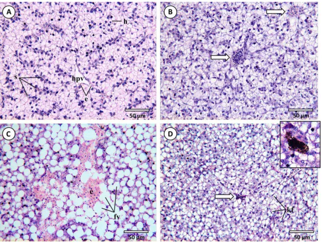
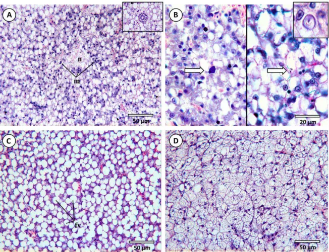
Fig. 4.1. Common histopathological lesions and alterations observed in the livers of D. Labrax (H&E). (A)
14
Fig. 4.2. Common histopathological lesions and alterations observed in the livers of D. Labrax– continuation
(H&E). (A) Necrotic foci (n) with macrophage defence cell intrusions (m) from a fish exposed to mixture treaments at T28. Inset: detail of a bile duct with fat vacuolation. (B) Left panel indicates early stage of apoptosis from an animal exposed to the “high” concentration of B[b]F after 28 days. Right panel: apoptotic bodies as a result of programmed cell death (PCD) (arrows). Inset: detail of a nuclear pleomorphism. (C) Fat vacuoles (fv) leading towards severe lipidosis, common in fish exposed to either PAHs, in this case, to the lowest concentration of Phe after 28 days. (D) Diffuse microvesicular fat vacuolation (steatosis) caused by excessive lipid accumulation in hepatocytes in a fish from the previously mentioned assay.
4.2. Hepatic histopathological condition indices
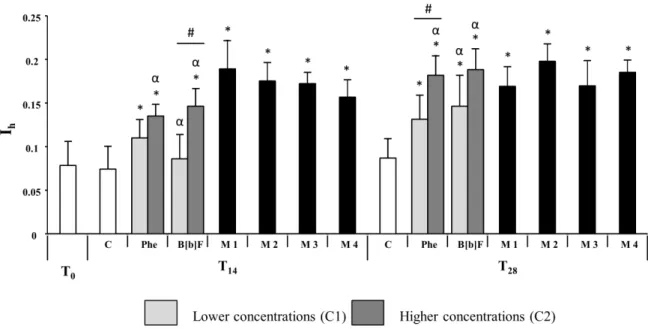
With the exception of individuals exposed to lower concentrations of benzo[b]fluoranthene at T14,
all tests caused an increase in the global hepatic histopathological indice Ih compared to T0 and
control fish (Fig 4.3). T0 and control fish presented no significant differences. The livers of fish
exposed to isolated contaminants yielded distinct Ih between exposures to higher and lower
concentrations, with animals subjected to the higher concentrations of either toxicant presenting higher Ih. This difference is visible at both sampling times T14 and T28, however, statistical
differences were only identified on fish exposed to benzo[b]fluoranthene at T14 and Phenanthrene
at T28 (Mann-Whitney U, p<0.05). No clear differences were detected in livers of animals exposed
15
regardless of concentrations. Also, fish subjected to these treatments revealed a resemblance to those exposed to higher concentrations of isolated contaminants at T28.
Fig. 4.3. Comparison of the average global hepatic histopathological indice (Ih) between fish exposed to isolated and mixture contaminated sediments at sampling times T0, T14 and T28; * means significant differences between contaminated and control assays, p < 0.05 (Mann-Whitney U test). α means significant differences between T14 and T28 assays, p < 0.05 (Mann-Whitney U test). # means significant differences between C1 and C2 concentrations in isolated assays, p < 0.05 (Mann-Whitney U test). Error bars indicate 95% confidence intervals.
All indices for each reaction pattern presented a similar variation to that of Ih (Fig. 4.4).
Accordingly, mixtures and higher concentrations of isolated assays commonly held higher scores of histopathological alterations for each reaction pattern, when comparing to control and T0 fish.
Inflammatory response (I1) presented statistical differences between fish exposed to both
concentrations of B[b]F for both sampling times (Fig. 4.4A). Regressive changes (I2), however,
revealed significant differences between sampling times of higher concentrations on isolated assays for both contaminants (Fig. 4.4B). Average histopathological indice score regarding progressive changes (I3) also exhibited no clear statistical variations between higher and lower concentrations,
16
17
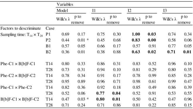
Through discriminant analysis (Table 4.1) it was observed that there were no reaction patterns that significantly contributed to differentiate between the isolated contaminant assays, when testing for concentration of exposure. On the other hand, discriminant analysis revealed that regressive (I2)
and progressive changes (I3) contributed the most to differentiate between sampling times. Also,
comparing mixture treatments, particularly M1M3 and M1M4, revealed circulatory disturbances/inflammatory response (I1) to be the most significant reaction pattern contributing to
the differentiation between tests, principally at sampling time T14 (Table 4.2). Inflammatory
response/circulatory disturbances (I1) was the most significant reaction pattern contributing to
differentiate between isolated and mixture assays at lower concentrations (Table 4.3). On the other hand, regressive changes (I2) contributed the most to differentiate between higher concentration
mixture (M2) and higher concentration (C2) isolated assays. Also, different concentration mixtures (M3 and M4) and corresponding concentrations of isolated contaminants (C1 and C2) displayed differences in progressive alterations (Table 4.3). The model that best described the differences between mixture and isolated contaminants was observed mainly at T14 Wilk’s λ = 0.17, p 0).
Table 4.1. Discriminant analysis results taking sampling time and concentration of exposure as grouping variables factors) for isolated assays. Lowest Wilk's λ statistic was employed to assess best model. F-tests determined the most significant variables (α = 0.05). The models' dependent variable is the hepatic histopathological condition indice (Ih) obtained for each individual.
*best model to assess discrimination between factors Bold figures indicate significant variables within the model
Factors to descriminate Case
P1 0.69 0.17 0.75 0.30 1.00 0.03 0.74 0.34
P2 0.44 0.01 * 0.45 0.68 0.83 0.00 0.58 0.06
B1 0.57 0.05 0.66 0.17 0.57 0.91 0.77 0.05
B2 0.36 0.01 0.38 0.88 0.63 0.02 0.71 0.01
Phe-C1 × B[b]F-C1 T14 0.80 0.33 0.86 0.31 0.83 0.52 0.96 0.10
T28 0.73 0.31 0.94 0.10 0.81 0.29 0.80 0.35
Phe-C2 × B[b]F-C2 T14 0.78 0.34 0.91 0.17 0.78 0.99 0.85 0.28
T28 0.95 0.89 0.96 0.71 0.98 0.61 0.99 0.47
Phe-C1 × Phe-C2 T14 0.82 0.36 0.92 0.18 0.85 0.49 0.86 0.39
T28 0.52 0.06 0.77 0.04 0.52 0.91 0.53 0.55
B[b]F-C1 × B[b]F-C2 T14 0.47 0.03 * 0.80 0.01 0.50 0.42 0.47 0.96
T28 0.71 0.24 0.71 0.86 0.81 0.22 0.85 0.15
p to
remove Wilk's λ
p to remove
Wilk's λ p to
remove Wilk's λ
p to
remove Wilk's λ
Sampling time: T14 × T28
Variables
18
Table 4.2. Discriminant analysis results taking sampling time and concentration of exposure as grouping variables factors) for mixture assays. Lowest Wilk's λ statistic was employed to assess best model. F-tests determined the most significant variables (α = 0.05). The models' dependent variable is the hepatic histopathological condition indice (Ih) obtained for each individual.
*best model to assess discrimination between factors Bold figures indicate significant variables within the model
Table 4.3. Discriminant analysis results when comparing between mixture and isolated assays with the corresponding concentration as grouping variable factor). Lowest Wilk's λ statistic was employed to assess best model. F-tests determined the most significant variables (α = 0.05). The models' dependent variable is the hepatic histopathological condition indice (Ih) obtained for each individual.
*best model to assess discrimination between factors Bold figures indicate significant variables within the model
Factors to descriminate Case
M1 0.93 0.78 0.95 0.55 0.93 0.96 0.96 0.51
M2 0.86 0.48 0.91 0.37 0.90 0.44 0.93 0.28
M3 0.81 0.42 0.90 0.25 0.92 0.21 0.82 0.69
M4 0.76 0.21 0.84 0.22 0.85 0.18 0.79 0.46
M 1 × M 2 T14 0.76 0.21 0.89 0.12 0.79 0.46 0.86 0.17
T28 0.74 0.22 0.76 0.54 0.78 0.38 0.93 0.08
M 1 × M 3 T14 0.51 0.03 * 0.76 0.03 0.59 0.20 0.55 0.38
T28 0.76 0.26 0.79 0.44 0.78 0.59 0.93 0.10
M 1 × M 4 T14 0.66 0.10 0.97 0.02 0.66 0.88 0.69 0.48
T28 0.67 0.11 0.11 0.38 0.72 0.31 0.92 0.03
M 2 × M 3 T14 0.73 0.24 0.90 0.11 0.82 0.23 0.79 0.32
T28 0.83 0.38 0.88 0.35 0.92 0.20 0.83 0.86
M 3 × M 4 T14 0.86 0.60 0.86 0.91 0.89 0.52 0.97 0.24
T28 0.88 0.52 0.90 0.63 1.00 0.15 0.89 0.67
Model I1
Wilk's λ p to
remove Wilk's λ p to
remove Wilk's λ p to
remove Wilk's λ p to remove
I2 I3
Sampling time: T14 × T28
Variables
Factors to descriminate Case
M 1 × Phe-C1 T14 0.37 0.00 * 0.64 0.00 0.38 0.42 0.47 0.05
T28 0.35 0.01 * 0.93 0.00 0.39 0.27 0.38 0.33
M 1 × B[b]F-C1 T14 0.26 0.00 * 0.46 0.00 0.29 0.22 0.37 0.03
T28 0.76 0.33 0.95 0.11 0.76 0.76 0.79 0.53
M 3 × Phe-C2 T14 0.17 0.00 * 0.19 0.28 0.44 0.00 0.55 0.00
T28 0.76 0.27 0.76 0.94 0.85 0.22 0.91 0.13
M 3 × B[b]F-C1 T14 0.24 0.00 * 0.29 0.14 0.24 0.92 0.46 0.01
T28 0.47 0.16 0.74 0.37 0.71 0.60 0.93 0.05
M 4 × Phe-C1 T14 0.54 0.02 * 0.67 0.08 0.68 0.07 0.60 0.23
T28 0.37 0.00 * 0.41 0.29 0.41 0.28 0.54 0.02
M 4 × B[b]F-C2 T14 0.53 0.05 0.64 0.15 0.54 0.83 0.90 0.02
T28 0.85 0.46 0.85 0.76 0.85 0.74 0.99 0.13
M 2 × Phe-C2 T14 0.35 0.00 * 0.35 0.73 0.54 0.01 0.71 0.00
T28 0.73 0.21 0.82 0.22 0.75 0.64 0.92 0.08
M 2 × B[b]F-C2 T14 0.29 0.00 * 0.34 0.18 0.41 0.04 0.87 0.00
T28 0.81 0.38 0.89 0.25 0.81 0.79 0.94 0.16
p to
remove Wilk's λ p to remove Wilk's λ p to
remove Wilk's λ p to
remove Wilk's λ Variables
19
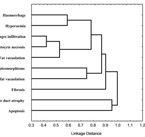
Cluster analysis based on correlation statistic 1-Pearson r indicated the degree of correlation between the observed individual histopathological changes (Fig. 4.5). The analysis indicated a high correlation of hepatocyte necrosis with macrophage infiltration and a high link with these and fat vacuolation. These alterations, together with haemorrhage and hyperaemia formed a distinct cluster. Nuclear pleomorphisms indicated a feeble correlation degree with steatosis. By their turn, fibrosis, bile duct atrophy and apoptosis, although showing some link between them, comprised a distinct cluster to that of preceding alterations.
21 5. Discussion
The present work revealed that sediments contaminated by ecologically-relevant concentrations of the two PAHs, either isolated or combined, caused significant histopathological alterations in the livers of exposed fish. However, basses exposed to the combination of Phe (considered non-carcinogenic to humans) and B[b]F (non-carcinogenic) endured the overall highest level of histopathological alterations, unlike for the isolated PAH assays, without a clear dose- or time-dependent pattern. Nevertheless, in any case, the histopathological alterations are consistent with chronic hepatic disease (rather than acute), given the overall moderate degree of severity and dissemination of changes to the hepatic parenchyma. On the other hand, progressive changes were the most significant contributors to differentiate between the hepatic condition indices of animals exposed to mixtures from those exposed to the isolated toxicants (refer to Fig. 4.2 and Table 4.3). It must be noted that the present study surveyed phenanthrene and benzo[b]fluoranthene concentrations between the range of two sediment quality guidelines (TEL and PEL), meaning within the boundaries of low and high risk to exert deleterious effects to the biota. Under this point of view, the current findings are in accordance with the expected moderate levels of histopathological alterations observed in the liver of animals exposed to the spiked sediments. However, the mixed PAHs caused distinctively higher levels of histopathological alterations and at earlier stages of exposure, which reveals that interactions between contaminants are not contemplated in the SQGs while antagonistic, synergistic or additive effects likely exist in sediments contaminated by mixtures of toxicants, especially organic, whose high hydrophobicity render sediments as the most important trap in aquatic environments.
The occurrence of liver lesions and alterations in fish exposed to both isolated contaminants augmented with exposure time, with Ih revealing a clear increase from animals collected at T14 to
T28 animals. However, B[b]F induced only marginally higher histopathological alterations
22
for its toxicity to aquatic organisms and for inducing oxidative stress by the production of ROS as well (Correia et al., 2007; Yin et al., 2007; Aimová and Poljaková, 2010). Some PAHs (such as B[a]P ) may promote the transcription of specific genes involved in bioactivation, since PAHs may bind to the aryl hydrocarbon receptor (AHR), which, in turn, binds to the aryl hydrocarbon nuclear translocator (ARNT) that can be transferred to the cell nucleus, where on its turn, it binds to the specific xenobiotic response element (XRE) of the gene, promoting transcription (Bucheli and Fent, 1995). B[a]P is known to express AHR-mediated effects, representing higher enzymatic activity and thus increased production of toxic metabolites, as opposed to Phe, which is a weak AHR agonist (Mu et al., 2012). Nevertheless, the differences between the toxicological pathways underneath exposure to these two PAHs are not well understood, especially regarding CYP-mediated activation and relative potency as toxic agents.
It is well known that polycyclic aromatic hydrocarbons in fish are metaboli ed “activated”) during Phase I by the cytochrome P450 (CYP1A) monooxygenases system, followed by Phase II enzymes, which are involved in the elimination or inactivation of toxic or hazardous metabolites. For instance, glutathione S-transferases (GST), conjugate glutathione (GSH) with highly toxic activated metabolites and also some forms of ROS (Pretti et al., 2001). The genotoxicity of activated PAHs in aquatic animals was already confirmed, including fish (see for instance, Gravato and Santos, 2002; Holth et al., 2009; Pacheco and Santos, 1997; Taylor et al., 2011) as well as mutagenicity (Pacheco and Santos, 1997) and potential carcinogenicity (Varanasi et al., 1986). In accordance, the global histopathological indice for each contaminant showed that, as expected, B[b]F exposure may lead to higher hepatic damage, when comparing to phenanthrene, confirming its higher toxicity. It is likely that B[b]F induces higher levels of hepatic histopathological alterations through a combination of factors resulting from its activation, from direct action of ROS to DNA damage, however, previous research attempting to link known PAH effects, such as DNA lesions, to specific histological traits is essentially absent.
The activation of PAHs triggers a series of metabolic defence mechanisms that may affect the integrity of tissues and organs, depending on exposure concentrations and duration, thus contributing to mask the toxicopathic effects of exposure. In fact, phase II defence systems such as the enzymes GST, superoxide dismutase (SOD), catalase (CAT), and glutathione peroxidase (GPx) can be induced even by moderate increases in intracellular oxidative radicals, as a compensatory response (Xu et al., 2009). However, severe oxidative stress will either suppress or saturate the activities of anti-oxidative enzymes and lead to increasing oxidative damage (Sun et al., 2006). This can explain higher Ih at T28, when comparing assays with either isolated contaminant. Also,
comparing T14 and T28 animals exposed to phenanthrene revealed an obvious, albeit unexpected,
23
B[b]F, as generally acknowledged for lower molecular weight PAHs. It must be noticed that, under severe toxicological challenge, hepatocytes are damaged by the production of ROS and other metabolites and may be unable to recover from these lesions to normal hepatic function (Yin et al., 2007). However, Sun et al (2006) showed that after some depuration time, fish exposed to waterborne phenanthrene (thus expectedly more bioavailable) would present levels of antioxidant enzyme activities similar to control level, returning cellular antioxidant defence to normal physiological conditions, even following acute exposures to this toxicant. Should this indicate reduced toxicity of Phe comparative to B[b]F, this information contradicts the reduced histopathological alterations induced by fish exposed to the latter after 14 (but not 28) days of exposure for the lowest concentration exposure (C1) (Fig. 4.3).
As previously stated, metabolites resulting from the activation of higher molecular weight PAHs are presumably more toxic. However, the high risk of B[a]P and other high molecular weight PAHs contradicts, at least partly, the reduced Ih obtained for the lowest concentration of B[b]F at T14,
which was not significantly raised over controls. The global Ih from B[b]F-C1 at 14 days of
exposure suggests that this contaminant at lower concentrations is metabolized by fish without harsh consequences to their hepatic health. On the other hand, benzo[b]fluoranthene assays with higher concentration (B[b]F-C2) revealed noticeable effects on progressive and regressive changes, where an increase of both reaction patterns is striking (see Table 4.1). The present results suggest the existence of cumulative effects of exposure to B[b]F (i.e. derived from ROS and activated metabolites), particularly at higher concentrations and more prolonged exposures, thus accordant with time- and dose-related effects. Altogether, it may be inferred that fish, even PAH-naïve, may be able to respond to B[b]F at lower concentrations, likely for being able to metabolize this compound within the ability to cope with ROS and other by-products of activation, resulting in reduced histopathological alterations. However, the effects of elevated phenanthrene-induced alterations at lower concentrations remain elusive.
24
classified as synergistic (meaning a different effect rather than simply the addition of isolated effects), antagonistic (when the mixture effect is less than the isolated effects) or agonistic/additive (where the mixture effects are lower than the sum of the individual effects) (Gonçalves et al., 2008). However, the lack of clear dose- and time-effect relationships indicates other factors modulating the cumulative toxicity of the two toxicants, which could include dose-dependent inhibition of toxicity caused by phase I enzyme impairment. In fact, Taylor et al (2012) noted that the activity of cytochrome CYP1A could be inhibited at higher concentrations of some inducers, which leads to hypothesize that the PAH mixture had an antagonistic effect.
Interaction effects between different contaminants have already been suggested, even in human toxicological studies, as well as the possible synergistic or antagonistic effects of CYP1A1 activity in mixed exposures (Tarantini et al., 2011). In fish, synergistic effects are mainly identified by the analysis of ethoxyresorufin-O-deethylase EROD activity, an indicator of phase I enzymes
biotransformation. Basu et al (2001) observed a synergistic effect in mixtures of PAHs, where numerous PAHs could increase suprea-additively the mutagenicity of benzo[a]pyrene. However, combination bioassays with CYP1A-activatable PAHs may yield an underestimation of effects, probably due to an inhibitory action of other PAHs (Willett et al., 2001; Wills et al., 2010). However, synergistic effects may also be responsible for the present findings, for some PAHs may disturb the metabolization pathway of others. Altogether, it is clear that further research is needed to fully disclose toxicokinetics of PAHs and their interactions.
25
contemplates the initial phase of homeostatic and metabolic disturbance, where various defence mechanisms are activated to deal with that disturbance. According to Steinberg et al. (2008), after that initial phase, with a longer exposure time, effects may come to decrease, since the presence of metabolites may induce, not only the activation of more specific defensive mechanisms, but also repair mechanisms. This means that the fish undergo an adaptive compensatory response following that initial disruption. Also, the term “adaptive response” implies that similar defence mechanisms are activated regardless concentration, which may explain no clear differences in reaction patterns between different mixtures.
In the environment, contaminants usually occur in mixtures, generally at low levels of individual components, and an analysis of single chemicals is likely to be misleading when comparing with mixtures (Altenburger et al., 2003). Mixtures are rarely addressed in mechanistic toxicological studies. In particular, studying PAH mixtures may unravel complicated mechanisms associated to distinct PAH detoxification pathways that may be hard either to identify or compare with the effects of isolated toxicants. For instance, in human cells, it was observed that B[k]F (another 5-ring PAH) inhibits metabolization of B[a]P, probably through competition to bind to monooxygenase active sites, whereas, B[b]F and B[k]F did not seem to compete for metabolization (Tarantini et al., 2011). In fact, the same authors state that the differential affinity towards CYP1A FO’s active sites and the competition between the contaminants for these sites as an important factor in the formation of reactive metabolites. In a study with rainbow trout (Onchrorhynchus mykiss) involving acute toxicity tests, Bols et al. (1999), showed that fluoranthene (a 4-ring PAH)
is a non-inducer EROD activity. In fact, in this same study, fluoranthene showed little or no evidence of differences between controls regarding the activity of this CYP enzyme often surveyed as biomarker of exposure, and, when in mixture with B[a]P, EROD activities were actually significantly lower than in fish treated only with B[a]P. Another rainbow trout study with different PAH mixture exposures presented an additive effect on EROD activity (Basu et al., 2001) . Synergistic effects following exposure to different classes of contaminants, such as PAH/metal mixtures have also been reported. For instance, a subacute combination of B[a]P and cadmium in soles (Solea senegalensis) revealed inhibited responses related to liver regeneration and apoptosis
(Costa et al., 2010). Also, studies with killifish (Fundulus heteroclitus) embryos exposed to
different waterborne PAH mixtures suggest that sites with PAH mixtures generally induce CYP1A activity, such as those containing B[a]P and B[k]F. However, environmental PAH mixtures may also contain compounds that can act as CYP1A inhibitors, like fluoranthene, which means that additive models currently used to estimate PAH toxicity may over or underestimate that same toxicity in PAH mixtures (Wassenberg and Di Giulio, 2004). In the present work, histopathological effects in mixtures yielded higher Ih values than in fish subjected to isolated assays, but the sum of
26
Interestingly, contrasting the effects of mixtures and isolated contaminants with their respective concentrations showed specific differences to reaction patterns for each mixture (see Table 4.3). Fish exposed to the mixture comprising the lowest concentrations of either PAH (M1) presented signs of greater inflammatory response, when compared to fish exposed to isolated contaminants (Table 4.1). Inflammatory response-related alterations (such as infiltration and hyperaemia) were highly correlated (see clusters), being this reaction pattern related to non-specific immunological responses. Inflammatory response is often associated with other reaction patterns and is the reaction pattern observed that is considered to have the less damaging potential to the organ. On the other hand, mixtures comprising combination of “high” and “low” concentrations of PAHs (M3 and M4) display an increase in progressive changes when comparing to isolated assays of each respective contaminant. Progressive changes regard alteration of hepatocyte functions, mainly fat vacuolation. This alteration, together with other progressive changes not observed in this work (like eosinophilic bodies), has also been observed in livers of juvenile soles (Solea senegalensis)
exposed to PAH-contaminated sediments (Costa et al., 2009). It is possible that fat vacuolation, alongside other progressive alterations may be a little or null-specificity biomarker, albeit able to progress into severe cirrhosis (Koehler, 2004). This is supported by a high correlation between fat vacuolation and hepatocyte necrosis observed (Fig. 4.3). The mixture comprising the highest concentrations of both PAHs (M2) revealed an increase of lesions considered of greater severity, when comparing to the respective isolated assays (Table 4.3). The results also suggest that histopathological alterations are evident sooner, where fish exposed to this mixture endured more alterations at T14 than fish subjected to isolated PAH assays, which means that PAH mixtures may
27 6. Conclusions
The current findings confirmed that sediments contaminated with PAHs, even in “low” and environmentally-relevant concentrations, are able of inducing hepatic lesions and alterations in a benthopelagic fish, consistent with sub-lethal toxicopathological effects. Also, this work showed that semi-quantitative indices based on the relative weights of lesions and quantitative data proved to be a useful and practical tool to assess fish health when exposed to xenobiotics. The data revealed that the histopathological alterations are consistent with chronic hepatic disease (rather than acute), given the overall moderate degree of severity and dissemination of changes to the hepatic parenchyma. Knowing that PAH concentrations used in this work were within the boundaries of low and high risk to exert deleterious effect to the biota, the current findings are in accordance with the expected moderate levels of histopathological alterations observed in the liver of animals exposed to the spiked sediments.
Individuals exposed to phenanthrene (considered non-carcinogenic to humans) presented lower liver histopathological alterations than benzo[b]fluoranthene (carcinogenic) especially at T28, thus
contributing to confirm a positive relation between the number of benzene rings and toxicity. Also, mixture treatments caused more hepatic damage, without a clear dose- or time-dependent pattern, which may suggest interactions between the two contaminants. However, there is still a lack of knowledge concerning these interactions and their connection to hepatic histopathological traits. Complementary histochemical biomarker analysis would be crucial in increasing knowledge of metabolic pathways and possible interactions because PAHs are usually present in the environment in complex mixtures and never isolated. Furthermore, additional histopathological analysis on a different organ, such as gills, due to their role as a main entry route for toxic agents, or kidneys as well and their high susceptibility to adverse environmental conditions, could prove useful in identifying the organs and mechanisms regarding apical entry and excretion of xenobiotics and their metabolites, respectively.
29 7. References
Aimová, D., Poljaková, J., 2010. Ellipticine and benzo(a)pyrene increase their own metabolic activation via modulation of expression and enzymatic activity of cytochromes P450 1A1 and 1A2. Interdisciplinary Toxicology 1, 160–168.
Akcha, F., Izuel, C., Venier, P., Budzinski, H., Burgeot, T., 2000. Enzymatic biomarker measurement and study of DNA adduct formation in benzo[a]pyrene-contaminated mussels. Aquatic Toxicology 49, 269–287.
Altenburger, R., Monika, N., Schüürmann, G., 2003. Mixture toxicity and its modeling by quantitative structure - Activity relationships. Environmental Toxicology and Chemistry 22, 1900– 1915.
Au, D.W.T., 2004. The application of histo-cytopathological biomarkers in marine pollution monitoring: a review. Marine Pollution Bulletin 48, 817–34.
Basu, N., Billiard, S., Fragoso, N., Omoike, A., Tabash, S., Brown, S., Hodson, P., 2001. Ethoxyresorufin-O-deethylase induction in trout exposed to mixtures of polycyclic aromatic hydrocarbons. Environmental Toxicology and Chemistry 20, 1244–51.
Bernet, D., Schmidt, H., Meier, W., Burkhardt-Holm, P., Wahli, T., 1999. Histopathology in fish: proposal for a protocol to assess aquatic pollution. Journal of Fish Diseases 22, 25–34.
Bols, N.C., Schirmer, K., Joyce, E.M., Dixon, D.G., Greenberg, B.M., Whyte, J.J., 1999. Ability of polycyclic aromatic hydrocarbons to induce 7-ethoxyresorufin-o-deethylase activity in a trout liver cell line. Ecotoxicology and Environmental Safety 44, 118–28.
Bucheli, T.D., Fent, K., 1995. Induction of cytochrome P450 as a biomarker for environmental contamination in aquatic ecosystems. Critical reviews in Environmental Science and Technology 25, 201–268.
30
Correia, A.D., Gonçalves, R., Scholze, M., Ferreira, M., Henriques, M.A.-R., 2007. Biochemical and behavioral responses in gilthead seabream (Sparus aurata) to phenanthrene. Journal of
Experimental Marine Biology and Ecology 347, 109–122.
Costa, P.M., Caeiro, S., Lobo, J., Martins, M., Ferreira, A.M., Caetano, M., Vale, C., DelValls, T.Á., Costa, M.H., 2011. Estuarine ecological risk based on hepatic histopathological indices from laboratory and in situ tested fish. Marine Pollution Bulletin 62, 55–65.
Costa, P.M., Caeiro, S., Vale, C., Delvalls, T.À., Costa, M.H., 2012. Can the integration of multiple biomarkers and sediment geochemistry aid solving the complexity of sediment risk assessment? A case study with a benthic fish. Environmental Pollution 161, 107–20.
Costa, P.M., Carreira, S., Costa, M.H., Caeiro, S., 2013. Development of histopathological indices in a commercial marine bivalve (Ruditapes decussatus) to determine environmental quality.
Aquatic Toxicology 126, 442–54.
Costa, P.M., Chicano-Gálvez, E., López Barea, J., DelValls, T.A., Costa, M.H., 2010. Alterations to proteome and tissue recovery responses in fish liver caused by a short-term combination treatment with cadmium and benzo[a]pyrene. Environmental Pollution 158, 3338–46.
Costa, P.M., Diniz, M.S., Caeiro, S., Lobo, J., Martins, M., Ferreira, A.M., Caetano, M., Vale, C., DelValls, T.A., Costa, M.H., 2009. Histological biomarkers in liver and gills of juvenile Solea senegalensis exposed to contaminated estuarine sediments: a weighted indices approach. Aquatic
Toxicology 92, 202–12.
Danion, M., Le Floch, S., Lamour, F., Guyomarch, J., Quentel, C., 2011. Bioconcentration and immunotoxicity of an experimental oil spill in European sea bass (Dicentrarchus labrax L.).
Ecotoxicology and Environmental Safety 74, 2167–74.
DelValls, T.A., Blasco, J., Sarasquete, M.C., Forja, J.M., Gomez-Parra, A., 1998. Evaluation of heavy metal sediment toxicity in littoral ecosystems using juveniles of the fish Sparus aurata.
Ecotoxicology and Environmental Safety 41, 157–67.
31
Eisler, R., 1987. Polycyclic aromatic hydrocarbon hazards to fish, wildlife, and invertebrates: A synoptic review. U.S. Fish and Wildlife Service Biological Report nº 85, 55p.
Ferreira, M., Caetano, M., Antunes, P., Costa, J., Gil, O., Bandarra, N., Pousão-Ferreira, P., Vale, C., Reis-Henriques, M.A., 2010. Assessment of contaminants and biomarkers of exposure in wild and farmed seabass. Ecotoxicology and Environmental Safety 73, 579–88.
Fleming, C.R., Di Giulio, R.T., 2011. The role of CYP1A inhibition in the embryotoxic interactions between hypoxia and polycyclic aromatic hydrocarbons (PAHs) and PAH mixtures in zebrafish (Danio rerio). Ecotoxicology 20, 1300–14.
Fragoso, N.M., Hodson, P. V, Zambon, S., 2006. Evaluation of an exposure assay to measure uptake of sediment PAH by fish. Environmental Monitoring and Assessment 116, 481–511.
Gonçalves, R., Scholze, M., Ferreira, A.M., Martins, M., Correia, A.D., 2008. The joint effect of polycyclic aromatic hydrocarbons on fish behavior. Environmental Research 108, 205–13.
Gravato, C., Guilhermino, L., 2009. Effects of benzo(a)pyrene on seabass (Dicentrarchus labrax L.): biomarkers, growth and behavior. Human and Ecological Risk Assessment: An International
Journal 15, 121–137.
Gravato, C., Santos, M. A., 2002. Juvenile sea bass liver P450, EROD induction, and erythrocytic genotoxic responses to PAH and PAH-like compounds. Ecotoxicology and Environmental Safety 51, 115–27.
Hibyia, T., (Ed.), 1982. An Atlas of Fish Histology: Normal and Pathological Features. Tokyo, Kodansha, 147p.
Holth, T.F., Beylich, B. A., Skarphédinsdóttir, H., Liewenborg, B., Grung, M., Hylland, K., 2009. Genotoxicity of environmentally relevant concentrations of water-soluble oil components in cod (Gadus morhua). Environmental Science and Technology 43, 3329–34.
32
Kerambrun, E., Le Floch, S., Sanchez, W., Thomas Guyon, H., Meziane, T., Henry, F., Amara, R., 2012. Responses of juvenile sea bass, Dicentrarchus labrax, exposed to acute concentrations of
crude oil, as assessed by molecular and physiological biomarkers. Chemosphere 87, 692–702.
Koehler, A., 2004. The gender-specific risk to liver toxicity and cancer of flounder (Platichthys flesus L.) at the German Wadden Sea coast. Aquatic Toxicology 70, 257–76.
Kopecka-Pilarczyk, J., Correia, A.D., 2009. Biochemical response in gilthead seabream (Sparus aurata) to in vivo exposure to pyrene and fluorene. Journal of Experimental Marine Biology and
Ecology 372, 49–57.
Lang, T., Wosniok, W., Barsiene, J., Broeg, K., Kopecka, J., Parkkonen, J., 2006. Liver histopathology in Baltic flounder (Platichthys flesus) as indicator of biological effects of
contaminants. Marine Pollution Bulletin 53, 488–96.
Logan, D.T., 2007. Perspective on ecotoxicology of PAHs to fish. Human and Ecological Risk Assessment: An International Journal 13, 302–316.
MacDonald, D.D., Carr, R., Calder, F., Long, E., Ingersoll, C., 1996. Development and evaluation of sediment quality guidelines for Florida coastal waters. Ecotoxicology 5, 253–278.
art n- a , .L., Blasco, ., Sales, ., el alls, T. A., 2004. Biomarkers as tools to assess sediment quality. Trends in Analytical Chemistry 23, 807–818.
Martins, M., Costa, P.M., Ferreira, A.M., Costa, M.H., 2013. Comparative DNA damage and oxidative effects of carcinogenic and non-carcinogenic sediment-bound PAHs in the gills of a bivalve. Aquatic Toxicology 142-143C, 85–95.
artoja, R., artoja, ., 1967. Initiation aux Techniques de l’Histologie Animal. Masson & Cie, Paris.
33
Mu, J., Wang, X., Jin, F., Wang, J., Hong, H., 2012. The role of cytochrome P4501A activity inhibition in three- to five-ringed polycyclic aromatic hydrocarbons embryotoxicity of marine medaka (Oryzias melastigma). Marine Pollution Bulletin 64, 1445–51.
Oliva, M., Garrido, M.C., Sales Márquez, D., González de Canales, M.L., 2009. Sublethal and lethal toxicity in juvenile Senegal sole (Solea senegalensis) exposed to copper: a preliminary
toxicity range-finding test. Experimental and Toxicologic Pathology : Official Journal of the Gesellschaft für Toxikologische Pathologie 61, 113–21.
Pacheco, M., Santos, M. A., 1997. Induction of EROD activity and genotoxic effects by polycyclic aromatic hydrocarbons and resin acids on the juvenile eel (Anguilla anguilla L.). Ecotoxicology
And Wnvironmental Safety 38, 252–9.
Palanikumar, L., Kumaraguru, A.K., Ramakritinan, C.M., 2011. Genotoxic assessment of anthracene and benzo[a]pyrene to milkfish Chanos chanos. Toxicological and Environmental
Chemistry 94, 350–363.
Pisoni, M., Cogotzi, L., Frigeri, A., Corsi, I., Bonacci, S., Iacocca, A., Lancini, L., Mastrototaro, F., Focardi, S., Svelto, M., 2004. DNA adducts, benzo(a)pyrene monooxygenase activity, and lysosomal membrane stability in Mytilus galloprovincialis from different areas in Taranto coastal
waters (Italy). Environmental Research 96, 163–75.
Pretti, C., Salvetti, A., Longo, V., Giorgi, M., Gervasi, P.G., 2001. Effects of beta-naphthoflavone on the cytochrome P450 system, and phase II enzymes in gilthead seabream (Sparus aurata).
Comparative biochemistry and physiology Part C: Toxicology and Pharmacology 130, 133–44.
Rodrigues, A.P., Lehtonen, K.K., Guilhermino, L., Guimarães, L., 2013. Exposure of Carcinus maenasto waterborne fluoranthene : Accumulation and multibiomarker responses. Science of the
Total Environment 443, 454–463.
Sanchez, B.C., Ralston-Hooper, K.J., Kowalski, K. A., Dorota Inerowicz, H., Adamec, J., Sepúlveda, M.S., 2009. Liver proteome response of largemouth bass (Micropterus salmoides)
![Table 3.1. Nominal PAH concentrations (ng g -1 ) used for spiking artificial sediments of isolated (Phe-C1, Phe-C2, B[b]F-C1, B[b]F – C2) and combined assays (M1, M2, M3, M4)](https://thumb-eu.123doks.com/thumbv2/123dok_br/16663126.742366/26.892.107.767.691.763/table-nominal-concentrations-spiking-artificial-sediments-isolated-combined.webp)