1
UNIVERSIDADE FEDERAL DE SÃO CARLOS
CENTRO DE CIÊNCIAS BIOLÓGICAS E DA SAÚDE
PROGRAMA DE PÓS-GRADUAÇÃO EM ECOLOGIA E
RECURSOS NATURAIS
VIVIANE DE CASSIA PEREIRA ABDALLA
Endophytic fungi from
Serjania lethalis
A.St-Hil leaves and their
phytotoxic potential
2
UNIVERSIDADE FEDERAL DE SÃO CARLOS
CENTRO DE CIÊNCIAS BIOLÓGICAS E DA SAÚDE
PROGRAMA DE PÓS-GRADUAÇÃO EM ECOLOGIA E
RECURSOS NATURAIS
VIVIANE DE CASSIA PEREIRA ABDALLA
Endophytic fungi from
Serjania lethalis
A.St-Hil leaves and their
phytotoxic potential
Orientadora:
Prof
a. Dr
a. Sonia Cristina Juliano Gualtieri
São Carlos- SP
2018
4 Agradecimentos
Agradeço a Deus, primeiramente, pela oportunidade de concluir mais uma etapa da minha vida.
Agradeço à minha família pelo apoio e confiança, à minha mãe Lourdes e meus irmãos Daniel, Fernando e Renaldo. À pequena Nena pela silenciosa e agradável companhia durante a escrita. Ao Gabriel Abdalla por todo o apoio e companheirismo.
Aos meus amigos por compartilharem momentos felizes e de desabafo, em especial à Grazi e Fernanda por compartilharem comigo muito mais do que um lar.
À profa Dra Sonia Cristina Juliano Gualtieri pela longa caminhada desde o mestrado. Pela paciência e companheirismo. Ao Dr José Luís pela ajuda e colaboração.
Aos professores e funcionários do Departamento de Botânica pela amizade. Aos colegas de laboratório pela agradável companhia.
Ao Prof Maurício Bacci e ao doutorando Sergio Kakazu pela ajuda com a biologia molecular.
Ao prof. Paulo Cezar Vieira (Departamento de Química) por toda a colaboração com a parte química. À doutoranda Ana Carolina (Carol) pela colaboração, paciência e dedicação.
Ao Fábio Kusumi Otsuka pelas agradáveis aulas de inglês, pela amizade e pela revisão da tese.
Á minha terapeuta Manoela por todo suporte psicológico.
Aos membros da banca examinadora pelas sugestões, críticas e colaboração. Ao Programa de Pós-Graduação em Ecologia e Recursos Naturais.
Ao Conselho Nacional de Desenvolvimento Científico e Tecnológico (CNPq) pela bolsa de estudos concedida.
5 RESUMO GERAL
Os fungos endofíticos vivem assintomaticamente no interior dos órgãos vegetais sem lhes causar prejuízo. Estes fungos vivem em associação simbionte com as plantas e seu metabolismo secundário produz compostos que são utilizados por elas como mecanismo de defesa ao ataque de predadores e patógenos. Neste trabalho foram isolados 29 fungos endofíticos de folhas sadias de Serjania lethalis, uma liana pertencente à família
Sapindaceae de ocorrência comum no cerrado brasileiro. Estes fungos foram caracterizados macro e microscopicamente e alguns gêneros puderam ser identificados com o auxílio de técnicas de biologia molecular. Este trabalho foi pioneiro no isolamento e identificação de endófitos desta espécie vegetal. Foram selecionados dois fungos para os estudos de avaliação do potencial fitotóxico, sendo uma Alternaria sp. e
um Diaporthe sp. Para estes estudos foram inicialmente preparados dois extratos de
cada fungo, um extrato micelial e um extrato de meio de cultura. O primeiro foi extraído com etanol e o segundo com acetato de etila. Também foi preparado um extrato de meio de cultura estéril. Inicialmente os extratos foram testados sobre o crescimento de coleóptilos de trigo (Triticum aestivum) a fim de selecionar os mais ativos e verificou-se
que o extrato de meio de cultura de Alternaria sp e o extrato micelial de Diaporthe sp.
foram os que apresentaram maior fitotoxicidade sobre o crescimento dos coleóptilos, quando comparados com o tratamento controle negativo (solução tampão). O extrato de meio de cultura estéril não foi ativo. Com os extratos mais ativos de cada fungo foram feitos ensaios de germinação de sementes, crescimento de plântulas e crescimento de células do metaxilema com as espécies invasoras Euphorbia heterophylla
(amendoim-bravo) e Megathyrsus maximus (capim-colonião). Verificou-se que o extrato de meio de
cultura de Alternaria sp foi fitotóxico para o crescimento de plântulas de E.
heterophylla. O extrato micelial do fungo Diaporthe sp. diminuiu a porcentagem de
germinação de sementes e o crescimento da raiz das plântulas de M. maximus. Também
foi fitotóxico para o crescimento das plântulas de E. heterophylla. Tais extratos contém
metabólitos secundários produzidos pelos fungos que são responsáveis por tais atividades. A fim de se conhecer os metabólitos presentes no extrato de meio de cultura de Alternaria sp., este extrato foi fracionado em coluna de sílica de fase normal e as
6
carboxílico e as dicetopiperazinas ciclo (Pro-Leu), ciclo (Pro-Val) e ciclo (Pro-Ile). Estes compostos foram testados sobre o crescimento de coleóptilos de trigo e os resultados indicaram que apenas o ácido indol-3- carboxílico (ICA) foi fitotóxico. A atividade fitotóxica do ICA foi melhor explorada realizando-se ensaios de germinação de sementes, crescimento de plântulas e de células do metaxilema de E. heterophylla e
M. maximus. Foram utilizados como controles uma solução tampão e o ácido
indoleacético (IAA), um hormônio vegetal que apresenta estrutura semelhante à do ICA. Verificou-se que o ICA afetou os parâmetros da germinação das duas espécies invasoras testadas, assim como o crescimento da parte aérea e raiz de E. heterophylla e da raiz de
M. maximus. Os controles não afetaram nenhum dos parâmetros estudados. Foram feitas
reações de esterificação do ICA com três diferentes álcoois, o metanol, etanol e hexanol, produzindo, respectivamente, os ésteres Methyl- carboxylate, Ethyl- Indol-3-carboxylate e Hexyl- Indol-3-Indol-3-carboxylate. A atividade fitotóxica dos três ésteres foi avaliada por meio do crescimento de coleóptilos de trigo e verificou-se que o aumento
da cadeia de alquil promove um aumento da fitotoxicidade. O IC50 e o coeficiente de
lipofilia foram medidos e juntos comprovam um menor valor de IC50 para os ésteres de
maior cadeia e um maior índice de lipofilia para os mesmos. Assim, concluímos que o ICA, um metabólito secundário produzido pelo fungo endofítico de folhas de S. lethalis,
foi fitotóxico para as espécies invasoras testadas e é um promissor agente fitotóxico natural.
Palavras-chave: fungos endofíticos; fitotoxicidade; herbicidas naturais; Serjania
7 GENERAL ABSTRACT
Endophytic fungi live asymptomatically within plant organs without causing them any harm. These fungi live in symbiotic association with plants, and their secondary metabolism produces compounds that are used by plants as a defense mechanism to attack predators and pathogens. In this work, 29 endophytic fungi were isolated from healthy mature leaves of Serjania lethalis, a liana belonging to the family Sapindaceae of
common occurrence in the Brazilian cerrado. These fungi were characterized macro and microscopically, and some genera could be identified with the aid of molecular biology techniques. This work was a pioneer in the isolation, and identification of endophytes in this plant species. Two fungi were selected for the studies of evaluation of the phytotoxic potential, being them an Alternaria sp, and a Diaporthe sp. For these studies two extracts
of each fungus were initially prepared, a mycelial extract, and an extract of culture medium. The former was extracted with ethanol, and the second with ethyl acetate. An extract of sterile culture medium was also prepared. Initially, the extracts were tested on the growth of wheat coleoptiles (Triticum aestivum) in order to select the most
active ones, and it was verified that the extract of culture medium of Alternaria sp and
the mycelial extract of Diaporthe sp. were those that presented greater phytotoxicity on
the growth of the coleoptilos, when compared with the negative control treatment. The extract of sterile culture medium was not active. The most active extracts from each fungus were seed germination, seedling growth, and metaxilema cell growth experiments with the invasive species Euphorbia heterophylla, and Megathyrsus
maximus. The extract of Alternaria sp culture medium was found to be phytotoxic for
the growth of E. heterophylla seedlings. The mycelial extract of the fungus Diaporthe
sp. decreased the percentage of seed germination, and growth of the root of M. maximus
seedlings. It was also phytotoxic for the growth of Euphorbia heterophylla seedlings.
Such extracts contain secondary metabolites produced by the fungi that are responsible for such activities. In order to know the metabolites present in the culture medium extract of Alternaria sp. This extract was fractionated on a normal phase silica column, and its
8
activity of ICA was better explored by carrying out tests on seed germination, seedling growth, and metaxilema cells of Euphorbia heterophylla and Megathyrsus maximus. A
buffer solution and indoleacetic acid (IAA), a plant hormone with a similar structure to ICA, were used as controls. It was verified that ICA affected the parameters of the germination of the two invasive species tested, as well as the growth of the aerial part and root of E. heterophylla, and the root of M. maximus. Controls did not affect any of the
parameters studied. ICA esterification reactions were performed with three different alcohols, methanol, ethanol and hexanol, producing, respectively, the methyl esters Methyl-carboxylate, Ethyl-carboxylate, and Hexyl- Indole-3-carboxylate. The phytotoxic activity of the three esters was evaluated by growth of wheat coleoptiles, and it was found by increasing the alkyl chain, it promotes an increase in phytotoxicity. The IC50 and the lipophilicity coefficient were measured, and together
they showed a lower IC50 value for the higher chain esters, and a higher lipophilicity
index for them. Thus, we conclude that ICA, a secondary metabolite produced by the endophytic fungus of Serjania lethalis leaves, was phytotoxic to the invasive species
tested, and it is a promising natural phytotoxic agent.
9 LIST OF FIGURES
Introduction
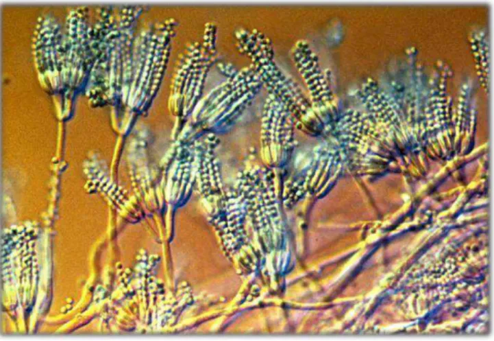
Figure 1.Penicillium fungus, representative of the Ascomycota Phylum ………….. 19 Figure 2. Mushroom of the genus Amanita, representative of the Phylum
Basidiomycota………20
Figure 3. Fungus of the genus Allomyces, representative of the Phylum
Blastocladiomycota ………20
Figure 4. Fungus spores of the genus Synchytrium, representative of the Phylum
Chytridiomycota ……….21
Figure 5. Spores of the fungus of the genus Acaulospora, representative of the Phylum
Glomeromycota ………..21
Figure 6. Spores of the fungus of the genus Nosema………22 Figure 7. Zoospores of Neocallimastix, representative of the Phylum
Neocallimastigomycota ………..22
Figure 8. Some compounds isolated from fungi of the genus Alternaria that present
phytotoxic activity. (a) Dextruxin B; (b) Tentoxin; (c) Altersolanol; (d) Bostrycin and 4-Deoxybostrycin ………...27
Figure 9. Some compounds isolated from fungi of the genus Cochliobolus that have
phytotoxic activity. (a) HC-Toxin; (b) T-Toxin; (c) Victorin ………....28
Figure 10. Secondary metabolites produced by fungi with phytotoxic activity. (a) cercosporin; (b) phleichrome; (c) herbarumin ………...………….28
Figure 11. Individuals of S. lethalis in the cerrado reserve of UFSCar
………...29
Chapter I
Figure 1. Growth of endophytic fungi of Serjania lethalis leaves in PDA culture
medium ……….………. 43
Figure 2. Microculture on slide for microscopic observation ………44
Figure 3. Blades stained with Lactophenol dye for microscopic observation ……...…45
Chapter II
Figure 1. Conidia of the fungus Alternaria sp. (a) and Diaporthesp ….………. 77 Figure 2. Percent inhibition of growth of wheat coleoptiles (Triticum aestivum) under
the effect of ethyl acetate and ethanolic extracts of endophytic fungi. (*) Indicates a significant difference in relation to the negative control. (A) Extract obtained from fungal culture medium only; (B) Ethyl acetate extract ofAlternaria sp.; (C) Ethanolic
extract of Alternaria sp.; (D) Ethyl acetate extract of Diaporthe sp.; (E) Ethanolic
extract of Diaporthe sp ………..………... 78 Figure 3. The effects of ethyl acetate extract of Alternaria sp. on M. maximus and E.
heterophylla seed germination. (a) Germinability of seeds, (b) mean germination time,
10 Figure 4. The effects of ethanolic extract of Diaporthe sp. on M. maximus and E.
heterophylla seed germination (a) Germinability of seeds, (b) mean germination time,
(c) Maguire's index, (d) synchrony……….………81
Figure 5. The effects of oxyfluorfen on M. maximus and E. heterophylla seed
germination. (a) Germinability of seeds, (b) mean germination time, (c) Maguire's index, (d) synchrony …………...………...85
Figure 6. Effect of the ethyl acetate extract of Alternaria sp on seedling growth of M.
maximus and E. heterophylla (a) mean shoot length (b) mean root length.……..…… 86 Figure 7. Effect of the ethanolic extract of Diaporthe sp on seedling growth of M.
maximus and E. heterophylla (a) mean shoot length (b) mean root length……….…... 87 Figure 8. Effect of oxyfluorfen on seedling growth of M. maximus and E. heterophylla
(a) mean shoot length (b) mean root length……….88
Figure 9. Micrographs of E. heterophylla metaxilem cells grown at different
concentrations of ethyl acetate extract of Alternaria sp. (a) Control (b) 5.0 mg.mL-1 (c)
2.5 mg.mL-1 (d) 1.25 mg .mL-1 (e) 0.625 mg.mL-1 ………...………..89 Figure 10. Micrographs of E. heterophylla metaxilem cells grown at different
concentrations of ethanolic extract of Diaporthe sp. (a) Control (b) 2.5 mg.mL-1 (c) 1.25
mg .mL-1 (d) 0.625 mg.mL-1 ….……….… 91
Chapter III
Figure 1. Percentage inhibition caused by the negative control of the Alternaria extract
(aseptic medium) (A); Ethyl acetate extract from culture medium (B); Ethanolic extract of mycelium from Alternaria sp; Positive control (herbicide Oxyfluorfen) (C) (*)
Indicates significant difference in relation to the negative control (buffer solution)...………..103
Figure 2. Percentage inhibition of the ethyl acetate fractions of the fungus Alternaria
sp. on wheat coleoptile growth (*) indicates a significant difference in relation to the negative control ………103
Figure 3. Secondary metabolites produced by the fungus Alternaria sp. a)
indole-3-carboxylic acid (b) Cyclo leucine (c) cyclo valine (d) cyclo proline-isoleucine ………...104
11 Figure 5. Main couplings gCOSY (double arrow) and gHMBC (single arrow) for compound 3c (left) and compound 3d (right) ………..………107
Figure 6. Percentage inhibition / growth of wheat coleoptiles (Triticum aestivum)
grown at different molar concentrations of indole-3-carboxylic acid (ICA). (*) indicate a significant difference in relation to the negative control (buffer solution) …………..108
Figure 7. Percentage inhibition / growth of wheat coleoptiles (Triticum aestivum)
grown at different molar concentrations of indole-3-carboxylic acid (ICA). (*) indicate a significant difference in relation to the negative control (buffer solution)………..….109
Chapter IV
Figure 1. Percentage inhibition caused by the negative control of the fungus (aseptic medium) (A); Ethyl acetate extract from Alternaria sp.(B); Ethanolic extract from
Alternaria sp. (C) and positive control (herbicide oxyfluorfen). (*) Indicate a significant
difference in relation to the negative control (buffer solution) ……… 123
Figure 2. Percentage inhibition of fractions of ethyl acetate from Alternaria sp. on the
growth of wheat coleoptiles (*) indicates a significant difference in relation to the negative control ………...……… 123
Figure 3. Indole-3- carboxylic acid ………..……….. 124
Figure 4. Percentage inhibition of growth of wheat coleoptiles in different concentrations of indole-3-carboxylic acid (ICA), indolacetic acid (IAA), and herbicide oxyfluorfen. (*) indicate a significant difference in relation to the negative control
(buffer solution) ………... 124
Figure 5. Germinability (a), mean germination time (b), germination speed (c), and synchrony (d) of E. heterophylla and M. maximus seeds grown at different ICA
concentrations ………..………...……. 126
Figure 6. Germinability (a), mean germination time (b), germination speed (c), and synchrony (d) of E. heterophylla and M. maximus seeds grown effects caused by IAA in
concentrations ………..…… 127
Figure 7. Germinability (a), mean germination time (b), germination speed (c), and synchrony (d) of E. heterophylla and M. maximus seeds grown at different molar
concentrations of oxyfluorfen ………... 128
Figure 8. Growth of shoot and root of E. heterophylla (a) and M. maximus (b) at
12 Figure 9. Seedlings of E. heterophylla (a) and M. maximus (b) grown at different
concentrations of ICA ………..….129
Figure 10. Growth of shoot and root of E. heterophylla (a) and M. maximus (b) at
different molar concentrations of IAA………..130
Figure 11. Seedlings of E. heterophylla (a) and M. maximus grown at different
concentrations of IAA ………...130
Figure 12. Growth of shoot and root of E. heterophylla (a) and M. maximus (b) in
different molar concentrations of the herbicide oxyfluorfen………...…..131
Figure 13. Seedlings of E. heterophylla (a) and M. maximus grown at different
concentrations of oxyfluorfen ………..… 131
Figure 14. Micrographs of E. heterophylla metaxilem cells grown at different
concentrations of ICA (a) control (b) 10-3 M (c) 3.10-4 M (d) 10-4 M (e) 3.10-5 M (f) 10-5
M ………..…… 132
Figure 15. Micrographs of E. heterophylla metaxylem cells grown at different
concentrations of IAA (a) control (b) 10-3 M (c) 3.10-4 M (d) 10-4 M (e) 3.10-5 M (f) 10-5
M……….. 133
Figure 16. Structure of the compounds Methyl-Indole-3-carboxylate (a), Ethyl-Indole-3-carboxylate (b), Hexyl-Indole-Ethyl-Indole-3-carboxylate (c) produced through Fischer esterification………....………..134
Figure 17. Percentage inhibition of carboxylic acid esters on wheat coleoptile growth (*) indicates significant difference in relation to the negative control (buffer
13 ANNEXES
Figure A. DNA Sequence Aligment of Alternaria sp. fungus ………....144
Figure B. DNA Sequence Aligment of Diaporthe sp. fungus ……… 145
Figure C. Cyclo proline- valine 1H NMR, MeOD, 400 MHz ……… 146
Figure D. Cyclo proline-isoleucine 1H NMR, MeOD, 400 MHz ………... 146
Figure E. Cyclo proline-leucine 1H NMR, MeOD, 400 MHz ……… 147 Figure F. Spectroscopic characteristics of diketopiperazines ………. 147 Figure G.Indole-3-carboxylic acid 1H NMR, MeOD, 400 MHz ………...….148
Figure H. Indole-3-methyl carboxylate 1H NMR, acetone-d 6, 400 MHz …...……….148 Figure I.Indole-3-ethyl carboxylate 1H NMR, acetone-d6, 400 MHz ………149
14 LIST OF TABLES
Introduction
Table 1. Fungus virulence vs. plant defense in a non-obligatory fungus-host interaction………... 24
Chapter I
Table 1. Symbiotic criteria used to classify endophytic fungal classes ……… 41
Chapter II
Table 1. Cell size classes of the Euphorbia heterophylla root metaxilem submitted to
the effect of the ethyl acetate extract of Alternaria sp……….…………89 Table 2. Cell size classes of the Euphorbia heterophylla root metaxilem submitted to
the effect of the ethanolic extract of Diaporthe sp.………...90 Chapter III
Table 1.Chromatographic method used in the analysis ………..101
Table 2. Coefficient of lipophilicity of compounds isolated from the culture medium extract of Alternariasp ………..………...109 Chapter IV
Table 1. Statistical comparison of average growth of coleoptiles (mm) between fractions and positive control (oxyfluorfen) at molar concentrations .…….………... 125
Table 2. Size classes of E.heterophylla roots metaxylem ………...……….132 Table 3. Size classes of E.heterophylla roots metaxylem ………133
Table 4. Esters obtained by Fischer esterification and their respective codes ...……. 134
Table 5. Statistical comparison of average growth of coleoptiles (mm) between esters and positive control (oxyfluorfen) at molar concentrations ………...………. 136
Table 6. Statistical comparison of average growth of coleoptiles (mm) between esters and Indole-3- carboxilic acid (ICA) at molar concentrations …………...…………... 136
Table 7. Inhibitory concentration of ICA, IAA, ICA esters derivatives and oxyfluorfen
………...137
15 SUMMARY
INTRODUCTION ……….……….. 19
GOALS ………..……… 30
General objectives ………...……….. 30
Specific objectives …….……… 30
HYPOTHESIS OF WORK ………... 31
REFERENCES ………...32
CHAPTER I ………....39
Endophytic fungi isolated from Serjania lethalis A.St-Hil (Sapindaceae) leaves ABSTRACT ……….. 40
1. Introduction ……….. 40
2. Goal ……….. 42
3. Material and methods ………... 43
3.1Isolation of fungi ……….. 43
3.2Morphological characterization ………... 44
3.3Sporulation induction of fungi ………. 45
3.4Molecular characterization ………....45
3.4.1 Extraction of genomic DNA ………45
3.4.2 Amplification and sequencing of the ITS region ……… 45
3.4.3 Purification of ITS region …………... 45
3.4.4 Sequencing of ITS genes ……….46
3.4.5 Purification and sequencing reaction ………...46
3.4.6 Sequence analysis ………....47
3.5 Preservation of fungi ………47
4. Results and discussion ………... 47
4.1 Morphological and molecular classification of endophytic fungi… 47 5. Conclusions ……….64
References ………...…65
CHAPTER II ………. 69
Phytotoxic potential of endophytic fungi extract from leaves of Serjania lethalis on wheat (Triticum aestivum L.) and two weeds (Euphorbia heterophylla and Megathyrsus maximus) ABSTRACT ………..……… 70
16
2. Goal ………. 71
3. Material and methods ……….. 72
3.1Isolation of microrganisms ... 72
3.2 Molecular characterization of endophytic fungi……….72
3.2.1 Genomic DNA extraction……….72
3.2.2 PCR amplification and sequencing of the internal transcribed spacer (ITS) region ………...…... 73
3.2.3 Purification of ITS amplicon.………... 73
3.2.4 Sequencing of ITS genes ……….………….73
3.2.5 Purification and sequencing reaction ………...73
3.2.6 Sequence analysis ………... 74
3.3 Preparation of extracts …………...……… 74
3.4 Wheat coleoptile growth bioassay………...74
3.5 Seed germination bioassay ………...…. 75 3.6 Seedlings growth bioassay ….………..……….. 75
3.7 Growth of metaxylem cells of wild poinsettia (E. heterophylla) ……...76
3.8 Statistical analysis …………..……… 76
4. Results and discussion………...……… 76 4.1 Identification and preservation of microrganisms ………... 76
4.2 Wheat coleoptile growth bioassay ………..77
4.3 Seed germination bioassay… ……….78
4.3.1Ethyl acetate extract of Alternaria sp., ethanolic extract of Diaporthe sp. and positive control with oxyfluorfen herbicide.………...……...78
4.4 Seedlings growth bioassay .……….……….. 85
4.4.1Ethyl acetate extract Alternaria sp., ethanolic extract of Diaporthe sp. and positive control with oxyfluorfen herbicide.………..85
4.5 Growth of metaxylem cells ……….. 88
4.5.1 Ethyl acetate extract of Alternaria sp. and ethanolic extract of Diaporthe sp. ………. 88
5. Conclusions ……… 91
References ………...92
17
Secondary metabolites from Alternaria sp., Endophytic fungus isolated from Serjania
lethalis A. St.Hil leaves
ABSTRACT ………...98
1. Introduction………98
2. Goal ………...99
3. Material and Methods ...99
3.1 Isolation of the fungus ………..…99
3.2 Genomic DNA extraction, PCR and sequencing of the ITS region………...……99
3.3 Purification of ITS amplicon, sequencing of ITS genes and purification of the sequencing reaction ……….………100
3.4 Sequence analysis ………100
3.5 Preparation of extracts, isolation and identification of secondary metabolites ………101
3.6 Growth bioassay of wheat coleoptiles with the fractions from the culture medium extract, franctions and compounds of the fungus Alternaria sp. ………102
4. Results and discussion ...102
4.1 Identification of the fungus ………...102
4.2 Growth bioassay of wheat coleoptiles (Triticum aestivum) with extracts of culture medium and mycelium ………102
4.3 Growth bioassay of wheat coleoptiles with the fractions from the culture medium extract of the fungus Alternaria sp………..103
4.4 Isolated substances and spectroscopic characteristics ……….104
4.5 Growth bioassay of wheat coleoptiles (Triticum aestivum) with the isolated substances ……….………..107
4.6Coefficient of lipophilicity ………..109
5. Conclusions ...111
References ...112
CHAPTER IV ………...115
The phytotoxic potential of indole-3-carboxylic acid produced by Alternaria sp, an
18
ABSTRACT ……….116
1. Introduction ……….117
2. Goal ……….117
3. Material and methods ………..118 3.1 General ………118 3.2 Isolation of the fungi Alternaria sp………..118
3.3 Molecular characterization of endophytic fungi …………...…...118
3.4 Preparation of extracts and fractionation ……….118
3.5 Isolation and identification of indole-3-carboxylic acid………...119
3.6 Growth bioassay of wheat coleoptiles (Triticum aestivum) …………119
3.7 Seed germination bioassay and growth of seedlings bioassay ………119
3.8 Growth of metaxylem cells of Euphorbia heterophylla………..119
3.9Obtaining the esters ICA-ME, ICA-ET and ICA-HE ………..120
3.10 Statistical analyses ………..121
4. Results and discussion ………..……….122
4.1 Identification of the fungus ………..…………...122 4.2 Growth bioassay of wheat coleoptiles (Triticum aestivum) with crude extracts and fractions ………122
4.3 Indole-3-carboxylic acid ……….123
4.4 Influence of ICA and IAA on the growth of wheat coleoptiles (Triticum aestivum) ………...124
4.5 Seed germination bioassay ………...……...125
4.5.1 Indole-3- carboxylic acid (ICA) ………...125
4.5.2 Indoleacetic acid (IAA) ………126
4.5.3 Oxyfluorfen ………..127
4.6 Seedlings growth assay ………128
4.6.1 Indole-3-carboxylic acid (ICA) ……….128
19
4.6.3 Oxyfluorfen ………...…...130
4.7 Growth of metaxylem cells ……….131
4.7.1 Indole-3-carboxylic acid (ICA) ………131
4.7.2 Indoleacetic acid (IAA) ………133
4.8 Fischer esterification reaction products ………...134
4.9 Effect of ICA esters derivatives on the growth of wheat coleoptiles (Triticum aestivum) ………...134
4.10 Inhibitory Concentration (IC50) ………136
4.11 Coefficient of lipophilicity ………...137
5 Conclusions ………....138
References ……….139 General conclusions ………..133
20 INTRODUCTION
The Kingdom Fungi
The fungi kingdom consists of mushrooms, yeasts, molds, truffles, and morels. They are an independent group of plants and animals. They are heterotrophs, and can be uni, or multicellular. The general characteristics of the fungi kingdom are: eukaryotic organisms; non-vascular; typically non-mobile; heterotrophs; chitin producers; they may be saprophytes, parasites, or symbionts; produce exoenzymes for digestion; sexual, and asexual reproduction (Trabulsi et al., 2008; Madigan et al., 2010).
The fungi kingdom contains seven phyla:
- Phylum Ascomycota: It is a monophyletic group (Bruns et al., 1992), and together with Basidiomycota form the Dikarya sub-kingdom (Schaffer, 1975). They are known as sac- fungi. They produce ascospores, which are nonmotile spores. It has commercial, medical, and agronomic importance (Lutzoni et al., 2004). They may be saproba, parasites or symbionts. The most common genera of ascomycetes are Aspergillus; Penicillium; Tuber;
Candida and Saccharomyces (Hanlin, 1998). Figure (1) below illustrates a fungus of the
genus Penicillium.
Figure 1. Penicillium fungus, representative of the Ascomycota Phylum (Source: http://www.airqualityassessors.com/Blog/TabId/256/Tag/penicillium/Default.aspx)
21 Figure 2. Mushroom of the genus Amanita, representative of the Phylum Basidiomycota (Source: http://www.mykoweb.com/CAF/photos/Amanita_muscaria(fs-03).jpg)
- Phylum Blastocladiomycota: These are aquatic environment fungi (Sharma, 2005), traditionally treated as a member of Chytridiomycota. They display alternation of sporophytic, and gametophyte generations. They are saprotrophs, as well as parasites of fungi, algae, plants, and invertebrates, and may be optionally anerobic (James et al., 2006). Figure 3 illustrates a fungus of the genus Allomyces.
Figure 3. Fungus of the genus Allomyces, representative of the Phylum Blastocladiomycota (Source: http://micol.fcien.edu.uy/atlas/Allomycesmeio.JPG)
22
the tropics to arctic regions (Powell, 1993). They can be parasites of vascular plants, such as chytridios of the genus Synchytrium that parasites potatoes (Abdullahi et al., 2005),
and vertebrate parasites such as Batrachochytrium dendrobatidis (Longcore et al., 1999).
Figure 4 shows a chytridium of the genus Synchytrium.
Figure 4. Fungus spores of the genus Synchytrium, representative of the Phylum Chytridiomycota (Source: http://comenius.susqu.edu/biol/202/fungi/chytridiomycota/synchytrium.htm
-Phylum Glomeromycota: Arbuscular mycorrhizal fungi (AMFs) were found in this phylum (Stürmer and Siqueira, 2006). In this case, company is welcome, especially phosphorus (P) that are promoted by fungal hyphae (Smith and Read, 2008). Their five known AMF families are Gigasporaceae, Glomeraceae, Acaulosporaceae,
Paraglomaceae, and Archaeosporaceae (Stürmer and Siqueira, 2006). Some of the main
genera are: Acaulospora, Gigaspora, Rizhophagus, Septoglomus and Cetraspora (IBG,
2018). Figure 5 shows a fungus of the genus Acaulospora.
23 -Phylum Microsporidia: This phylum owns the responsibility over intracellular parasitic fungi of animals, and they are triggers of diseases known as microsporidioses (Franzen and Müller, 1999). Some known genera are Enterocytozoon, Encephalitozoon,
Nosema, Pleistophora, Vittaforma, Trachipleistophora, and Brachiola. Figure 6 shows
spores of the fungus of the genus Nosema, which causes disease in bees.
Figure 6. Spores of the fungus of the genus Nosema (Source: https://www.wur.nl/en/Expertise- Services/Research-Institutes/plant-research/Biointeractions-Plant-Health/Bees-1/Bee-
diseases/Nosema.htm
-Phylum Neocallimastigomycota: Comprised of anaerobic fungi that live in the digestive system of ruminants. Some genera belonging to this phylum are: Cyllamyces,
Piromyces, Anaeromyces, and Neocallimastix (Fliegerová et al., 2010). Figure 7 presents
zoospores of the fungus of the genus Neocallimastix.
24 Endophytic fungi
The first definition of endophytic fungi was given by De Barry in 1966, who called endophytes the fungi that live within plant tissues. In 1986, Caroll proposed a new definition, in which he considered as endophytes all the endosymbionts that cause asymptomatic reactions in host plants, being excluded from this definition mycorrhizal, and pathogenic. Petrini, in 1991, called endophytes all the microorganisms that at some point in their life cycle colonize a plant without causing damage to it. The most recent definition calls endophytes all microorganisms that have or do not have the capacity to grow in culture medium, and that colonize the interior of plants in an asymptomatic, and non-visible way, besides being unable to produce external structures such as gall (Azevedo and Araújo, 2007). These microorganisms can be transmitted to future generations through seed, or wind action (Rodriguez et al., 2009). They are considered systemic fungi, once they can be present in all parts of the plant, feeding on the nutrients present in the vegetable sap (Sherwood & Carroll, 1974; Carroll, 1988; Stone et al., 2004). Over 100 years of research suggest that virtually all plants living in natural environments live in symbiosis with mycorrhizal or, endophytic fungi (Petrini, 1986). Mycorrhizal fungi grow within the roots, and colonize only the rhizosphere, whereas endophytes can colonize all plant organs, and emerge to sporulate when the plant is senescent (Sherwood and Carroll, 1974; Carroll, 1988; Stone et al., 2004)
25 Table 1. Fungus virulence vs. plant defense in a non-obligatory fungus-host interaction
Source: Schulz and Boyle (2005)
Fungi must first cross the plant defense barriers, and cause some injury later (Schulz and Boyle, 2005). Pathogens do this with the aid of their secondary metabolites, and exoenzymes (Agrios, 1997). And these metabolites produced by the fungi both in the endophytic state and in the pathogenic state have been the target of several biological studies. It is worth noting that we call here the fungi in the "endophytic state", and "pathogenic state" because we know that the same fungus species can be present in the plant of both forms, producing different metabolites which have been studied as promising alternatives to synthetic products that are often harmful to living things and the environment.
Biological activities of secondary metabolites of endophytic fungi
From the more than 20,000 secondary metabolites produced by fungi (Owley et al., 2010), 80% are bioactive (Paranagama et al., 2007, Yang et al., 2012). Some compounds, such as Taxol ® used in cancer treatment, are produced by the fungi, and plants from which it has been isolated. Taxol® is a compound produced by the fungus
Taxomyces andreanae, isolated from the Taxus brevifolia plant, which is also a source of
26
nodulisporic acid A; tert-butyl nodulisporin, and chrysogenamide A (Aly et al., 2012; Ondeyka et al., 1997). Neuroprotective activity of chrysogenamide A (Aly et al., 2010). Antioxidant activity of the compounds isopestacin, pestacin (Jalgaonwala et al., 2011), luteolin (Zhao et al., 2014), and palmarumicin C3 (Mou et al., 2013; Aly et al., 2010). In addition, the cytotoxic activity of the cytotoxic compounds A and B (Strobel and Daisy, 2003; Guo et al., 2000). The antineoplastic activities of camptothecins and anthraquinones (Zhang et al., 2006; Sweta et al., 2010), and anticholinesterase α-hydroxy- 5-N-acetylardimine (Aly et al., 2011) are also known.
As mentioned earlier, some compounds are produced by both the host plants, and their endophytes. However, some compounds are synthesized only by fungi, as it is the case of Griseofulvim polyketides, isolated from Peniccilium sp. and Xylaria sp. which
has antifungal activity (Park et al., 2005), Brefeldim A produced by the fungi of the genera Curvularia, Alternaria, Ascochyta, Phyllosticta, Penicillium, and Cercospora, and
which presented antifungal, antiviral and anticancer activity. Seedlings, isolated from
Aspergillus parasiticus, an endophytic fungus of Sequoia sempervirens, which was active
in inhibiting the growth of tumor cells in humans (Stierle, et al., 1999), and Nodulisporins, produced by fungi of the genus Nodulisporium sp. which are antifungal. Terpenes are also
part of the classes of secondary metabolites produced by endophytic fungi. Among them, we can highlight the sesquiterpenes 5- (hydroxymethyl) -2- (20-trimethyltetrahydro-2H- pyran-2-yl) -phenol produced by the fungus Lophodermium sp, which has antifungal
activity (Sumarah et al. 2011), and phomadecalinos 8α-acetoxyphomadecalin C, and phomadecalin E, isolated from the fungus Microdiplodia sp. KS 75-1 were active against
the bacterium Pseudomonas aeruginosa ATCC 15442 (Hatakeyama et al., 2010). Other
known bioactive terpenes are Xylarionas (Hu et al., 2008), Tuberculariols (Xu et al., 2009), and Peniconicins (Shin et al., 2005). A thorough review of these compounds allows us to conclude that almost all of them have antimicrobial activity. Next, we will present some compounds of fungal origin that act as controllers of the growth and development of vegetal species.
Secondary metabolites of fungi with phytotoxic activity
27
particular species of plants (Wolpert et al., 2002), while the other refers to active phytotoxins over a range of species (Berestetskiy, 2008).
The phytotoxic activity of SM from endophytic and pathogenic fungi has been studied, and proven for different compounds, such as tenuazolic acid (Stierle et al., 1988), maculosin (Liebermann et al., 1996), and tentoxin (Sanodiya et al. , 2010), all from fungi of the genus Alternaria sp. (Lou et al., 2013).
Fungi belonging to the class Dothideomycete, largest class of fungi of the phylum Ascomycota, produce phytotoxic SM belonging to the classes polyketides, nonribosomal peptides, alkaloids, terpenes, and mix metabolites of biosynthetic origin (Collemare and Lebrun, 2011), being the largest producers of these SM fungi from the genera Alternaria, and Cochliobolus (Stergiopoulos et al., 2012).
The genus Alternaria has been studied as a source of promising phytotoxins,
such as Destruxin B (Fig. 8a), a cyclopeptide isolated from Alternaria brassicae, causing
chlorosis, and necrosis, and is believed to be responsible for the aggressiveness of A.
brassicae on host, and non-host plants (Buchwaldt and Green, 1992). Other known
phytotoxins are tentoxin (Fig. 8b), a cyclic tetrapeptide that inhibits the development of chloroplast, and chlorosis in cucumber, and pumpkin (Halloin et al., 1970), altersolanol A (Fig. 8c), a tetrahydroanthraquinone isolated from Alternaria solani that inhibits the
growth of Nicotiana rustica cells (Haraguchi et al., 1996), and bostricin (Fig. 8d), and
4- Deoxybostricin isolated from Alternaria eichhorniae, a pathogen of the aquatic plant
Eicchornia crassipes (Chatudattan and Rao, 1982).
a)
28 d)
Figure 8. Some compounds isolated from fungi of the genus Alternaria that present phytotoxic activity. (a) Dextruxin B; (b) Tentoxin; (c) Altersolanol; (d) Bostrycin and 4-Deoxybostrycin.
The Cochliobolus genus produces HC-toxin (Fig. 9a), a cyclic tetrapeptide that
suppresses host plant defenses (Brosch et al., 1995; Walton, 2006). It also produces T- toxin (Fig. 9b), a linear polyketide that causes disruption of plant mitochondrial activity (Walton, 1996), and Victorin (Fig. 9c), a cyclic pentapeptide that induces PCD (Meehan and Murphy, 1946; Meehan, 1947; Stergiopoulos et al., 2012).
a)
b)
29 c)
Victorin
Figure 9. Some compounds isolated from fungi of the genus Cochliobolus that have phytotoxic activity. (a) HC-Toxin; (b) T-Toxin; (c) Victorin
Other genotypes of fungi Dothideomycetes also produce known phytotoxins, such as cercosporine (Fig 10a), produced by fungi of the genus Cercospora, which is
phytotoxic to various plant species, among them Nicotiana tabacum, and Phaseolus
vulgaris (Fajola, 1978). The phleicrome (Fig. 10b), produced by Cladosporium phlei that
causes purple dots disease (Araki and Shimanuki, 1983), and herbarumin (Fig. 10c), produced by Phoma herbarum, which inhibits the germination and growth of Amaranthus
hypochondriacus (Furstner et al., 2002).
a) b) c)
Cercosporin Phleicrome Herbarumin
Figure 10. Secondary metabolites produced by fungi with phytotoxic activity. (a) cercosporin; (a) phleichrome; (c) herbarumin
The host plant Serjania lethalis
Serjania lethalis is a liana belonging to the family Sapindaceae, popularly
30 Figure 11. Individuals of S. lethalis in the cerrado reserve of UFSCar (Photo: Personal archive)
31 GENERAL CONCLUSIONS
- This work reported for the first time the endophytic fungi present in Serjania lethalis
leaves. The species or genera found are endophytesm and plant pathogens;
- To identify the other fungi found it will be necessary to use different molecular markers because only with the primers ITS1, and ITS4 it was not possible to identify many genera. - The extracts of culture medium of Alternaria sp, and mycelium of Diaporthe sp. have
compounds that act as inhibitors of the development of invasive plants Euphorbia
heterophylla and Megathyrsus maximus. Compounds responsible for such activities are
present in these extracts;
- The growth of E. heterophylla metaxilem cells as well as their growth were affected
by the culture medium extract of Alternaria sp.
- Mycorrhizal extract from Diaporte sp. affected the seed germination of Megathyrsus
maximus.
- For having presented the best results in the tests of phytotoxicity it was decided to investigate better the compounds present in the extract of culture medium of Alternaria
sp.
- From the culture medium extract of Alternaria sp. cycloproline valine, cyclo proline
isoleucine and cyclo prolinaleucine, and indole-3-carboxylic acid (ICA) were isolated, being the only compound with phytotoxic activity on the growth of wheat coleoptiles (Triticum aestivum)
- Investigation of the phytotoxic potential of ICA showed that this compound was phytotoxic for the growth, and development of the abovementioned invasive species when tested pure, as well as for the growth of Euphorbia heterophylla metaxilema cells.
- The esterification reactions made with the ICA and the three different alcohols (methanol, ethanol and hexanol) showed that the esters of greater alkyl chain were the most phytotoxic for the growth of wheat coleoptiles (Triticum aestivum). This can also
be demonstrated by means of the IC50, and lipophilic index values.
- From the beginning of the work the extract of culture medium of Alternaria sp. was
shown to be more promising in phytotoxicity assays, leading us to believe that there was a compound in it that would produce such effect. We have verified that the ICA was responsible for such activities.
32
phytotoxic products derived from the secondary metabolism of the endophytic fungus of leaves of Serjania lethalis, Alternaria sp.
References (Introduction)
-Acevedo-Rodríguez, P.1990.Distributional patterns in Brazilian Serjania
(Sapindaceae). Acta Botanica Brasilica, v.4: 69-82.
- Barr, D.J.S. 1990. Phylum Chytridiomycota. In: Margulis L, Corliss JO, Melkonian M,
Chapman DJ, eds. Handbook of Protoctista. Boston: Jones & Bartlett. p 454–466. - Barr, D.J.S. 2001. Chytridiomycota. In: McLaughlin DJ, McLaughlin EG, Lemke PA, eds. The Mycota. Vol. 7A. New York: Springer-Verlag. p 93–112.
- Berestetskiy, A.O. 2008. A review of fungal phytotoxins: from basic studies to practical use. Applied Biochemistry and Microbiology, v.44: 453–465.
- Abdullahi, I.; Koerbler, M.; Stachewicz, H.; Winter, S. 2005. The 18S rDNA sequence of Synchytrium endobioticumand its utility in microarrays for the
simultaneous detection of fungal and viral pathogens of potato. Applied Microbiology
and Biotechnology, v. 68:368-375.
-Agrios, G. H. 1997. Plant Pathology. Academic Press, London.
- Aly, A. H.; Debbad, A.; Kjer, J.; Proksch, P. 2010. Fungal endophytes from higher plants: a prolific source of phytochemicals and other bioactive natural products
Fungal Diversity, v.41: 1-16.
- Aly, A. H.; Debbad, A.; J.; Proksch, P. 2011. Fungal endophytes: unique plant inhabitants with great promises Applied Microbiology and Biotechnology V. 90: 1829- 1845.
-Araki, T.; Shimanuki, T. 1983. Phleichrome, a non-host specific toxin produced by the causal organism of timothy purple spot, Cladosporium phlei, and its toxic effect on
timothy leaf blades and leaf surface microorganisms. Nippo Sochi Gakkaishi, v. 28: 427–
431.
33
34
- Brosch, G.; Ramsom, R.; Lechner, T.; Walton, J.D.; Loidl, P. 1995. Inhibition of maize histone deacetylases by HC-toxin, the host-selective toxin of Cochliobolus carbonum.
Plant Cell, v.7: 1941–1950.
-Bruns, T. D., R. Vilgalys, S. M. Barns, D. Gonzalez, D. S. Hibbett, D. J. Lane, L. Simon, S. Stickel, T. M. Szaro, W. G. Weisburg, AND M. L. Sogin. 1992. Evolutionary relationships within the fungi: analyses of nuclear small subunit RNA sequences.
Molecular Phylogenetics and Evolution, v.1: 231–241.
- Carroll, G. 1988. Fungal endophytes in stems and leaves – from latent pathogen to mutualistic symbiont. Ecology, v. 69: 2–9.
- Chapla, V. M.; Biasetto, C. R.; Araujo, A. R. 2013. Fungos Endofíticos: Uma Fonte Inexplorada e Sustentável de Novos e Bioativos Produtos Naturais. Revista Virtual de
Química, v. 5: 421-437.
- Charudattan, R.; Rao, K.V. 1982. Bostrycin and 4-deoxybostrycin: Two nonspecific phytotoxins produced by Alternaria eichhorniae. Applied Environmental Microbiology,
v. 43: 846–849.
- Collemare, J.; Lebrun, M.H. 2011. Fungal secondary metabolites: ancient toxins and novel effectors in plant– microbe interactions. Effectors in Plant–Microbe Interactions (F Martin and S Kamoun, eds), pp. 377–400.
- Fajola, A.O.1978. Cercosporin, a phytotoxin from Cercospora Spp. Physiological Plant
Pathology, v.13: 157–164.
- Fernandes, G. W.; Negreiros, D. 2001. The occurrence and effectiveness of hypersensitive reaction against galling herbivores across host taxa. Ecological
Entomology, v.26: 46–55.
- Frazen, C.; Müller, A. 1999. Molecular techniques for detection, species differentiation,
and phylogenetic analysis of microsporidia. Clinical Microbiology Review, v.12: 243-
285.
- Furstner, A.; Radkowski, K.; Wirtz, C.; Goddard, R.; Lehmann, C.W.; Mynott, R. 2002. Total syntheses of the phytotoxic lactones herbarumin I and II and a synthesis-based solution of the pinolidoxin puzzle. Journal of American Chemical Society, v.124: 7061–
7069.
- Fliegerová, K., Mrázek, J., Hoffmann, K.; Zábranská, J.; Voigt, K. 2010. Diversity of anaerobic fungi within cow manure determined by ITS1 analysis. Folia Microbiologica,
35
- Guo, B.; Dai, J.; Ng, S.; Huang, Y.; Leong, C.; Ong, W.; Carte, B. K. 2000. Cytonic acids A and B: novel tridepside inhibitors of hCMV protease from the endophytic fungus Cytonaema species. Journal of Natural Products, v. 63: 602- 604.
- Halloin, J.M.; De Zoeten, G.A.; Gaard, G.R.; Walker, J.C. 1970. Effects of tentoxin on chlorophyll synthesis and plastid structure in cucumber and cabbage. Plant Physiology,
v. 45: 310–314.
- Hanlin, R.T. Combined Keys to Illustrated Genera of Ascomycetes is a compilation of the 200 genera covered in Illustrated Genera of Ascomycetes, Volumes 1 and 2. 113 p.
1998.
- Haraguchi, H.; Abo, T.; Fukuda, A.; Okamura, N.; Yagi, A. 1996. Mode of phytotoxic action of altersolanols. Phytochemistry, v.43: 989–992.
- Hatakeyama, T.; Koseki, T.; Murayama, T.; Shiono Y. 2010. Eremophilane sesquiterpenes from the endophyte Microdiplodia sp. KS 75-1 and revision of the
stereochemistries of phomadecalins C and D. Phytochemistry Letters, v.3: 148–151.
- Hu, Z.Y.; Li, Y.Y.; Huang, Y.J.; Su, W.J.; Shen, Y.M. 2008. Three new sesquiterpenoids from Xylaria sp. NCY2. Helvetica Chimica Acta, v. 91:46–52.
- IBG- The International Bank for the Glomeromycota. Disponível em < http://www.i- beg.eu/> Acesso em 03/01/2018
- Liebermann, B.; Ellinger, R.; Pinet, E. 1996. Isotentoxin, a conversion product of the phytotoxin tentoxin. Phytochemistry, v.42, 1537–1540.
- Lou, J.; Fu, L.; Peng, Y.; Zhou, L. 2013. Metabolites from Alternaria Fungi and Their
Bioactivities. Molecules 18: 5891-5935.
- Stergiopoulos, I.; Collemare, J.; Mehrabi, R.; De Wit, P.J.G.M. 2013. Phytotoxic secondary metabolites and peptides produced by plant pathogenic Dothideomycete fungi, FEMS Microbiology Reviews, v. 37, 67:93.
- Stierle, A.C.; Cardellina II, J.H.; Strobel, G.A. 1988. Maculosin, a host-specific phytotoxin for spotted knapweed from Alternaria alternata. Proceedings of the National.
Academy of Sciences of the United States of America, v.85, 8008–8011.
36
- James, T.Y.; Letcher, P.M.; Longcore, J.E.; Mozley-Standridge, S.E.; Powell, M.J.; Griffith, G.W.; Vilgalys, R. 2006. A molecular phylogeny of the flagellated fungi (Chytridiomycota) and description of a new phylum (Blastocladiomycota). Mycologia,
v.98: 860-871.
- Longcore, J.E.; Pessier, A.P.; Nichols, D.K. 1999. Batrachochytrium dendrobatidis gen.
et sp. nov., a chytrid pathogenic to amphibians. Mycologia, v. 91:219–227.
- Lutzoni,F.; Kauff, F.; Cox, C.J.; McLaughlin, D.; Celio, G.; Dentinger, B.; Padamsee, M.; Hibbett, D.; James, T.Y.; Baloch, E.; Grube, M.; Reeb, V.; Hofstetter, V.; Schoch, C.; Arnold, A.E.; Miadlik,owska, J.; Spatafora, J.; Johnson, D.; Hambleton, S.; Crockett, M.; Shoemaker, R.; Sung, G.H.; Lücking, R.; Lumbsch, T.; O'Donnell, K.; Binder, M.; Diederich, P.; Ertz, D.; Gueidan, C.; Hansen, K.; Harris, R.C.; Hosaka, K.; Lim, Y.W.; Matheny, B.; Nishida, H.; Pfister, D.; Rogers, J.; Rossman, A.; Schmitt, I.; Sipman, H.; Stone, J.; Sugiyama, J.; Yahr, R.; Vilgalys R. 2004. Assembling the fungal tree of life: progress, classification, and evolution of subcellular traits. American Journal of Botany, v. 91: 1446–1480.
- Madigan, M.T.; Martinko, J.M.; Dunlap, P.V.; Clark, D.P. Microbiologia de Brock. Editora Artmed. 12ª edição. 2010. 1160 p.
- Meehan, F. 1947. Differential phytotoxicity of metabolic byproducts of Helminthosporium victoriae. Science, v.106: 270–271.
- Meehan, F.; Murphy, H.C. 1946. A new Helminthosporium blight of oats. Science, v. 104: 413.
- Mou, Y.; Meng, J.; Fu, X.; Wang, X.; Tian, J.; Wang, M.; Peng, Y.; Zhou, L. 2013.
Antimicrobial and Antioxidant Activities and Effect of 1-Hexadecene Addition on Palmarumycin C2 and C3 Yields in Liquid Culture of Endophytic Fungus Berkleasmium
sp. Dzf12. Molecules, v. 18: 15587–15599.
- Ondeyka, J. G.; Helms, G. L.; Hensens, O.D.; Goetz, M. A.; Zink, D. L.; Tsipouras, A.; Shoop, W. L.; Slayton, L.; Dombrowski, A. W.; Polishook, J. D.; Ostlind, D. A.; Tsou, N.N.; Ball, R. G.; Singh, S. B. 1997. Nodulisporic Acid A, a Novel and Potent Insecticide from a Nodulisporium Sp. Isolation, Structure Determination, and Chemical
37
- Paranagama, P. A.; Wijerante, E. M. K.; Gunatilaka, A. A. L. 2007. Uncovering Biosynthetic Potential of Plant-Associated Fungi: Effect of Culture Conditions on Metabolite Production by Paraphaeosphaeria quadriseptata and Chaetomium chiversii
Journal of Natural Products, v. 70: 1939- 1945.
- Park, J.H.; Choi, G.J.; Lee, S.W.; Lee, H.B.; Kim, K.M.; Jung, H.S.; Jang, K.S.; Cho, K.Y.; Kim, J.C. 2005. Griseofulvin from Xylaria sp. strain F0010, an endophytic fungus
of Abies holophylla and its antifungal activity against plant pathogenic fungi. Journal of
Microbiology and Biotechnology, v.15:112–117.
- Pereira, V.C.; Rodrigues Filho, E.; Canedo, E. M.; Santos, A. C. A.; Grisi, P.U.; Imatomi, M.; Gualtieri, S. C. J. 2016. Phytotoxic potential of Serjania lethalis A. St.-Hil.
Journal of Allelochemical Interactions, v. 2: 17-24
- Petrini, O. Taxonomy of endophytic fungi of aerial plant tissues. In: Fokkema NJ, van den Huevel J, eds. Microbiology of the phyllosphere. Cambridge, UK: Cambridge
University Press, 1986, 175–187.
- Powell, M.J. 1993. Looking at mycology with a Janus face: a glimpse of Chytridiomycetes active in the environment. Mycologia, v. 85:1–20.
- Rodriguez R.J.; White Jr, J.F.; Arnold, A.E.; Redman, R.S. 2009. Fungal endophytes: diversity and functional roles. New Phytologist, 182:314-30.
- Sanodiya, B.S.; Thakur, G.S.; Baghel, R.K.; Pandey, A.K.; Bhogendra, G.; Prasad, K.S.; Bisen, P.S. 2010. Isolation and characterization of tenuazonic acid produced by
Alternaria alternata, a potential bioherbicidal agent for control Lantana camara. Journal
of Plant Protection Research, v.50: 133–139.
-Schaffer, R. L. 1975. The major groups of Basidiomycetes. Mycologia, v. 66: 1–18.
- Schulz, B.; Boyle, C. 2005. The endophytic continuum. Mycology Research, v. 109 :
661–686.
- Sharma, P.D. Fungi and Allied Organisms Alpha Science International. pp. 145–6.
2005.
38
- Sherwood, M.; Carroll G. 1974. Fungal succession on needles and young twigs of old- growth Douglas fir. Mycologia, v. 66: 499–506.
- Shin, D.S.; Oh, M.N.; Yang, H.C.; Oh, K.B. 2005. Biological characterization of periconicins, bioactive secondary metabolites, produced by Periconia sp. OBW-15.
Journal of Microbiology and Biotechnology, v. 15: 216–219.
- Smith, S.E.; Read, D.J. Mycorrhizal symbiosis. 3.ed. London, Academic Press, 2008. 785p.
- Stierle, A.; Stierle, D.; Strobel, G.; Bignami, G.; Grothaus, P. 1994. Bioactive Metabolites of the Endophytic Fungi of Pacific Yew, Taxus brevifolia. CS Symposium
Series, v.583: 81–97.
- Stierle, A.A.; Stierle, D.B.; Bugni, T. 1999. Sequoiatones A and B: novel antitumor metabolites isolated from a redwood endophyte. Journal of Organic Chemistry, v. 64:
5479–5484.
- Stone, J.K.; Polishook, J.D.; White, J.R.J. Endophytic fungi. In: Mueller G, Bills GF, Foster MS, eds. Biodiversity of fungi: inventory and monitoring methods. Burlington,
MA, USA: Elsevier, 2004, p.41–270.
- Strobel, G.; Daisy, B. 2003. Bioprospecting for Microbial Endophytes and Their Natural Products Microbiology and Molecular Biology Reviews, v. 67: 491- 502.
- Stürmer, S.L.; Siqueira, J.O. Diversity of arbuscular mycorrhizal fungi in Brazilian ecosystems. In: Moreira, F.M.S.; Siqueira, J.O.; Brussaard, L., eds. Soil biodiversity in Amazonian and other Brazilian ecosystems. Wallingford, CABI-Pub., 2006. p. 206-236. - Sumarah, M.W.; Kesting, J.R.; Sorensen, D.; Miller, J.D. 2011. Antifungal metabolites from fungal endophytes of Pinus strobus. Phytochemistry, v.72:1833–1837.
- Shweta, S.; Zuehlke, S.; Ramesha, B. T.; Priti, V.; Mohana, K. P.; Ravikanth, G.; Spiteller, M.; Vasudeva, R. 2010. Endophytic fungal strains of Fusarium
solani, from Apodytes dimidiata E. Mey. ex Arn (Icacinaceae) produce camptothecin, 10-
hydroxycamptothecin and 9-methoxycamptothecin Phytochemistry, v.71: 117-122.
39
- Xu, R.; Wang, M.Z.; Lu, C.H.; Zheng, Z.H.; Shen, Y.M. 2009. Tuberculariols A–C, new sesquiterpenes from the mutant strain M-741 of Tubercularia sp. TF 5. Helvetica
Chimica Acta, v. 92:1514–1519.
- Zhao, J.; Ma, D.; Luo, M.; Wang, W.; Zhao, X.; Zu, Y.; Fu, Y.; Wink, M. In vitro antioxidant activities and antioxidant enzyme activities in HepG2 cells and main active compounds of endophytic fungus from Pigeon pea [Cajanus cajan (L.) Millsp.]. Food
Research International, Essex, v. 56, p. 243–251, 2014.
- Zhang, H. W.; Song, Y. C.; Tan, R. X. 2006. Biology and chemistry of endophytes.
Natural Product Reports, v. 23: 753-751.
- Wang, J.; Huang, Y.; Fang, M.; Zhang, Y.; Zheng, Z.; Zhao, Y.; Su, W. 2002. Brefeldin A, a cytotoxin produced by Paecilomyces sp. and Aspergillus clavatus isolated from
Taxus mairei and Torreya grandis. FEMS Immunology and Medical Microbiology, v.
34:51–57.
-Yang, X.; Zhang, J.; Luo, D. 2012. The taxonomy, biology and chemistry of the fungal
Pestalotiopsis genus. Natural Product Reports, v. 29: 622-641.
-Wang, L.; Xu, B.; Su, Z.; Lin, F.; Zhang, C.; Kubicek, C. P. 2012. Bioactive metabolites from Phoma species, an endophytic fungus from the Chinese medicinal plant Arisaema
erubescens. Applied Microbiology and Biotechnology , v. 93, 1231: 1239.
-Walton, J.D. 1996. Host-selective toxins: agents of compatibility. Plant Cell, v.8: 1723–
1733.
-Walton, J.D. 2006. HC-toxin. Phytochemistry , v.67: 1406–1413.
-Wolpert, T.J.; Dunkle, L.D.; Ciuffetti, L.M. 2002. Host-selective toxins and avirulence
40 References (Chapter 1)
- Aly, A. H.; Debbad, A.; J.; Proksch, P. 2011. Applied Microbiology and Biotechnology,
90, 1829p.
- Anjos, J.R.N.; Charchar, M.J.A.; Akimoto, A.K. 2002. Ocorrência de antracnose causada por Colletotrichum acutatum em pequizeiro no Distrito Federal. Fitopatologia
brasileira, 27: 96-98.
- Arnold AE, Lutzoni F. 2007. Diversity and host range of foliar fungal endophytes: are tropical leaves biodiversity hotspots? Ecology 88: 541–549.
- Bacon, C.W. and J.F. White Jr. 2000. Microbial Endophytes. CRC Press, Roca Baton,
FL, 500 pp.
- Bailey, A. J.; Jeger, J. M. Colletotrichum: biology, pathology and control .Oxford:
British Society for Plant Pathology, 1992. 388p.
- Breen, J. P. 1994. Acremonium endophyte interactions with enhanced plant resistance
to insects. Annual Review of Entomology, 39: 401–423.
- Bush, L. P.; Wilkinson, H. H.; Schardl, C. L. 1997. Bioprotective alkaloids of grass- fungal endophyte symbioses. Plant Physiology, 114: 1–7.
- Cota, L.V.; Silva, D.D.; Costa, R.V. 2013. Helmintosporiose Causada por Exserohilum
turcicum na Cultura do Milho. Circular técnica p.195.
- Chowdhary, A.; Agarwal, K.; Kathuria, S.; Singh, P.K.; Roy, P.; Gaur, S.N.; de Hoog, G.S.; Meis, J.F. 2013 Clinical significance of filamentous basidiomycetes illustrated by isolates of the novel opportunist Ceriporia lacerata from the human respiratory tract.
Journal of Clinical Microbiology, 51:585-590.
41
- Fernandes, G. W.; Negreiros, D. 2001. The occurrence and effectiveness of hypersensitive reaction against galling herbivores across host taxa. Ecological
Entomology, 26: 46–55.
- Ferro, M.; Antonio, E.A.; Souza, W.; Bacci. M. Jr. 2014. ITScan: a web-based analysis tool for Internal Transcribed Spacer (ITS) sequences. BMC Research Notes 7:857-860.
- Firáková, S.; Šturdíková, M.; Múčková, M. 2007. Bioactive secondary metabolites produced by microorganisms associated with plants. Biologia. 62: 251–257.
- Garcia, A.; Rhoden, S.A.; Bernardi-Wenzel, J.; Orlandelli, R. C.; Azevedo, J.L.; Pamphile, J.A. 2012. Antimicrobial Activity of Crude Extracts of Endophytic Fungi Isolated from Medicinal Plant Sapindus saponaria L. Journal of Applied Pharmaceutical
Science, 10: 035-040.
- Gilbertson RL, Ryvarden L. 1986. North American polypores, Vol. 1. Fungiflora, Oslo
- Gomes, R.R.; Glienke, C.; Videira, S.I.R.; Lombard, L.; Groenewald, J.Z.; Crous, P.W. 2013. Diaporthe: a genus of endophytic, saprobic and plant pathogenic
fungi. Persoonia : Molecular Phylogeny and Evolution of Fungi, 31:1-41.
- Hall TA. 1999. BioEdit: a user-friendly biological sequence alignment editor and analysis program for Windows 95/98/NT. Nucleic Acids Simposium Series, 41: 95-98.
- Hiruma, K.; Gerlach, N.; Sacristán, S.; Nakano, R. T.; Hacquard, S.; Kracher, B.; Neumann, U.; Ramirez, D.; Bucher, M.; O’Connell, R.J.; Schulze-Lefert, P. 2016. Root Endophyte Colletotrichum tofieldiae Confers Plant Fitness Benefits that Are Phosphate
Status Dependent. Cell, 165: 464–474.
- Jia, M.; Chen, L.; Xin, H.L.; Zheng, C.J.; Rahman, K.; Han, T.; Qin, L.P. 2016. A Friendly Relationship between Endophytic Fungi and Medicinal Plants: A Systematic Review. Frontiers in Microbiology, 7: 906.
- Jumpponen A, Trappe JM. 1998. Dark septate endophytes: a review of facultative biotrophic root colonizing fungi. New Phytologist 140: 295–310.
- Jumpponen A. 2001. Dark septate endophytes – are they mycorrhizal? Mycorrhiza 11:
207–211.
- Kriel WM, Swart WJ, Crous PW. 2000. Foliar endophytes and their interactions with host plants, with specific reference to the gymnospermae. Advanced in Botany Research
42
- Kuldau, G.; Bacon, C. 2008. Clavicipitaceous endophytes: Their ability to enhance resistance of grasses to multiple stresses. Biological Control, 46: 57-71.
- Lou, J.; Fu, L.; Peng, Y.; Zhou, L. 2013. Metabolites from Alternaria Fungi and Their
Bioactivities. Molecules 18: 5891-5935.
- Kwon-Chung, K.J.; Bennett, J.E. - Medical mycology. Philadelphia, Lea & Febiger, 1992. 866p.
- Macia’-Vicente JG, Jansson HB, Mendgen K, Lopez-Llorca LV. 2008. Colonization
of barley root by endophytic fungi and their reduction of take-all caused by
Gaeumannomyces graminis var. tritici. Canadian Journal of Microbiology 54: 600–609.
- Petrini, O.; Sieber, T.N.; Toti, L.; Viret O. 1993. Ecology, metabolite production, and substrate utilization in endophytic fungi. Natural Toxins 1:185-196.
- Pinheiro, E.A.A.; Borges, F.C.; Pina, J.R.S.; Ferreira, L.R.S.; Cordeiro, J.S.; Carvalho, J.M.; Feitosa, A.O.; Campos, F.R.; Barison, A.; Souza, A.D.L.; Marinho, P.S.B.; Marinho, A.M.R. 2016. Annularins I and J: New Metabolites Isolated from Endophytic Fungus Exserohilum rostratum, Journal of Brazilian Chemical Society, 27, 1432-1436.
- Poltronieri L.S.; Verzignassi, J.R.; Benchimol, R.L.; Francisco Chagas Oliveira Freire, 2008. Primeiro registro de Exserohilum rostratum (anamorfo de Setosphaeria rostrata)
causando manchas foliares em açaizeiro no Brasil. Summa phytopathologica, 34, p. 195. - Revankar, S.G.; Sutton, D.A. 2010. Melanized fungi in human disease. Clinical Microbiology Reviews, v. 23: 884–928.
- Ribeiro, I.J.A. Doenças da mangueira. In: Kimati, H., Amorim, L., Bergamim Filho, A.,
Camargo, L.E.A. & Rezende, J.A.M. (Eds.) Manual de Fitopatologia; Doenças de Plantas. 3 a ed. São Paulo. Agronômica Ceres. 1997. pp.511-524.
- Rodriguez R.J.; White Jr, J.F.; Arnold, A.E.; Redman, R.S. 2009. Fungal endophytes: diversity and functional roles. New Phytologist, 182:314-30.
- Saikkonen K.; Ion D.; Gyllenberg M. 2002. The persistence of vertically transmitted fungi in grass metapopulations. Proceedings of the Royal Society B: Biological Sciences
269: 1397–1403.
43
- Song, S.; Otkur, M.; Zhang, Z.; Tang, Q. 2007. Isolation and characterization of endophytic microorganisms in Glaycyrrhiza inflat Bat. from Xinjiang. Microbiology 5:
867–870.
- Suryanarayanan, T. S.; Wittlinger, S. K.; Faeth, S. H. 2005. Endophytic fungi associated with cacti in Arizona. Mycology Research, 109: 635–639.
- Töfoli, J.G.; Domingues, R.J.; Ferrari, J.T. 2014. Requeima e mancha de alternaria nas culturas da batata e tomate. Biológico, São Paulo, 76, p.41-50.
- Zakaria L.; Yaakop, A.S.; Salleh, B.; Zakaria M. 2010. Endophytic Fungi from Paddy.
Tropical Life Sciences Research, 21, 101-107.
- Zoberi, M.H. Tropical Macrofungi. 1ª edição, London, The MacMillan Press, 1972.
- Wang, L.W.; Xu, B.G.; Wang, J.Y.; Su, Z.Z.; Lin, F.C.; Zhang, C.L.; Kubicek, C.P. 2012 Bioactive metabolites from Phoma species, an endophytic fungus from the Chinese medicinal plant Arisaema erubescens. Applied Microbiology Biotechnology, 93:1231-
1239.
- Wang, Y.; Yang, M.H.; Wang, X.B.; Li, T.X.; Kong, L.Y. 2014 Bioactive metabolites from the endophytic fungus Alternaria alternate. Fitoterapia 99:153-8.
- Ying, Y.M.; Shan, W.G.; Zhang, L.W.; Zhan, Z.J. 2013. Ceriponols A-K, tremulane sesquitepenes from Ceriporia lacerata HS-ZJUT-C13A, a fungal endophyte of Huperzia
serrata. Phytochemistry, 95:360-367.
References (Chapter 2)
- Aly, A.H., Debbad, A.J. and Proksch, P. 2011. Fungal endophytes: unique plant
inhabitants with great promises. Applied Microbiology and Biotechnology , v. 90: 1829-
1845.
- Aly, A.H., Debbad, A., Kjer, J. and Proksch, P. 2010. Fungal endophytes from higher plants: A prolific source of phytochemicals and other bioactive natural products. Fungal
Diversity, v.41: 1-16.
- Andolfi, A., Boari, A., Evidente, M., Cimmino, A., Vurro, M., Ash, G. and Evidente, A. 2015. Gulypyrones A and B and Phomentrioloxins B and C Produced by Diaporthe
gulyae, a Potential mycoherbicide for Saffron Thistle (Carthamus lanatus). Journal of
Natural Products, v.78: 623-629.