RESUMO.-
[
Investigação sorológica e PCR na detecção de
leptospiras patogênicas em serpentes
.] A detecção de
Lep-tospira
pela técnica de PCR não havia sido descrita em
ser-pentes. Este estudo investigou pelo teste de aglutinação
mi-croscópica (MAT) e PCR, a presença de anticorpos anti-
Lep-tospira
spp. e
Leptospira
spp., respectivamente, em
serpen-tes peçonhentas e não peçonhentas de vida livre e de
cativei-ro. As serpentes foram divididas em três grupos para
comparação: Grupo 1 (serpentes recémchegadas da natureza
WS); Grupo 2 (serpentes em regime de cativeiro intensivo
-IC) e Grupo 3 (serpentes em regime de cativeiro coletivo
semi-extensivo - CC). Do total de 147 serpentes estudadas, 52
(35,4%) foram positivas para leptospirose pelo MAT, as quais
8 (15,4%) pertenciam ao Grupo 1 (WS), 34 (65,4%) ao
Gru-po 2 (IC) e 10 (19,2%) ao GruGru-po 3 (CC). Das espécies
estuda-das, a jararaca (
Bothrops jararaca
) apresentou maior
soro-positividade (66,7%, N=22/33). O sorovar mais prevalente
foi o Hardjo prajtino (88,5%, N=46/52) e os títulos variaram
de 100 a 3200.
Leptospira interrogans
foi revelada por PCR
nos rins e no fígado de caiçaca (
Bothrops moojeni)
e de
jara-raca-pintada (
Bothrops pauloensis
)
,
mostrando 100% e 93%
de identidade, respectivamente. Futuros estudos devem ser
realizados para melhor compreensão do papel das
serpen-tes como reservatório de leptospiras na natureza.
TERMOS DE INDEXAÇÃO:PCR, MAT, Leptospira spp., serpentes, Bothrops jararaca, Bothrops moojeni, Hardjo prajtino.
INTRODUCTION
Leptospirosis is a worldwide zoonotic disease with high
oc-currence in tropical countries. Rats are considered the most
significant reservoirs. In tropical countries other animals may
Serological investigation and PCR in detection of pathogenic
leptospires in snakes
1
Natália P. Biscola
2,3, Felipe Fornazari
4, Eduardo Saad
2,3,Virginia B. Richini-Pereira
4,
Michelle V. Campagner
2,3, Helio Langoni
4, Benedito Barraviera
2,3and Rui S. Ferreira Junior
2,3*ABSTRACT.-
Biscola N.P., Fornazari F., Saad E., Richini-Pereira V.B., Campagner M.V., Langoni
H., Barraviera B. & Ferreira Junior R.S. 2011.
Serological investigation and PCR in detection
of pathogenic leptospires in snakes.
Pesquisa Veterinária Brasileira 31(9):806-811.
Centro
de Estudos de Venenos e Animais Peçonhentos, Universidade Estadual Paulista, Fazenda
Ex-perimental Lageado, Rua José Barbosa de Barros 1780, Botucatu, SP 18610-307, Brazil.
E-mail: [email protected]
Detection of
Leptospira
by PCR had not yet been described in snakes. This study
investi-gated, by microscopic agglutination test (MAT) and PCR, the presence of antibodies to
Lep-tospira
spp. and
Leptospira
spp., respectively, in venomous and non-venomous wildlife and
captivity snakes. All snakes were divided into three groups to be compared: Group 1 (wildlife
snakes - WS); Group 2 (snakes in intensive captivity - IC), and Group 3 (collective
semi-extensive captivity -CC). Of the 147 snakes studied, 52 (35.4%) were positive for leptospirosis
by MAT, 8 (15.4%) belonging to Group 1 (WS), 34 (65.4%) to Group 2 (IC) and 10 (19.2%) to
Group 3 (CC). Jararaca (
Bothrops jararaca
) presented the highest average titer (66.7%, N=22/
33) among the three group studied, and Hardjo prajtino was the most prevalent serovar
(88.5%, N=46/52), with titers varying from 100 to 3200.
Leptospira interrogans
was revealed
by PCR in kidney and liver of caiçaca (
Bothrops moojeni)
and jararaca-pintada (
Bothrops
pauloensis
)
,
showing 100% and 93% identity respectively. Future studies should be carried
out for better understanding of the role of snakes as a reservoir of
Leptospira
in nature.
INDEX TERMS:PCR, MAT, Leptospira spp., snakes, Bothrops jararaca, Bothrops moojeni, Hardjo prajtino.
1 Received on October 28, 2010.
Accepted for publication on June 21, 2011.
2 Centro de Estudos de Venenos e Animais Peçonhentos (CEVAP), Univer-sidade Estadual Paulista (Unesp), Fazenda Experimental Lageado, Rua José Barbosa de Barros 1780, Botucatu, SP 18610-307, Brazil. *Corresponding author: [email protected]
3 Escola de Medicina, Universidade Estadual Paulista (Unesp), Campus de Botucatu, Distrito de Rubião Júnior s/n, Botucatu, SP18618-970.
also be important sources of infection, such as cattle, horses,
swine, dogs and wildlife (Weekes et al. 1997, Blancou 2005,
Aguiar et al. 2006, Ghneim et al. 2007, Hashimoto et al. 2007).
Among wildlife animals, venomous and non-venomous
snakes may be considered natural reservoirs, mainly due to
their diet, which is based on rodents (Hyakutake at al. 1980).
In Brazil,
Leptospira
in snakes were first investigated in
1976 by Hyakutake et al. that verified remarkable
predominance of serovar Andamana (80.6%). Abdulla &
Karstad (1962) showed that captive snakes may acquire
leptospirosis due to their food and water intake and also by
direct contact with each other.
Leptospira
is detectable through urine or tissues by culture,
dark field microscopy, immuno-staining or polymerase chain
reaction (PCR) (Adler and Moctezuma 2010). The most widely
used microscopic agglutination test (MAT) has the advantage
of being specific for serovars and serogroups. Several PCR
protocols for detection of
Leptospira
DNA in clinical material
have been developed since the 1990s and most of them
repor-ted a high sensitivity. Compared with dark field microscopy
and immuno-staining, polymerase chain reaction (PCR) has
several advantages as rapidity, simplicity and low-cost
(Romero & Yasuda 2006, Adler & Moctezuma 2010). However,
no protocol for
Leptospira
detection in snakes by PCR has been
described up to this moment.
The aim of this study was to investigate the presence of
Leptospira
spp. and
Leptospira
spp. antibodies in venomous
and non-venomous snakes using MAT and PCR methods to
understand the role of wildlife and captive snakes as
reservoirs in nature.
MATERIALS AND METHODS
Blood samples (n=147) were collected from the caudal vein of 127 venomous and 20 non-venomous snakes distributed into three groups: Group 1 (G1): 35 wildlife snakes (WS) including six jararacas (Bothrops jararaca), three jararacas-pintada(Bothrops pauloensis), 20 cascavéis(Crotalus durissus terrificus), four jibóias (Boa constrictor amarali), one jararaca-da-seca (Bothrops leucurus), andone caiçaca (Bothrops moojeni). Group 2 (G2): 64 snakes in intensive captivity (IC) kept in individual plastic boxes adapted to their size under controlled temperature and humidity. The boxes were cleaned daily, water was given “ad libitum” and the snakes were fed with one mouse every 30 days. This group comprised 15 caiçacas (Bothrops moojeni), 15 jararacas(Bothrops jararaca), 14 jararacas-pintada(Bothrops pauloensis)and 20 cascavéis(Crotalus durissus terrificus). Group 3 (G3): 48 snakes kept in semi-extensive collective captivity (CC) in heated stalls with running water and solarium. This group comprised 12 jibóias (Boa constrictor amarali), 12 jararacas(Bothrops jararaca), 20 cascavéis (Crotalus durissus terrificus)and four caninanas (Spilotes pullatus). The G2 and G3 snakes were maintained in captivity for more than one year until tests were carried out.
Blood samples were collected and centrifuged at 1000G for 10 min. Serum was stored in 1.5 mL microtubes at -200C for batch
testing. This study was developed after receiving authorization from the Instituto Chico Mendes de Conservação da Biodiversidade (ICMBio) and Animal Research Ethics Committee (CEEA) number 778/2010.
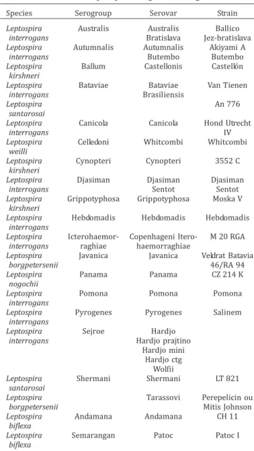
Samples were analyzed using MAT as recommended by the World Organization for Animal Health Animal (OIE) (Faine 1982; Office World Organization for Animal Health Animal 2009). Samples
were screened at 1:100 dilution using a battery of 29 live antigen serovar strains (Table 1).
Three animals died of natural causes during the study. Fragments of the heart, spleen, pancreas, intestine, liver and kidney were collected and processed for DNA extraction from these snakes by PCR: one jararaca-pintada (Bothrops pauloensis) from G1 and two caiçacas (Bothrops moojeni) from G2.
Genomic DNA was extracted from tissue samples using a com-mercially available DNA extraction (llustra Tissue & Cells genomic Prep Mini Spin® kit, GE Healthcare) and evaluated in
spectropho-tometer (NanoVue®, GE Healthcare).
The amplification of Leptospira spp. DNA was performed utilizing primersLEP-1 (5’-GGCGGCGCGTCTTAAACATG-3’) and LEP-2 (5’-TTCCCCCCATTGAGCAAGATT-3’) resulting in 330 pb (Mérein 1992). The process carried out in a 25 μL solution containing 1·Taq polymerase buffer (10mM Tris HCl pH 8.0, 50mM KCl), 1.5mM MgCl2, 200 μM dNTP, 10 pmol of each primer (LEP-1
and LEP-2), 10 ng of purified DNA and 0.2 U/μl of Taq Platinum DNA polimerase system (Invitrogen). A MasterCycler gradient (Eppendorf) with initial denaturation at 94oC for 3 min, followed
Table 1. Species, serogroups and respective serological variants for Leptospira diagnosis using MAT
Species Serogroup Serovar Strain
Leptospira Australis Australis Ballico
interrogans Bratislava Jez-bratislava
Leptospira Autumnalis Autumnalis Akiyami A
interrogans Butembo Butembo
Leptospira Ballum Castellonis Castellón
kirshneri
Leptospira Bataviae Bataviae Van Tienen
interrogans Brasiliensis
Leptospira An 776
santarosai
Leptospira Canicola Canicola Hond Utrecht
interrogans IV
Leptospira Celledoni Whitcombi Whitcombi
weilli
Leptospira Cynopteri Cynopteri 3552 C
kirshneri
Leptospira Djasiman Djasiman Djasiman
interrogans Sentot Sentot
Leptospira Grippotyphosa Grippotyphosa Moska V
kirshneri
Leptospira Hebdomadis Hebdomadis Hebdomadis
interrogans
Leptospira Icterohaemor- Copenhageni Itero- M 20 RGA
interrogans raghiae haemorraghiae
Leptospira Javanica Javanica Veldrat Batavia
borgpetersenii 46/RA 94
Leptospira Panama Panama CZ 214 K
nogochii
Leptospira Pomona Pomona Pomona
interrogans
Leptospira Pyrogenes Pyrogenes Salinem
interrogans
Leptospira Sejroe Hardjo
interrogans Hardjo prajtino
Hardjo mini Hardjo ctg
Wolfii
Leptospira Shermani Shermani LT 821
santarosai
Leptospira Tarassovi Perepelicin ou
borgpetersenii Mitis Johnson
Leptospira Andamana Andamana CH 11
biflexa
Leptospira Semarangan Patoc Patoc I
by 30 cycles at 94oC for 1 min, then at 63oC for 1 min and 30 sec
and a final extension at 72oC for 2 min was used.
Aliquots of the PCR products, along with a 100 bp DNA ladder (Invitrogen), were loaded into 2% agarose gel (Invitrogen) stained with SYBR® safe (Invitrogen) and submitted to electrophoresis in
TBE buffer (0.09 M Tris–HCl, 0.09 M boric acid, 2mM EDTA, pH 8.3) for 60 min at 80V (Electrophoresis Power Supply Model EPS 301; GE Healthcare). The amplified DNA fragments were observed through GelDoc-IT™ Imaging System, using Vision Works® LS
Software.
The amplicons were purified by using ExoSap (USB) and the sequencing reactions were carried out on both strands in a MegaBace™1000 (GE-Healthcare). Sequences were analyzed using Chromas 2.3 and Mega 4 software and compared with NCBI database by using BLAST search in GenBank (National Center for Biotechnology Information, Washington, D.C., http://www.ncbi. nlm.nih.gov/BLAST).
Tukey test was employed to compare proportions with a = 0.05 (Zar 1999) and to study epidemiological variables and serological test for leptospirosis.
RESULTS
Of the 147 snakes studied, 52 (35.4%) were seropositive for
leptospirosis by MAT, with eight (15.4%) in G1 (WS), 34
(65.4%) in G2 (IC) and ten (19.2%) in G3 (CC), according
Table 2. Table 3 shows the number of snakes positive for
Lep-tospira
using MAT according to group, specie, serovar and
titers. Titers varied from 100 to 3200 and Hardjo prajtino
was the most prevalent serovar (88.5%, N=46/52), as shown
in Table 3. Jararaca (
Bothrops jararaca
) (66.7%, N=22/33)
showed the highest average titer among the three groups.
Positive amplifications of predictive amplicons of
Leptos-pira
spp.
were detected in the kidney of one jararaca-pintada
(
B. pauloensis
) (G1) with titer of 200 Hardjo prajtino and two
caiçacas (
B. moojeni
)
(G2) with titers of 200 and 1600 Hardjo
prajtino, as shown in Fig. 1. When submitted to BLASTn
analysis, all the purified and sequenced amplicons revealed
100% identity with
Leptospira interrogans
sequences
deposited in the GenBank, producing unambiguous fragments
Fig.1. Electrophoresis of the PCR products using primers Lep1 and Lep2 with products of 331 pb in samples of kidney and liver of Bothrops pauloensis and Bothrops moojeni. Lane 1-7. 1) 100bp DNA ladder (Invitrogen), 2) kidney Bothrops moojeni (Group 2), 3) kidney Bothrops moojeni (Group 2), 4) kidney Bothrops pauloensis (Group 1), 5) liver Bothrops pauloensis (Group 1), 6) positive control; 7) negative control.
Table 3. Number of snakes positive for Leptospira using MAT according to group, species, serovar and titers
Group N Serovar Titer (1/)
100 200 400 800 1600 3200
1 (WS) 34 Hardjo 2 Bp 1 Bl 2 Bj
prajtino
Sentot 2 Cdt Andamana 1 Cdt
2 (IC) 64 Hardjo 4 Bj 5 Bj 3 Bm 1Bj 3 Bm prajtino
1 Bm 3Bm 3Bm
4 Bp 4Bp 1Bp
1Cdt Hardjo mini 2 Bj 3 Bj 1 Bj
Patoc 1 Bj
Gryppo- 1 Bm
thyphosa
Pyrogenes 1 Bp
3 (CC) 48 Hardjo 1 Bj 1Bj 3 Bj 1 Bj 2 Bj
prajtino
Panama 1 Bca
WS = wild snakes; IC = intensive captivity; CC = collective captivity; N = number of snakes per group. Cdt = Crotalus durissus terrificus; Bm = Bo-throps moojeni; Bj = BoBo-throps jararaca; Bp = BoBo-throps pauloensis; Bl = Bothrops leucurus; Bca = Boa constrictor amarali.
from 364 bases (Table 4). Liver samples from
jararaca-pinta-da (
B. pauloensis-
G1) showed one lower band than expected
and 93% sequence identity with
Leptospira
spp. (Table 4).
DISCUSSION
Many animals are
Leptospira
spp. hosts, and each serotype
has one or more hosts with different adaptation degrees. The
Table 2. Comparison of antibodies to Leptospira seropositivityin the three groups and in the different species studied
Group N Species Non Reactive Total
reactive
1 (WS) 35 b Bothrops jararaca 4 2 A 6
Bothrops pauloensis 1 2 A 3
Crotalus durissus terrificus 17 3 B 20
Bothrops moojeni 1 0 B 1
Bothrops leucurus 0 1 B 1
Boa constrictor amarali 4 0 B 4
2 (IC) 64 a Bothrops jararaca 4 11 A 15
Bothrops moojeni 2 13 A 15
Bothrops pauloensis 5 9 A 14
Crotalus durissus terrificus 19 1 B 20
3 (CC) 48 b Boa constrictor amarali 11 1 B 12
Bothrops jararaca 3 9 A 12
Crotalus durissus terrificus 20 0 B 20
Spilotes pullatus 4 0 B 4
Total 86 52 147
studied snakes came from farms and pastures, where they
were probably infected by urine of affected cattle. According
to Correa et al. (1965/1967) the predominant serovar in
bovines is Hardjo prajtino. The seroprevalence of Hardjo
serovar is observed in slaughterhouses and farm employees,
suggesting that the exposure risk is related to direct and
indirect contact with infected animals (Santa Rosa et al. 1980).
Seroagglutination tests showed that bovines reacted
positively (45.6%) to serovar Hardjo according to Langoni
et al. (2008).
Homem et al. (2001), researching humans and other
ani-mals in Amazonia, observed that serovar Hardjo was the most
prevalent. Bovine leptospirosis is endemic in Brazil and may
cause abortions, infertility, anorexia, jaundice,
hemoglobinu-ria, mastitis and even death (Santa Rosa et al. 1972, Silva 1976,
Simpson 2002, Langoni et al. 2004). Out of four pregnant
jararacas (
Bothrops
)
included in this study, three layed atresic
eggs suggesting a possible influence of
Leptospira
on them.
These findings indicate that egg atresia may be related to
se-ropositivity to serovar Hardjo, however experimental studies
Table 4. Nucleotide sequence alignment and percentage of identity on liver and kidneysamples of Bothrops pauloensis and Bothrops moojeni
Sample Species/ Nucleotide sequence alignment % identity/
Group GenBank access
Liver Bothrops CGGAAAGGCTCAGACAGCGACTACTGTCCAG 93% (237/265)
pauloensis GCGTGCGACACTGCGCGGTCGATGTAACATCT AY461867.1
G1 GTGGCGGATAAAGCAATGGCCGACATGAGTC TCTGGGACTAACTTTCCAGTAACGGTGAAGCG TAACTACATGGATTGGTCCCGTAGGAGAGTCA ATAAGGATTTTTCGGGTAAAGATTTATTGCTC GGAGATGAGCCCGCGTCCGATTAGCTAGCTTG GTGATGTGTAACAGGCTCACCAAGGCGACGA TCGGTAGCCTGTGCCTGAGAGTGGTGTTCGGC CACAATGGAACTGAGACACGGTCCATACTCCT ACGGCGAGGCATGCAGTTAAGAATCTTGCTCA ATGGGGGGAAACATGG
Kidney Bothrops CCCTTCTTGGCGGCGCGTCTTAAACATGCAA 100% (364/364)
pauloensis GTCAAGCGGAGTAGCAATACTCAGCGGCGA AE010300.2
G1 ACGGGTGAGTAACACGTGGGTAATCTTCCTC TGAGTCTGGGATAACTTTCCGAAAGGGAAGC TAATACTGGATGGTCCCGAGAGATCATAAGA TTTTTCGGGTAAAGATTTATTGCTCGGAGAT GAGCCCGCGTCCGATTAGCTAGTTGGTGAGG TAAAGGCTCACCAAGGCGACGATCGGTAGC CGGCCTGAGAGGGTGTTCGGCCACAATGGA ACTGAGACACGGTCCATACTCCTACGGGAGG CAGCAGTTAAGAATCTTGCTCAATGGGGGGA ACCCTGAAGCAGCGACGCCGCGTGAACGAT GAA
Kidney Bothrops CTTCTTGGCGGCGCGTCTTAAACATGCAAGT 100% (364/364)
moojeni CAAGCGGAGTAGCAATACTCAGCGGCGAAC AE010300.2
G2 GGGTGAGTAACACGTGGGTAATCTTCCTCTG
AGTCTGGGATAACTTTCCGAAAGGGAAGCTA ATACTGGATGGTCCCGAGAGATCATAAGATT TTTCGGGTAAAGATTTATTGCTCGGAGATGA GCCCGCGTCCGATTAGCTAGTTGGTGAGGTA AAGGCTCACCAAGGCGACGATCGGTAGCCG GCCTGAGAGGGTGTTCGGCCACAATGGAACT GAGACACGGTCCATACTCCTACGGGAGGCA GCAGTTAAGAATCTTGCTCAATGGGGGGAAC CCTGAAGCAGCGACGCCGCGTGAACGATGA AATTCCT
and isolation of the agent are needed to corroborate this
hypothesis.
Several studies have been carried out to determine the
occurrence of
Leptospira
spp.
and antibodies to
Leptospira
spp.
in wild animals, but few include venomous or non
venomous snakes. In Brazil this research was first performed
by Hyakutake et al. (1976), in which agglutination tests
detected Icterohaemorrhagiae, Ballum, Canicola, Guaicurus,
Brasiliensis, Andamana and Javanica serovars. The
predominant serovar founded by this authors was Andamana
(80.6%, N=25/31) and do not corroborate with our results
that shown Hardjo prajtino as serovar predominant (88.5%).
This observation can be due the snakes came from different
locations, and also the inclusion of new serovars, but few
studies have been conducted until now.
Calle et al. (2001) found low-level
Leptospira
titers in
free-ranging Venezuelan anacondas (
Eunectes murinus
) and
sho-wed that this may represent previous infection, antibody
res-ponses to
Leptospira
antigens present in ingested prey, or
antibodies to
Leptospira
serovars that were not included in
the serologic panel.
Stanchi et al. (1986) analyzed 18 urutus-cruzeiro (
Bo-throps alternatus
) for the presence of antileptospiral
antibodies using a microscopic agglutination test, and
observed 72.2% (N=13/18) of positive results to Patoc,
Andamana, Wolffi, Tarassovi, Pomona, Pyrogenes and
Shermani serovars.
The high prevalence of positive isolates in G2, when
snakes were kept in individual boxes, may be explained by
Abdulla & Karstad (1962). These authors showed that snakes,
both free-ranging and captive, may be infected by ingesting
rodents and contaminated water. They also concluded that
snakes might transmit leptospirosis by direct contact. The
same authors, working with experimental serovar Pomona
infection, found spirochetes in the kidneys of these animals
six months after inoculation; interstitial nephritis was
observed in one of them. Therefore, snakes in captivity may
be infected by consuming infected mice or other food or
water contaminated with their urine. This fact is
demonstrated by the statistical difference between this group
and the other G2, even in the collective captivity group, which
has access to sunlight, water and more space per animal, as
previously indicated by Ferreira Junior et al
.
(2010).
Ferris et al. (1961) isolated
Leptospira interrogans
serovar
Ballum in an Eastern Hognose Snake (
Heterodon platgrhinus
).
According to Hyakutake et al. (1976) the isolation of serovar
Andamana in jararaca (
Bothrops pradoi
)
may contribute to
the transmission of leptospirosis to humans and other
animals. Andrews et al. (1965) observed the presence of
Lep-tospira ballum
agglutinins in the serum of snakes of Southern
Illinois, USA, as well as in other areas of the country. They
concluded that these findings demonstrate the feeding habits
of these animals.
According to Valentina & Fratini (2005), there are no data
of reptiles as source of infection of
Leptospira
to humans,
though they may have direct contact with spirochetes in the
environment. In the present study, there were animals with
high titers such as 3200, but without any clinical signs.
Reptiles rarely show clinical disease when infected with
Lep-tospira
spirochetes, although seropositive snakes of various
species have been reported with over 6400 titers (Santa Rosa
et al. 1972, Cordeiro et al.1981).
MAT showed that snakes came into contact with the
pathogen, with titers ranging from 100 to 3200, PCR
demons-trated that these animals harbored
Leptospira interrogans
.
Several studies have been conducted employing the use of
PCR for detection of
Leptospira
spp. in urine, serum and tissue
of animals and humans (Gravekamp et al. 1993, Wagenaar et
al. 2000, Bonfim et al. 2008). However, PCR had never been
used to detect
Leptospira
spp. in snake tissues. Moreover, the
present work showed that both wildlife and captivity snakes
may maintain the pathogen without clinical signs.
Acknowledgements.- To Centro de Raízes Tropicais (Cerat)/ Unesp-Botu-catu, SP, for facilities; to FAPESP for financial support (2008/51364-4); to Prof. Dr. Lidia Raquel de Carvalho for statistical analyses; and to Juliana S. Simionato for English support.
REFERENCES
Abdulla P.K. & Karstad L. 1962. Experimental infections with Leptospira pomona in snakes and turtles. Zoonoses Res. 1:295-306.
Adler B. & Moctezuma A.P. 2010. Leptospira and leptospirosis. Vet. Microbiol. 140:287-296.
Aguiar D.M., Gennari S.M., Cavalcante G.T., Labruna M.B., Vasconcellos S.A., Rodrigues A.A.R., Moraes Z.M. & Camargo L.M.A. 2006. Seroprevalence of Leptospira spp. in cattle from Monte Negro municipality, western Amazon. Pesq. Vet. Bras. 26:102-104.
Andrews R.D., Reilly J.R., Ferris D.H. & Hanson L.E. 1965. Leptospiral agglutinins in sera from southern Illinois herpetofauna. Bull. Wild. Dis. Assoc. 1:55-59.
Bonfim M.R.Q., Barbosa-Stancioli E.F. & Koury M.C. 2008. Detection of pathogenic leptospires in urine from naturally infected cattle by nested PCR. Vet. J. 178:251-256.
Blancou J., Chomel B.B., Belotto A. & Meslin F.X. 2005. Emerging or re-emerging bacterial zoonoses: factors of emergence, surveillance and control. Vet. Res. 36:507-522
Calle P.P., Rivas J., Muñoz M., Thorbjarnarson J., Holmstrom W. & Karesh W.B. 2001. Infectious disease serologic survey in free-ranging Venezuelan anacondas (Eunectes murinus). J. Zoo Wildl. Med. 32:320-323.
Cordeiro F., Sulzer C.R. & Ramos A.A. 1981. Leptospira interrogans in seve-ral wildlife species in Southeast Brazil. Pesq. Agropec. Bras., Sér. Vet. 1:19-29.
Correa M.O.A., Hyakutake S., Natale V., Galvão P.A. & Aguiar H.A. 1965/1967. Estudos sobre a Leptospira wolffi em São Paulo. Revta Inst. Adolfo Lutz, São Paulo, 25/27:11-25.
Faine S. 1982. Guideline for the Control of Leptospirosis. 2nd ed. WHO,
Geneva, Offset Publication 67:171.
Ferreira Junior R.S., Biscola N.P., Campagner M.V. & Barraviera B. 2010. How to raise snakes in captivity? Vet. Microbiol. 141:189.
Ferris D.H., Rhoades H.E., Hanson L.E., Galton M. & Mansfield M.E. 1961. Research into the nidality of Leptospira ballum in campestral hosts inclu-ding the hog-nosed snake (Heterodon platgrhinus). Cornell Vet. 51:405-419.
Ghneim G.S., Viers J.H., Chomel B.B., Kass P.H., Descollonges D.A. & Johnson M.L. 2007. Use of a case-control study and geographic information systems to determine environmental and demographic risk factors for canine leptospirosis. Vet. Res. 38:37-50
Gravekamp C., Van de Kemp H., Franzen D., Carrington D., Schoone G.J., Van Eys G.J.J.M., Everard C.O.R., Hartskeerl R.A. & Terpstra W.J. 1993. Detection of seven species of pathogenic leptospires by PCR using two sets of primers. J. Gen. Microbiol. 139:1691-1700.
Lep-tospira spp. in horses of the urban area of Londrina, Paraná, Brazil. Revta. Inst. Med. Trop. 49:327-330.
Homem V.S.F., Heinemann M.B., Moraes Z.M., Vasconcellos S.A., Ferreira F. & Ferreira Neto J.S. 2001. Estudo epidemiológico da leptospirose bovina e humana na Amazônia oriental brasileira. Revta. Inst. Med. Trop. 34:173-180.
Hyakutake S., Biasi P.D., Santa Rosa C.A. & Belluomini H.E. 1976. Contribui-ção ao estudo epidemiológico das leptospiroses em serpentes do Brasil. Rev. Inst. Med. Trop. 18:10-16.
Hyakutake S., Biasi P.D., Belluomini H.E. & Santa Rosa C.A. 1980. Leptospi-roses in Brazilian snakes. Int. J. Zoonoses 7:73-77.
Langoni H., Da Silva A.V., Pezerico S.B. & De Lima V.Y. 2004. Anti-leptospire agglutinins in equine sera, from São Paulo, Goias, and Mato Grosso do Sul, Brazil. 1996-2001. J. Venom. Anim. Toxins incl. Trop. Dis. 10:207-218.
Langoni H., Souza L.C., Da Silva A.V., Cunha E.L.P. & Silva R.C. 2008. Epide-miological aspects in leptospirosis: Research of anti-Leptospira spp. an-tibodies, isolation and biomolecular research in bovines, rodents and workers in rural properties from Botucatu, SP, Brazil. Braz. J. Vet. Res. Anim. Sci. 45:190-199.
Mérien F., Amouriaux P., Perolat P., Baranton G. & Saint Girons I. 1992. Po-lymerase chain reaction for detection of Leptospira spp. in clinical samples. J. Clin. Microbiol. 30:2219-2224.
Romero E.C., Yasuda P.H. 2006. Molecular characterization of Leptospira sp. strains isolated from human subjects in São Paulo, Brazil using a po-lymerase chain reaction-based assay: A public health tool. Mem. Inst. Oswaldo Cruz 101:373-378.
Santa Rosa C.A., Sulzer C.R. & Castro A.F.P. 1972. A new leptospiral serotype in the Bataviae group, isolated in São Paulo, Brazil. Am. J. Vet. Res. 33:1719-1721.
Santa Rosa C.A., Sulzer C.R., Castro A.F.P., Yanaguita R.M. & Giorgi W. 1980. Two new serovars in the hebdomadis group isolated from cattle in Brazil. Int. J. Zoonoses 7:158-163.
Silva I. 1976. A new leptospiral serotype isolated in Salvador, Bahia State. Revta Microbiol., São Paulo, 73:5-37.
Simpson V.R. 2002. Wild animals as reservoirs of infectious diseases in the UK. Vet. J. 163:128-146.
Stanchi N.O., Grisolía C.S., Martino P.E. & Peluso F.O. 1986. Presence of antileptospira antibodies in ophidia in Argentina. Revta Argent. Microbiol. 18:127-130.
Valentina V.E. & Fratini F. 2005. Bacterial zoonoses among domestic reptiles. Zoonosi batteriche trasmesse da rettili domestici. Ann. Fac. Med. Vet. 58:85-91.
Wagenaar J.A., Zuerner R.L., Alt D. & Bolin C.A. 2000. Comparison of poly-merase chain reaction assays with bacteriologic culture, immunofluo-rescence, and nucleic acid hybridization for detection of Leptospira borgpetersenii serovar hardjo in urine of cattle. Am. J. Vet. Res. 61:316-320.
Weekes C.C., Everard C.O.R. & Levett P.N. 1997. Seroepidemiology of canine of leptospirosis on the island of Barbados. Vet. Microbiol. 51:215-222. World Organization for Animal Health Animal, OIE. Downloaded in April
2010 from http://www.oie.int/.