Low-dose effects of Bisphenol A on human primary
vascular endothelial cells and colon cancer cells
TESE APRESENTADA PARA OBTENÇÃO DO GRAU DE DOUTOR EM BIOLOGIA
EDNA SORAIA GREGÓRIO RIBEIRO VARANDAS
ORIENTADORA: Doutora Maria Margarida Cabrita Xavier Delgado COORIENTADORA: Doutora Maria Wanda Sarujine Viegas
JÚRI:
Presidente: Reitor da Universidade de Lisboa
Vogais: Doutor José Alexandre de Gusmão Rueff Tavares Professor Catedrático
Faculdade de Ciências Médicas da Universidade Nova de Lisboa Doutora Elizabeth da Costa Neves Fernandes de Almeida Duarte Professora Catedrática
Instituto Superior de Agronomia da Universidade de Lisboa Doutora Maria da Graça Ribeiro Campos
Professora Auxiliar com agregação
Faculdade de Farmácia da Universidade de Coimbra Doutora Catarina Isabel Guerra Rodrigues Mansilha Investigadora Auxiliar
Instituto Nacional de Saúde Doutor Ricardo Jorge Doutora Maria Margarida Cabrita Xavier Delgado Investigadora Auxiliar
Instituto Superior de Agronomia da Universidade de Lisboa
LISBOA 2014
À minha filha;
aos meus pais;
ao meu marido;
aos meus amigos.
Low-dose effects of Bisphenol A on human primary vascular
endothelial cells and colon cancer cells
Abstract
Abstract
Abstract
Abstract
Bisphenol A (BPA) is an extensively utilized endocrine disruptor for which human exposure is considered generalized through ingestion. Information regarding BPA effects on vascular and digestive tract tissues is scarce. Therefore, in this work primary Human Umbilical Vein Endothelial Cells (HUVEC) and human colon adenocarcinona cell line HT29 were used to evaluate BPA effects at two distinct low-dose concentrations relevant in terms of human health risk assessment.
BPA differentially affects the cell types studied, with more pronounced aneugenic effects, nucleolar disruption and transcriptional deregulation observed in HUVEC. Prolonged BPA exposure affects aging processes in senescent HUVEC. Interaction experiments involving expression of key cancer related genes shows that BPA antagonizes transcriptional effects of the chemotherapeutic agent doxorubicin in HT29. Additionally BPA aneugenic effects are enhanced by co-exposure with Eupatorium cannabinum L. ethanolic extract, a medicinal plant, for which a potent cytotoxic activity against HT29 cells is also demonstrated here.
Altogether these results support increasing concerns regarding harmful effects of BPA at low-dose on human health and draw attention to the importance of a deeper understanding of BPA potential interactions with other chemicals.
Key-words: Bisphenol A (BPA), HUVEC, HT29, Gene transcription, Drug interaction.
Efeitos de doses baixas de Bisfenol A em células endoteliais
vasculares primárias e em células de cancro de cólon humanas
Resumo
Resumo
Resumo
Resumo
O bisfenol A (BPA) é um desregulador endócrino amplamente utilizado para o qual a exposição humana é considerada generalizada através de ingestão. A informação sobre os efeitos do BPA nos tecidos vasculares e digestivos é no entanto reduzida. Assim, neste trabalho foram utilizadas células endoteliais da veia do cordão umbilical humanas (HUVEC) e uma linha celular de adenocarcinoma de cólon HT29 para avaliar os efeitos de duas concentrações de BPA, consideradas doses baixas e relevantes para avaliação de risco para a saúde humana.
No que respeita à indução de aneugenia, alterações nucleolares e desregulação da transcrição génica foram observados efeitos mais pronunciados do BPA em HUVEC comparativamente com HT29. A exposição prolongada ao BPA interfere com o processo de envelhecimento em células HUVEC. A avaliação de interações em HT29 revelou um efeito antagonista do BPA, na transcrição de genes associados ao cancro, relativamente ao agente quimioterapêutico doxorrubicina. Também em HT29 foi observado um aumento dos efeitos aneugénicos do BPA após exposição conjunta com extrato etanólico da planta medicinal Eupatorium cannabinum L., para o qual foi também demonstrada uma potente atividade citotóxica.
Os resultados obtidos suportam a crescente preocupação com os efeitos nocivos do BPA na saúde humana e acentuam importância de uma avaliação aprofundada das possíveis interações do BPA com outros compostos químicos.
Palavras-chave: Bisfenol A (BPA), HUVEC, HT29, Transcrição génica, Interação entre compostos químicos.
Preamble
Preamble
Preamble
Preamble
Bisphenol A (BPA) is one of the greatest endocrine disruptor chemicals produced worldwide, employed in a wide variety of consumer products including food and drink containers. Human exposure to BPA through oral intake is considered generalized, at least in developed countries. However the levels of BPA internal exposure and its potential harmful outcomes to human health are still controversial. Extensive research in human cell lines, particularly from sex hormone responsive tissues, show that BPA can endorse very distinct cellular responses through estrogen receptor signalling pathways. Although BPA is absorbed in the gut and enters blood circulation, its effects on cells from digestive or vascular systems are largely unknown.
Therefore, in this research project two distinct cell types: Human Umbilical Vein Endothelial Cells (HUVEC), an in vitro model for vascular cells, and the human cell line HT29, originated from a colon adenocarcinona, the most common type of gastrointestinal cancer were utilized. Two BPA concentrations (44 nM and 4.4 µM), relevant in the context of risk assessment, were selected for evaluation of cellular proliferation and viability, aneugenic capacity, nucleolar organization and gene transcription in both cell types. Considering that distinct epidemiological studies correlate circulating BPA levels with age-related vascular diseases HUVEC were used to assess the effects of BPA prolonged exposure on aging cells. Furthermore there are some evidences that the action of chemotherapeutic drugs can be antagonized by BPA. Hence this work aimed also to evaluate in HT29 cells potential interactions of BPA with Doxorubicin (DOX), commonly used in chemotherapy, as well as with the extract of
Eupatorium cannabinum L., a plant utilized in alternative medicine for treatment
of several pathologies including cancer.
______________________________________________________________________
______________________________________________________________________ 1
List of contents
1 INTRODUCTION ... 3
1.1 HUMAN EXPOSURE AND BIOMONITORING ... 4
1.2 METABOLISM AND TOXICOKINETICS ... 6
1.3 MECHANISMS OF ACTION AS AN EDC ... 7
1.4 LOW DOSES EFFECTS ON HUMAN CELLS ... 8
1.4.1 Cell proliferation and viability ... 9
1.4.2 Transcriptional and epigenetic effects ... 11
1.5 BPA INTERACTIONS ... 11
2 BISPHENOL A AT CONCENTRATIONS FOUND IN HUMAN SERUM INDUCES ANEUGENIC EFFECTS IN ENDOTHELIAL CELLS ... 13
2.1 ABSTRACT ... 14
2.2 INTRODUCTION ... 14
2.3 MATERIALS AND METHODS ... 16
2.4 RESULTS AND DISCUSSION ... 20
2.4.1 Low BPA concentrations do not affect cell proliferation or viability independently of GPR30. 20 2.4.2 Low BPA concentrations induce micronuclei formation without affecting DNA integrity. . 23
2.4.3 BPA differentially affects genes related with chromosome segregation. ... 26
2.4.4 Low concentrations of BPA disrupt mitotic stability and organization in HUVEC. ... 28
2.5 CONCLUSION ... 30
2.6 REFERENCES ... 30
3 THE ENVIRONMENTAL POLLUTANT BISPHENOL A INTERFERES WITH NUCLEOLAR STRUCTURE .. 33
3.1 ABSTRACT ... 34
3.2 INTRODUCTION ... 34
3.3 MATERIALS AND METHODS ... 35
3.4 RESULTS ... 37
3.4.1 Nucleolar organization is affected by BPA exposure. ... 37
3.4.2 BPA effects on nucleolin transcription are cell specific and independent of rRNA levels. .. 39
3.4.3 Bisphenol A alters H3 epigenetic marks at the nucleolar region. ... 39
3.5 DISCUSSION ... 40
3.6 REFERENCES ... 42
4 BISPHENOL A DISRUPTS TRANSCRIPTION AND DECREASES VIABILITY IN AGING VASCULAR ENDOTHELIAL CELLS ... 43
4.1 ABSTRACT ... 44
4.2 INTRODUCTION ... 44
4.3 MATERIALS AND METHODS ... 46
4.4 RESULTS AND DISCUSSION ... 50
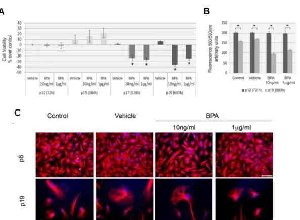
4.4.2 Continuous BPA exposure reduces viability in aging HUVEC. ... 53
4.4.3 Continuous BPA exposure induces differential gene expression in aging HUVEC. ... 56
4.5 CONCLUSION ... 58
4.6 REFERENCES ... 59
5 BISPHENOL A AT THE REFERENCE LEVEL COUNTERACTS DOXORUBICIN TRANSCRIPTIONAL EFFECTS ON CANCER RELATED GENES IN HT29 CELLS ... 62
5.1 ABSTRACT ... 63
5.2 INTRODUCTION ... 63
______________________________________________________________________
______________________________________________________________________ 2
5.4 RESULTS ... 69
5.4.1 BPA at reference level increases transcription of bcl-xl. ... 69
5.4.2 BPA does not affect gene expression alterations induced by low DOX concentration. ... 69
5.4.3 Gene expression alterations induced by DOX at therapeutic concentration are reverted by BPA. 70 5.4.4 DOX sub-therapeutic and therapeutic concentrations have differential effects on gene transcription. ... 70
5.4.5 BPA decreases DOX-induced apoptosis without immediate effects on cell viability. ... 71
5.4.6 BPA does not affect mitotic disruption induced by DOX. ... 73
5.5 DISCUSSION ... 74
5.6 CONCLUSION ... 78
5.7 REFERENCES ... 78
6 CYTOTOXICITY OF EUPATORIUM CANNABINUM L. ETHANOLIC EXTRACT AGAINST COLON CANCER CELLS AND INTERACTIONS WITH BISPHENOL A AND DOXORUBICIN ... 81
6.1 ABSTRACT ... 82
6.2 INTRODUCTION ... 83
6.3 MATERIALS AND METHODS ... 84
6.4 RESULTS ... 88
6.4.1 E. cannabinum ethanolic extract decreases HT29 cell viability. ... 88
6.4.2 E. cannabinum ethanolic extract induces alterations in nuclear structure and mitotic disruption. ... 89
6.4.3 E. cannabinum ethanolic increases Bisphenol A induced mitotic disruption. ... 92
6.4.4 Cytotoxic effects of Doxorubicin are enhanced by E. cannabinum. ... 93
6.5 DISCUSSION ... 95
6.6 CONCLUSION ... 98
6.7 REFERENCES ... 99
7 GENERAL DISCUSSION ... 102
8 REFERENCES TO INTRODUCTION AND GENERAL DISCUSSION ... 106
9 LIST OF ABBREVIATIONS ... 113
______________________________________________________________________
______________________________________________________________________ 3
1 Introduction
Bisphenol A (BPA), 2,2-bis(4-hydroxyphenyl) propane is an organic compound and one of the greatest volume industrial chemicals utilized in the world, with a production 4.6 million tonnes in 2012 (MRC 2014). BPA was first synthesized by A. P. Dianin in 1891 and its estrogenic properties were hypothesized in the search for synthetic estrogens in the 1930s, however BPA was abandoned for pharmaceutical use when Diethylstilbestrol (DES) was found to be more efective (reviwed in Vandenberg et al. 2009). Since 1940, BPA has been extensively used in the production of a variety of polymers such as polycarbonate plastics and epoxy resins and therefore employed in the manufacture of a variety of indoor applications and consumer products (Geens et al. 2011). In the 60´s several studies emerged focused on the hypersensitivity and metabolism of BPA in different model systems (Fregert and Rorsman 1962; Gaul 1960; Knaak and Sullivan 1966) and since then extensive research has been conducted on the effects of this endocrine disrupting chemical (EDC) in both animals and humans (reviewed in Rochester 2013; Vandenberg et al. 2009). Importantly, in 1993 Krishnan and coworkers accidentally discovered that BPA leaches from autoclaved polycarbonate flasks and showed for the first time its positive effect on the proliferation rate of the human cell line MCF-7 (Krishnan et al. 1993). In 2001 the United States National Toxicology Program’s Report of the Endocrine Disruptors Low-Dose Peer Review recognized that there was credible but not conclusive evidence that low doses of BPA can cause effects on specific endpoints (NTP 2001). Since 2006, the European Food Safety Authority (EFSA) conducted several scientific assessments on BPA, repeatedly concluding that there is no concern for human health (EFSA 2014). However in 2011 the European Legislation banned the use of BPA in the manufacture of baby bottles (EC 2011) and in 2012 EFSA decided to carry out a new risk assessment of BPA that is currently in progress (EFSA 2014).
______________________________________________________________________
______________________________________________________________________ 4
1.1 Human exposure and biomonitoring
Environmental human exposure to BPA is considered a generalized phenomenon, at least in developed countries, since analysis of tissue and fluid samples reveal the presence of BPA in the majority of the individuals analysed (Geens et al. 2012). Ingestion of contaminated food is estimated to contribute with more than 90% to the overall BPA environmental exposure for all age groups and a recent intervention study showed that the removal of packed food from diet results in a significant reduction of urine BPA levels (Rudel et al. 2011). Non-dietary sources, such as air or contact, are generally considered to be significant only for occupationally exposed individuals (Geens et al. 2012). In 2004 the European Legislation established the specific migration limit (SML) for BPA on plastic materials and articles intended to come into contact with food at 0.6 mg/kg food (EC 2004). This SML value was based on an adult average body of 60 kg, the consumption 1 kg of food, and a tolerable daily intake (TDI) of 0.01 mg/kg bw (EC 2002). Although the TDI value was later raised to 0.05 mg/kg bw due to the elimination of an uncertainty factor of 5 concerning reproduction and development (EFSA 2006, 2010), the SML value was not altered. The actual TDI for BPA was derived from a comprehensive tree-generation study in rats with a No Observable Adverse Effect Level (NOAEL) of 5 mg/kg body weight/day and the application of uncertainty factor of 100 (10 for inter-species differences, 10 for inter-individual differences) (Tyl et al. 2002). This value corresponds also to BPA oral reference dosage (RfD) established by the U.S. Environmental Protection Agency (EPA) (EPA 1993) based on a rat chronic toxicity study regarding mean body weight with a Lowest Observable Adverse Effect Level (LOAEL) of 50 mg/kg body weight/day considering an uncertainty factor of 1000 (10 for inter-species differences, 10 for inter-individual differences and 10 for the uncertainty of duration of the toxic effects) (NTP 1982).
Human exposure to BPA has been estimated either by direct measurement on human biological samples or from dietary studies where concentrations in food and beverages were measured and aggregate exposure derived from food and water consumption data (reviewed in Sekizawa 2008). For the past years
______________________________________________________________________
______________________________________________________________________ 5
urinary concentrations of total (free and conjugated) BPA have been evaluated in order to assess BPA exposure as the matrix of choice for biomonitoring studies (Calafat et al. 2008; Vandenberg et al. 2010). The most recent worldwide biomonitoring study base on data from urinary BPA concentrations estimated human exposure to be 0.27 µg/kg body weight/day for the general population, 0.78 µg/kg body weight/day for children and 0.45-1.61 µg/kg body weight/day for infants (WHO 2010). Moreover the scientific EFSA panel on “food additives, flavourings, processing aids and materials in contact with food” assumed more conservative scenarios and estimated that human exposure to BPA is in average of 1.5 µg/kg body weight/day (EFSA 2006). These results correspond to approximately 100 to 10 fold less than the TDI. Accordingly data from urinary excretion also estimate the daily intake of BPA at levels considerably lower than the TDI. Urinary excretion data analysis on US population survey NHANES 2005-2006 resulted in median intake levels of 0.032–0.036 µg/kg body weight/day with a upper level of 0.255 µg/kg body weight/day (Lakind and Naiman 2010). Identical values where also obtained through the compilation of distinct independent studies (Geens et al. 2012). However, these values are still controversial since biomonitoring data from non-urinary human samples point to higher human BPA exposure. Considerably high concentrations of BPA have been measured in human placental samples as well as in fetal serum indicating that the placenta does not work as a barrier to BPA and therefore that human developing fetuses are chronically exposed to BPA (Schonfelder et al. 2002). Accordingly, several inconsistencies have also been described between the estimate intake levels and biomonitoring studies performed in human blood/serum samples. These studies demonstrate that the long term daily intake of BPA leads to a steady-state presence of unmetabolized BPA in the range of 0.5–3 ng/ml (2–13 nM) (Vandenberg et al. 2007; 2010), which is about 10-fold higher than the worst case predictions for daily human exposure to BPA (Völkel et al. 2002). Also physiologically based model studies estimate that for adult humans these levels of unmetabolized BPA in blood would require dosages considerably higher than the TDI (Edginton and Ritter 2009; Fisher et al. 2011). Overall this suggests that either BPA intake is higher than estimated or that this chemical bioaccumulates in the body.
______________________________________________________________________
______________________________________________________________________ 6
On the other hand, for the past years epidemiological studies have established positive correlations between urinary or blood BPA concentrations and the prevalence of recurrent human diseases. These include thyroid hormone disruption (Wang et al. 2013); reproductive malfunctions (Ehrlich et al. 2012; Li et al. 2011), type-2 diabetes mellitus (Silver et al. 2011), obesity (Trasande et al., 2012) and pathogenesis of age-related diseases such as coronary and carotid atherosclerosis (Lind and Lind 2011; Melzer et al. 2010; Melzer et al. 2012).
1.2 Metabolism and toxicokinetics
The metabolism and toxicokinetics of BPA has been studied in rodents (Doerge et al. 2010a, 2011), non-human primates (Doerge et al. 2010b; 2011) and humans (Völkel et al. 2002; 2005). After ingestion, BPA is absorbed from the gastrointestinal tract and undertakes a rapid phase II metabolism in the gut and liver. BPA is extensively conjugated with glucuronic acid to BPA-glucuronide resulting in loss of estrogenic activity and consequent low circulating levels of active (free) BPA (Hengstler et al. 2011). Although BPA glucuronidation is the primary mode of phase II metabolism in both rodents and primates there are some differences regarding excretion. In primates, including humans, blood BPA clearance occurs in the kidney and BPA-glucuronide represents the major BPA component in urine (Fisher et al. 2011; Tominaga et al. 2006). In contrast in rodents the conjugated BPA undergoes enterohepatic recirculation and fecal excretion via bile (Pottenger et al. 2000). It has been suggested that this difference can result in higher internal exposure in rodents due to longer elimination time and this argument was used to maintain the TDI level based on NOAEL obtained in rats since an uncertainty factor of 100 was considered conservative (EFSA 2008). Nevertheless there are evidences that regardless of the route of excretion the bioavailability of BPA is similar in rodents and primates (Taylor et al. 2011; Uchida et al. 2002).
Evaluation of BPA excretion kinetic profile in a limited number of human volunteers subjected to a single oral dose of deuterated BPA revealed a rapid peak and a terminal half-life of less than 6 hours (Völkel et al. 2002; 2005). This data has been interpreted as indicating a rapid and complete BPA clearance in
______________________________________________________________________
______________________________________________________________________ 7
humans. However, large-scale biomonitoring data revealed that urine BPA levels do not decline rapidly with fasting time suggesting a longer BPA clearance (Stahlhut et al. 2009) what is also supported by the fact that in the 2002 Volkel study no significant removal of conjugated BPA was detected beyond 20 h of exposure (Teeguarden et al. 2005). The occurrence of BPA-glucuronide deconjugation by β-glucuronidases in specific organs and consequent conjugation–deconjugation cycling has been suggested as an explanation for the delayed excretion (reviewed in Ginsberg and Rice 2009; Vandenberg et al. 2009). Additionally, single oral bolus experiments may underestimate exposure to bioactive form of BPA. Steady state internal BPA concentrations in humans were predicted to occur in continuous exposure trough diet (Mielke and Gundert-Remy 2009) and in mice it was shown that BPA bioavailability for equivalent BPA dosages is lower for a single oral bolus administration than in continuous exposure through food (Sieli et al. 2011).
1.3 Mechanisms of action as an EDC
Although BPA estrogenic activity has long been acknowledged, it has been considered a weak estrogen due to the fact that its binding affinity to classical estrogen receptors α and β (ERα and ERβ) is 10,000- and 1,000-fold lower than that of endogenous estrogen estradiol (E2) for ERα and ERβ, respectively (Routledge et al. 2000). However, distinct studies have shown that BPA can promote estrogen-like effects similar or stronger than E2. BPA at a dose range of 0.1-1 nM was shown to be equally effective, or even more effective, than equimolar concentrations of E2 in suppressing adiponectin release from human adipose tissues (Hugo et al. 2008). In BG-1 ovarian cancer cells both BPA and E2 induce cellular proliferation by promoting the interaction between ERα and IGF-1R signaling pathways (Hwang et al. 2013b). BPA induction of alternative signaling pathways is as well a possible explanation for the paradigm of E2 and BPA equivalent effects. Interestingly, BPA binds to the orphan nuclear receptor estrogen-related receptor-γ (ERR-γ) (Matsushima et al. 2007) with 80-100 times higher affinity than for ERα or ERβ (Takayanagi et al. 2006; Thomas and Dong 2006). Also activation of membrane-bound variants of ERs, particularly ERα, that act outside the nucleus was been suggested as a possible BPA mode of
______________________________________________________________________
______________________________________________________________________ 8
action (reviewed in Alonso-Magdalena et al. 2012). Additionally, cellular responses to BPA at low concentrations have been correlated with the trans-membrane estrogen receptor G protein-coupled estrogen receptor (GPR30 or GPER) for which BPA has relative high affinity corresponding to 2.8% of that of E2 (Shanle and Xu 2010; Thomas and Dong 2006). BPA induces rapid activation of ERK signaling pathway through GPR30 in breast cancer cells and cancer-associated fibroblasts (Dong et al. 2010) resulting in increased cell proliferation and migration (Pupo et al. 2012). Hence BPA can affect gene transcription trough nuclear and membrane-bound estrogen receptors. On the other hand, it is worth noting that hormones and EDCs, including BPA, responses do follow non-monotonic dose responses (NMDR) curves. In the case of BPA NMDR were demonstrated to emerge on exposed pituitary, prostate and pancreatic cultured cells, since very low doses can induce significant effects that are not detectable at higher concentrations (reviewed in Vandenberg et al. 2012).
1.4 Low doses effects on human cells
BPA concentrations (equal or greater than 100 µM) are genotoxic and result in a severe decrease of cellular proliferation and viability (Bolli et al. 2008; Kim et al. 2007). Additionally BPA in a range of 50-200 µM directly interferes with mechanisms of cell division by targeting tubulin promoting microtubular polymerization, which results in the formation of multiplolar spindles in HeLa cells (George et al. 2008). BPA genotoxic aneugenic properties characterized by micronuclei formation was also reported in human lymphoblastoid cell lines such as AHH-1 with a dose range of 54.12-162.8 µM (Johnson and Parry 2008) as well as in MCL-5 at 22-132 µM and further associated with chromosome non-disjunction for the concentration range of 22-88 µM (Parry et al. 2002). On the other hand, similar genotoxic effects have been describe for both BPA and E2 in MCF-7 cells although achieved with 1000-fold higher doses of BPA than the natural hormone (Iso et al. 2006). Also in CHO-K1 cells E2 induces chromosome aberrations and aneuploidgenic effects at 250 µM and 50 µM concentrations respectively, whereas BPA had the same effects for 400 µM and 500 µM (Tayama et al. 2008).
______________________________________________________________________
______________________________________________________________________ 9
However, and considering the level of BPA exposure on human cells in vivo, the “low-dose” effects have been a focal point of numerous studies (reviewed in Sekizawa 2008; Teeguarden and Hanson-Drury 2013; Vandenberg et al. 2009; 2012). BPA “low-dose” effects were defined as biological changes occuring in the concentration range of typical human exposures or lower than the expected NOAEL level of 5000 µg/kg/day, used to establish the oral reference dosage (RfD) (NTP 2001). However a recent review demonstrated that the majority of in
vivo and in vitro toxicity studies have been performed within the BPA
concentration range of 0.1 nM to 10 µM (Teeguarden and Hanson-Drury 2013).
In vitro cell systems have been widely used in the assessment of low-dose BPA
effects however most studies have been directed to human cancer cell lines, particularly breast cancer revealing effects on proliferation and gene expression (reviewed in Vandenberg et al. 2012; Wetherill et al. 2007). It has been recently demonstrated that BPA have direct effects in vascular endothelial function (Andersson and Brittebo 2012) and acts as an E2 antagonist in colon cancer cells (Bolli et al. 2010). Nonetheless, the effects of environmental relevant levels of BPA in human vascular endothelial cells and digestive tract cells are mostly unknown, although these types of cells are in direct contact to BPA in vivo. 1.4.1 Cell proliferation and viability
Several studies that evaluate proliferation and viability effects of low BPA concentrations in human cells cultures reveal cell type specificities. While some cell types show a positive response to BPA (LaPensee et al. 2009; Ptak et al. 2011; Ricupito et al. 2009) in others no measurable effect is detected (Bolli et al. 2008; Kim et al. 2007; LaPensee et al. 2009). The divergence of the results has been correlated with distinct signaling pathways. BPA proliferative effects at concentrations as low as 1 nM, have been reported in ERα-positive human cell lines such as breast cancer MCF-7cell lines (Ricupito et al. 2009), human ductal breast epithelial tumor cell line (T47D) (LaPensee et al. 2009) and human ovarian carcinoma cell line (OVCAR-3) (Ptak et al. 2011). Accordingly, unresponsiveness to BPA was observed in ERα-negative ERβ-positive cell lines such as cervical cancer HeLa cells (Bolli et al., 2008) or breast cancer MDA-MB-468
cells
(Bolli et al. 2008; LaPensee et al. 2009). However, in SK-N-SH______________________________________________________________________
______________________________________________________________________ 10
neuroblastoma cells, also ERα-negative and ERβ-positive, increased proliferation induced by BPA (10 µM) was shown to be mediated by ERβ and associated with altered transcriptional levels of cell cycle-related genes (Zhu et al. 2009). On the other hand, in ERβ positive and ERα negative
human
testicular cancer cell line
JKT-1, signaling through the membrane boundGPR30 receptor results in enhanced cell proliferation for BPA concentrations ranging from 1 pM to 10 µM, (Bouskine et al. 2009).
Cell proliferation can be naturally disrupted in the aging process with loss of replicative capacity which is associated with alterations in cell structure and physiology (Debacq-Chainiaux et al. 2008; Hwang et al. 2009). A similar phenotype can also be observed after cell exposure to subcytotoxic doses of stressful agents in a process described as stress-induced premature senescence (Dumont et al. 2002; Toussaint et al. 2000) characterized by cell-type dependent alterations in cellular gene expression profiles (Debacq-Chainiaux et al. 2008; Shelton et al. 1999). A recent study revealed that BPA (10-100 nM) may enhance senescence in normal human mammary epithelial cells (HMEC), associated to deregulation of cell cycle regulatory genes (Qin et al. 2012). However, despite the knowledge that humans are in continuous contact to BPA in vivo, its effects on cellular aging processes remain largely unknown. On the other hand, EDCs and particularly BPA exposure has been positively correlated with decreased semen quality and male infertility, for which induction of germ cell apoptosis is considered a primary contributing factor (reviewed in Lagos-Cabre and Moreno 2012). BPA-induced apoptosis was also observed in human trophoblastic cells in vitro for a concentration of 1 µM (Morice et al. 2011) as well as in rat ovarian cells in vivo after prolonged exposure (Lee et al. 2013), indicating a potential negative role in reproductive function. Conversely, exposure to BPA results in apoptosis inhibition in human breast cancer cell line MCF-7 for a concentration range of 10 nM – 10 µM (Diel et al. 2002) as well in non-malignant epithelial breast cells from high risk donors for a concentration of 100 nM (Dairkee et al. 2013).
______________________________________________________________________
______________________________________________________________________ 11
1.4.2 Transcriptional and epigenetic effects
The effect of BPA exposure, in a low-dose concentration range on global gene expression profiles was evaluated in distinct cell lines including ER-positive and ER-negative Ishikawa endometrial cancer cells (Boehme et al. 2009; Naciff et al. 2010), breast cancer cells lines MCF-7 (Buterin et al. 2006), and T47D (Buterin et al. 2006) or human peripheral blood cells (PBMCs) (Wens et al. 2013). In general these studies demonstrate that BPA affects the transcriptional pattern of hundreds of genes involved in key cellular processes such as proliferation, division and apoptosis in a cell type and concentration dependent manner.
Furthermore, BPA-induced alterations on transcriptional patterns have been correlated to DNA methylation. The epigenetic effect of BPA was first demonstrated in mice after maternal exposure to BPA which resulted in decreased DNA methylation upstream of the Agouti gene what was prevented by maternal dietary supplementation with folic acid (Dolinoy et al. 2007). On human primary breast epithelial cells BPA low-dose exposure (4 nM) resulted in repressed expression and increased DNA methylation at CpG islands of LAMP3
(lysosomal-associated membrane protein 3) gene (Weng et al. 2010). Also BPA
exposure of normal-like human breast epithelial cells (MCF-10F) was shown to alter DNA methylation patterns of several genes including those involved in apoptosis and DNA repair (Fernandez et al. 2012). Although there is significant evidence that BPA is able to influence DNA methylation patterns, the available data regarding BPA effects on histone modifications is almost inexistent (reviewed in Singh and Li 2012). Still, in human breast cancer cell line MCF-7 it was shown that BPA exposure increases the expression of the histone methyltransferase EZH2 (enhancer of Zeste Homolog 2) and the overall level of histone H3 trimethylation at lysine 27 (H3K27me3) (Doherty et al. 2010).
1.5 BPA interactions
In addition to the direct cellular effects induced by BPA, this chemical is also capable to interact with other compounds including natural estrogenic hormones
______________________________________________________________________
______________________________________________________________________ 12
such as E2. In MCF-7 breast cancer cell line combined exposure to BPA at low dose and E2 at physiological concentration induces cell proliferation and decreases apoptosis (Mlynarcikova et al. 2013). Also in colon cancer cell line DLD-1 E2 pro-apoptotic action is antagonized by BPA through inhibition of cascade 3 activation (Bolli et al. 2010). Interestingly, it was also shown that BPA at environmental relevant dosages (0.01 – 10 nM) is as effective as E2 in antagonizing cytotoxicity of the chemotherapeutic agent cisplantin in breast cancer cells T47D and MDA-MB-468 (LaPensee et al. 2010). Moreover, in the same cells lines low BPA concentrations (0.1-10 nM) were also reported to antagonize the cytotoxicity of vinblastine and doxorubicin (DOX), also commonly used in cancer chemotherapy. Relevantly, in the case of BPA/DOX interaction the same study demonstrated that the BPA antagonistic effects were independent from classical estrogen receptors and potentially associated with increased expression of antiapoptotic proteins (LaPensee et al. 2009).
BPA interactions with plant-derived compounds have also been demonstrated. A few studies have suggested that some plants extracts may effectively antagonize BPA induced cytotoxicity. In hepatoma cells HepG2 two distinct medicinal plant combinations were able to antagonize the cytotoxicity of a Bisphenol-A/Atrazine mixture by restoring cellular viability up to 24-28% (Gasnier et al. 2011). In human red blood cells BPA-induced hemolysis was decreased by black tea extract or the flavonoid quercetin (Verma and Sangai 2009). Also genistein, a soy phytoestrogen was reported to efficiently suppress BPA ERα mediated proliferation trough inhibition of cell cycle progression in ovarian cancer cell line BG-1(Hwang et al. 2013a).
______________________________________________________________________
______________________________________________________________________ 13
2
Bisphenol A at concentrations found in
human serum induces aneugenic effects in
endothelial cells
Ribeiro-Varandas E, Viegas W, H Sofia Pereira, Delgado M. 2013. Bisphenol A at concentrations found in human serum induces aneugenic effects in endothelial cells. Mutation Research. 751: 27-33.
______________________________________________________________________
______________________________________________________________________ 14
2.1 Abstract
Bisphenol A (BPA) is an endocrine disrupting chemical to which humans are exposed. Continuous environmental exposure to BPA leads to its detection in the majority of individuals from developed countries with serum concentrations ranging from 0.5 to 10 ng/ml in the general population and at much higher concentrations associated to occupational exposure. In this work, Umbilical Vascular Endothelial Cells (HUVEC) and Human Colon Adenocarcinona (HT29) cell lines were utilized to represent endothelial and digestive tract tissues which are in direct contact to BPA in vivo. Our results demonstrate that BPA has cell type differential effects. Relevantly, BPA concentrations commonly found in humans induce micronuclei formation and interfere in cell division processes in endothelial cells, resulting in mitotic abnormalities. We also found a BPA induced up-regulation of two genes encoding for proteins associated with chromosome segregation, namely borealin/cell division cycle A8 (CDCA8) and shugoshin-like2 (SGOL2). Taken together, the aneugenic effects observed in endothelial cells (HUVEC) substantiate increasing concerns of BPA exposure in levels currently detected in humans.
2.2 Introduction
Endocrine disrupting chemicals (EDCs) are exogenous agents that have the capacity to behave as biological signals and interfere with/or mimic estrogenic hormones. Exposure to these compounds can therefore simultaneously and differentially trigger specific signaling pathways responsible for the nature and magnitude of biological responses in diverse cell types (Shanle and Xu 2010). Bisphenol A (BPA) is a widely utilized EDC, employed in the manufacture of a variety of consumer products such as polycarbonate plastic and resins, medical tubing, toys, water pipes, and dental sealants. BPA has been detected in biological tissues and fluids of the majority of individuals in developed countries, including amniotic fluid, placenta, urine and blood. A recent extensive revision on BPA detection in humans indicates levels of internal exposure ranging from 0.5–10 ng/ml (Vandenberg et al. 2010).
______________________________________________________________________
______________________________________________________________________ 15
Tissue specific as well as developmentally related BPA induced alterations are thought to be mediated by nuclear and/or non-nuclear estrogen receptors, which in turn are involved in various cell-signaling pathways (Welshons et al. 2006; Wetherill et al. 2007). BPA promotion of cell proliferation has been associated with nuclear estrogen receptor alpha (ERα) in distinct human cell lines (LaPensee et al. 2009; Ptak et al. 2011; Ricupito et al. 2009). On the other hand, the transmembrane estrogen receptor (GPR30) that holds much higher affinity for BPA than nuclear ER has been implicated in low dose response (Shanle and Xu 2010; Thomas and Dong 2006). BPA induced cell proliferation has been related with activation of PKA and PKG pathways via GPR30 (Bouskine et al. 2009). In a recent publication, it was also shown that BPA can induce Erk1/2/c-fos signaling through GPR30 (Dong et al. 2010).
Significantly, BPA has been characterized as an aneugenic chemical (Parry et al. 2002), and it has been suggested it directly interferes with the mechanisms of cell division (George et al. 2008). BPA induced effects include aberrations in spindle morphology, congression of chromosomes malfunctions at metaphase, nondisjunction at anaphase and abnormal microtubule organization in both cultured somatic cells and oocytes (Can et al. 2005; Lenie et al. 2008; Nakagomi et al. 2001; Pacchierotti et al. 2008; Parry et al. 2002). Furthermore, the expression of genes involved in mitotic processes also appear to be affected by BPA exposure in a variety of cell lines (Bouskine et al. 2009; Bredhult et al. 2009; Buterin et al. 2006; Naciff et al. 2010).
The aim of the present work was to investigate potential aneugenic effects of low concentration BPA exposure in two human cell types, Human Umbilical Vascular Endothelial Cells (HUVEC) and Human Colon Adenocarcinona cell line (HT29). HUVEC are primary vascular endothelial cells, and therefore models of cells that are in permanent contact with BPA in vivo. On the other hand, HT29 originated from the digestive tract, which is also directly exposed to BPA since exposure in humans is generally by ingestion (Vandenberg et al. 2007). The BPA concentrations used were 10 ng/ml, that is within the range detected in human blood for environmental exposure (Vandenberg et al. 2010), and 1 µg/ml within the range detected for occupational exposure (He et al. 2009; Li et al. 2010). Cytotoxic and genotoxic effects of BPA were evaluated
______________________________________________________________________
______________________________________________________________________ 16
through analysis of cell viability, DNA integrity and micronuclei induction. Furthermore we evaluated BPA effects on mRNA levels of specific chromosome segregation related genes and performed a cytological analysis of microtubule organization and mitotic abnormalities.
2.3 Materials and methods
Cell cultures, reagents and treatments
HT29 human cell line was purchased from European Collection of Cell Cultures (ECACC,UK) and cultivated in 75cm2 flasks with RPMI media containing GlutaMAX™ I, 25 mM HEPES (Gibco), supplemented with 10% (v/v) fetal bovine serum, 100 U/ml penicillin, 100 mg/ml streptomycin and 2 mM L-glutamine. HUVEC cell line was kindly offered by Dr Ana Costa from Instituto Português de Oncologia (IPO), and cultivated in 75cm2 flasks coated with 0.2% gelatin. HUVEC were grown in EGM- 2 media (LONZA # CABRCC-3162) supplemented with 5% (v/v) fetal bovine serum, 0.2% (v/v) Bovine Brain Extract (LONZA #CABRCC-4098), 0.5 ml epidermal growth factor, 0.5 ml R3 insulin-like growth factor-1, 2 ml human fibroblast growth factor, 0.5 ml endothelial growth factor, 0.5 ml ascorbic acid, 0.2 ml hydrocortisone, 0.5 ml heparin, and 0.5 ml gentamicin, for each 500 ml EGM-2. All cell cultures were maintained in a humidified 5% (v/v) CO2 atmosphere at 37°C. For treatments and experiments, HT29 cells were used between passages 2 and 15 and HUVEC between passages 3 and 7 post-confluence. BPA was freshly diluted in ethanol and added to the culture media to the final concentration of 10 ng/ml (44 nM) or 1 µg/ml (4.4 µM). After 24h cultivation, cells were incubated in medium supplemented with BPA for 24 h, 48 h or 72 h. BPA is highly stable in solution (Kang et al. 2004) and population studies have established that BPA concentrations in humans are stable over extended periods of time (Stahlhut et al. 2009). For all experiments, negative controls included cells grown in standard culture media as well as cells grown in standard culture media supplemented with a final concentration of 0.17 mM ethanol corresponding to the final concentration of ethanol in BPA treated cultures. BPA treated and control cells were analyzed simultaneously for all treatments.
______________________________________________________________________
______________________________________________________________________ 17
Cell viability assay
Cell viability was evaluated by CellTiter-Blue assay (Promega), which is a fluorometric method that assesses cell metabolic capacity. For this, cells were plated on 96-well dishes at a density of 3.2 × 10⁴ cells/well. Following overnight incubation, cells were treated with BPA at the specific dosages. After 24 h, 48 h and 72 h incubation, CellTiter-Blue Solution Reagent was added to each well according to manufacturer’s instructions, cells incubated for 4 h and viability determined by measuring fluorescent emission at 590 nm using a Synergy HT Bio-Tek plate-reader. Experiments were repeated three times with at least three replicates per treatment.
Micronuclei analysis
Cells were cultivated on Petri dishes containing glass coverslips coated with 0.2% (v/v) gelatin (Sigma). After treatments, cells where fixed with 4% paraformaldeyde, DAPI stained and mounted on glass slides with antifade AF1 (Citifluor) mounting medium for evaluation of micronuclei formation. Images were captured using the appropriate excitation and emission filters and recorded using an epifluorescence microscope Zeiss Axioskop2 equipped with a Zeiss AxioCam MRc5 digital camera. The analysis was performed on results of at least two independent experiments with at least two replicates.
TUNEL assay
For HT29 and HUVEC cell death assessment, cells were plated on Petri dishes with 0.2% (v/v) gelatin (Sigma) coated glass coverslips. After BPA treatments, cells were fixed in 4% (v/v) paraformaldehyde for 10 min at room temperature and subsequently treated with TUNEL assay kit (in Situ Cell Death Detection Kit Fluorescein- cat # 11684795910 Roche) according to manufacturers´ instructions. Fluorescence images were recorded for each fluorochrome using the appropriate excitation and emission filters in an epifluorescence microscope Zeiss Axioskop2 equipped with a Zeiss AxioCam MRc5 digital camera. Analysis was performed on two independent experiments with two replicates per treatment.
______________________________________________________________________
______________________________________________________________________ 18
Immunofluorescence
Cells were plated on Petri dishes containing glass coverslips coated with 0.2% (v/v) gelatin (Sigma). After BPA treatments cells were fixed in 4% (v/v) paraformaldehyde for 10 min at room temperature and permeabilized with 0.25% (v/v) Triton X-100 for 15 min. Fixed cells were then incubated in 5% (w/v) BSA/PBS solution for 60 min. For immunodetection cells were incubated independently with the primary antidodies anti-acetyl-histone H3 (Lys 56) (ab76307, Abcam) or anti- GPR30 (ab39742, Abcam) or simultaneously with anti-αTubulin (T9026, Sigma) and anti-λTubilin (T3559, Sigma). Antibodies were diluted 1:200 in 1% (w/v) BSA/PBS and incubation was carried out at 37ºC for 1:30 h. After washing with PBS, conjugated anti-rabbit-FICT IgG (1:200, Abcam) or anti-mouse-Cy3 IgG (1:200, Sigma) secondary antibodies were added at 1% (w/v) BSA/PBS and incubated for 60 min at 37°C. Cells were then washed three times with PBS, and DAPI stained, coverslips were mounted on glass slides with antifade AF1 (Citifluor). Immunofluorescence was recorded using an epifluorescence microscope Zeiss Axioskop2 equipped with a Zeiss AxioCam MRc5 digital camera. Images were captured for each fluorochrome using the appropriate excitation and emission filters and merged with Adobe Photoshop 7.0 (Adobe Systems) software. Two replicates for per treatment were analyzed for two independent experiments.
Protein extraction and western blotting analysis
Cells were washed once with PBS and collected by trypsinization and centrifugation. For total protein lysate, pellets were resuspended in 500 µl SDS buffer (0.125 M Tris-HCL, 10% 2-mercaptoethanol, 2% SDS and 10% Sucrose) (Dong et al. 2010) and sonicated on ice 2 x 15 sec 20%. After centrifugation (14000 g), the supernatant was transferred to a fresh centrifuge tube and stored at -20ºC. Protein concentrations were determined through the Bradford method (Protein assay cat. # 500-0006 BioRad). Western blotting electrophoresis on polyacrylamide gel were performed as described in (Laemmli 1970) using 50 µg of protein samples transferred on to PVDF membranes and stained by Ponceau S reagent. The immunoblots were blocked with 3% (w/v) dry milk in PBST (0.05% (v/v) Tween 20, 137 mM NaCl, 1.5 mM KH2PO4, 8.1 mM Na2HPO4, 2.7
______________________________________________________________________
______________________________________________________________________ 19
mM KCl) and incubated with primary antibodies: anti-acetyl-histone H3 (Lys 56) (ab76307), anti- GPR30 (ab39742), anti-ERb (ab3577) (dilution 1:1000, 1:400, 1:1500 respectively, Abcam) or anti-αTubulin (T9026) (dilution 1:2000, Sigma) Peroxidase-conjugated rabbit (cat # 32460 Pierce Biotechnology) and anti-mouse (cat # 32430 Pierce Biotechnology) antibodies were used at dilutions 1:1250. Detection was performed with SuperSignal West Femto Maximum Sensitivity Substrate (cat # 34094 Thermo Scientific) according to the manufacturer’s instructions. Immunoreactive protein bands were detected by BioRad Chemidoc XRS. Intensity levels of immunoreactive protein bands were analyzed by ImageJ software (http://rsbweb.nih.gov/ij/). Two independent protein extractions for each treatment were performed and at least three western blots were analyzed.
cDNA isolation and real-time quantitative PCR
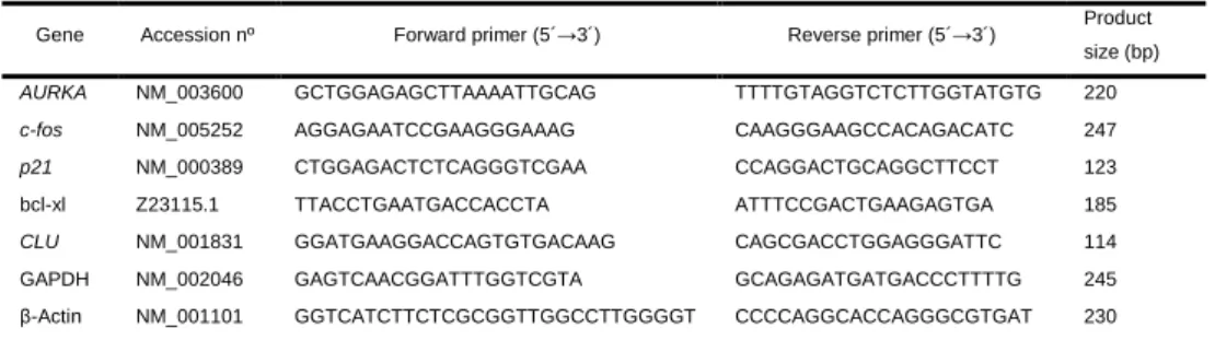
Transcriptional analysis of cell division genes CDCA8, SGOL2, Aurora A, γ -tubulin and the control gene GAPDH was performed by quantitative real-time PCR with gene specific primers as listed in table 1. Total RNA was extracted from HT29 and HUVEC cells exposed to both concentrations of BPA for 24 h and 72 h as well as non-treated and control culture to which ethanol alone was added to the culture media. Cells were collected at 80% confluence by trypsinization, washed in PBS, centrifuged at 1000 g, and RNA isolated with the RNAqueous Kit (Cat # AM1912 Thermo Scientific) following manufactures´ instructions. After verifying concentration and integrity, 3 µg of total RNA was utilized for RNase free DNase digestion (RQ1 RNase free DNase, cat # M6101 Promega) and first strand cDNA synthesis was completed with random primers following manufacturers´ instructions (DYNAmo cDNA syntesis Kit, ca t# F-470L Thermo Scientific). The resulting cDNA was utilized for qRT-PCR with the BIO-RAD SsoFast Eva Green Supermix (BIO-BIO-RAD Cat # 172-5201) utilizing the following conditions; 95 3 min, 35 cycles (95 30 sec, 55 30 sec, 72 °C-40 sec), and 72 °C-5 min. To ensure that genomic DNA was completely absent prior to cDNA synthesis, PCRs were performed with 18S primers and 250 ng of DNase digested RNA. Control PCRs were also performed for both primer combinations without template. Denaturation curves were calculated by
______________________________________________________________________
______________________________________________________________________ 20
measuring single stranded product at 0.5oC intervals from 55oC to 95oC. After denaturation curves were observed to ensure correct amplification products, threshold cycles (Ct) were equilibrated with mean GAPDHto calculate ∆Ct (∆Ct = Ct of interest – mean GAPDH Ct). Cell cycle associated genes expression levels were analyzed by calculating ∆∆Ct (∆∆Ct = ∆Ct a – mean ∆Ct b, where a and b are being compared), which in turn was used to determine mean fold change (2-∆∆Ct) ± standard deviation between treatments. Experiments were repeated three times with at least three replicates per cell treatment/primer combination in each experiment.
Table 1 Primers used for qRT-PCR.
Gene Forward primer (5´→3´) Reverse primer (5´→3´)
CDCA8 AAGGTAATACAGGTAGATGAA GTTCTCTTCTTGGATGGA
SGOL2 TACATTCACCTAACATACAA GCTCATCATCACTTACTT
AURKA GCTGGAGAGCTTAAAATTGCAG TTTTGTAGGTCTCTTGGTATGTG γ-Tubulin CTCAAGAGGCTGACGCAGAAT CTGGCTGACATGATGGTAGACAC
GAPDH GAGTCAACGGATTTGGTCGTA GCAGAGATGATGACCCTTTTG
a
GenBank accession numbers (National Center for Biotechnology).
Statistical analysis
Statistic analysis of was performed comparing cells grown in standard medium (control) to cells exposed to vehicle EtOH or BPA using Student’s t Test.
2.4 Results and discussion
2.4.1 Low BPA concentrations do not affect cell proliferation or viability independently of GPR30.
Potential cytotoxic and genotoxic effects of BPA exposure were evaluated by analyzing cell viability and proliferation in HT29 and HUVEC cells using two BPA concentrations 10 ng/ml (44 nM) and 1 µg/ml (4.4 µM). The CellTiter-Blue assay was utilized to measure viability at three time periods; two corresponding to non-confluent cultures (24 h and 48 h) and one where the culture is at 80% confluence (72 h) (Figure 1). Our results show no significant differences in viability or proliferation associated with BPA exposure. Previous studies have demonstrated that high BPA concentrations (equal or greater than 100 µM) are
______________________________________________________________________
______________________________________________________________________ 21
genotoxic, and resulted in decreased cellular proliferation as well as viability (Bolli et al. 2008; Kim et al. 2007). However, the literature shows that the effects of exposure to low BPA concentrations (in the range of 1 nM to 10 µM) are variable. Whereas no detectable effects have been reported in some cell types (Bolli et al. 2008; Kim et al. 2007; LaPensee et al. 2009), an increase in cell viability and proliferation was observed in others (LaPensee et al. 2009; Ptak et al. 2011; Ricupito et al. 2009). This discrepancy in the effects of BPA may be dependent on cell type and expression of estrogen receptors, as discussed in detail below.
Positive effects of BPA concentration as low as 1 nM on cell proliferation have been described in estrogen receptor alpha (ERα) positive human cell lines MCF-7, T47D and OVCAR-3 (LaPensee et al. 2009; Ptak et al. 2011; Ricupito et al. 2009). In fact, the lack of responsiveness in HeLa and MDA-MB-46 has been correlated with the absence of this receptor in these human cell lines (Bolli et al. 2008; LaPensee et al. 2009). Both HT29 and HUVEC cells used in this study express ERβ but not ERα (Campbell-Thompson et al. 2001; Toth et al. 2008), which could explain the lack of BPA effects on cell proliferation. However, increased proliferation was observed in ERβ positive and ERα
negative cell line JKT-1 when exposed to BPA concentrations ranging from 1 pM to 10 µM (Bouskine et al. 2009). This study showed that BPA effects in JKT1 cells are mediated through a G protein-coupled receptor 30 (GPR30). Interestingly, this membrane-bound estrogen receptor has a higher affinity for BPA than ERα or ERβ (Thomas and Dong 2006) and has been proposed as a mediator of BPA effects at low concentrations (Bouskine et al. 2009; Shanle and Xu 2010; Thomas and Dong 2006). In relation to the cells used here, the expression of GPR30 had not been analyzed in HT29 and is only expressed in HUVEC cultured in specific conditions (Takada et al. 1997).
Western immunoblotting was utilized to test GPR30 expression in HT29 and HUVEC in the culture conditions utilized in this study. The expression of ERβ
was confirmed for both cell lines as positive controls, evident as a specific immunoreactive protein band at 55 kDa (Figure 2A). As expected, GPR30 expression is not detected in HUVEC (Figure 2A), in agreement with pervious results showing that these cells express GPR30 exclusively under stream flow
______________________________________________________________________
______________________________________________________________________ 22
conditions (Takada et al. 1997). Conversely, a clear GPR30 immunoreactive protein band with the expected size of approximately 55 kDa is detected in HT29 cells (Figure 2A). Immunocytofluorescence shows that GPR30 was restricted to the cytoplasm in HT29 cells (Figure 2B), with identical distribution to that previously observed in MDA-MB231 and HEC50 cells and characteristic of its endoplasmic reticulum association (Otto et al. 2008). To the best of our knowledge, this is the first time that the expression of GPR30 is demonstrated in this cell line. More importantly, the presence of GPR30does not result in a positive response in cell proliferation in HT29 cells, in contrast with the results obtained in JKT-1 cells (Bouskine et al. 2009). This indicates that although GPR30 may have a role in mediating the responses to BPA exposure in some cell types, its expression does not result in induced cell proliferation in all GPR30 positive cells.
Figure 1 - BPA does not affect cell viability. Viability assay of HUVEC (A) and HT29 (B) cells after 24 h, 48 h and 72 h culture in control (control medium), vehicle (control
media supplemented with 0.17 mM ethanol) and 10 ng/ml or 1 µg/ml of BPA.
Cultivation and viability assays were performed simultaneously for all growth
conditions. Results are presented as mean ± standard deviation for fluorescence
______________________________________________________________________
______________________________________________________________________ 23
Figure 2 - GPR30 is present in HT29 cell line. (A) Western immunoblot confirmed the
presence of Estrogen Receptor beta (ERβ) in both HUVEC and HT29 cell lines (upper
panel). GPR30 receptor is detected in HT29 cells but not in HUVEC (lower panel). The detection of both estrogen receptors where performed on the same PVDF membrane. (B) Immunocytofluorescence images of representative interphase HT29 cells showing GPR30 distribution signals (left) and corresponding DNA DAPI staining (right), bar = 5
µm.
2.4.2 Low BPA concentrations induce micronuclei formation without affecting DNA integrity.
Potential cytotoxic and genotoxic effects of BPA exposure were evaluated by analyzing DNA integrity and micronuclei induction. Effects of BPA exposure on DNA stability and damage repair were evaluated through TUNEL assay as well as analysis of histone H3 acetylated on lysine 56 (H3K56ac). This post-translation modification of H3 was recently described to be crucial for genomic stability (Yuan et al. 2009), increasing in response to DNA damage and repair (Das et al. 2009). The TUNEL assay did not detect DNA double strand breaks or apoptotic cells associated to BPA exposure (Figure 3A). There was also no detectable variation in nuclear pattern of H3K56ac, where a dispersed distribution is observed throughout the nucleus and nucleoli regardless of cell line or BPA treatments (Figure 3B). Accordingly, western immunoblotting revealed an identical low intensity H3K56ac immunoreactive protein band with the expected size of approximately 17 kDa in all conditions tested (Figure 3C). These results indicate that the BPA concentrations utilized in this work do not affect DNA integrity or the expression of histone H3 acetylated on lysine 56 in HUVEC or HT29 cells.
______________________________________________________________________
______________________________________________________________________ 24
Micronucleus (MT) formation occurs due to chromosome segregation errors and/or chromosome fragmentation, and is therefore a relevant assay for genotoxicity. In this context, we evaluated the frequency of micronucleated cells in the distinct growth conditions, as demonstrated in Figure 4. Our results show marked differences in sensitivity to BPA between the two cell lines. Although a high level of micronucleated cells is observed in HT29 cultured in control conditions (4.45%), there is no alteration in the percentage of these cells with micronuclei after BPA exposure (4.6% for 10 ng/ml; 4.4% for 1 µg/ml). In contrast, micronucleated HUVEC cells are significantly less frequent in control conditions (1.1%), but both BPA concentrations resulted in a significant increase in the proportion of micronucleated cells (1.6% for 10 ng/ml, t-test, p = 0.0045; 2.2% for 1 µg/ml, t-test, p = 0.0002) (Figure 4).
BPA has been described as a genotoxic aneugenic chemical capable of inducing aneuploidy and resulting in centromere positive micronuclei (Kabil et al. 2008; Parry et al. 2002; Pfeiffer et al. 1997) in a dose dependent manner. However, the concentrations for which effects are detectable vary depending on cell type. In human cells lines AHH-1 (Johnson and Parry 2008) and MCL-5 (Parry et al. 2002) induction of micronuclei was observed only for concentrations at least ten times greater than the highest concentration utilized in this work, while for MCF-7 cell line a significant increase in the proportion of micronucleated cells was observed at a concentration equivalent to the higher concentration used here (Kabil et al. 2008). Our results with HT29 and HUVEC cell lines support previously described variation in BPA induced micronuclei formation between cell types. More importantly, we show for the first time that exposure to BPA at low concentrations increase the proportion of micronuclei in the HUVEC primary cell line, establishing the potential genotoxic effects of exposure to BPA.
______________________________________________________________________
______________________________________________________________________ 25
Figure 3 - BPA does not induce DNA double strand breaks or alter H3K56ac patterns. (A) TUNEL assay of HUVEC (left) and HT29 (right) cells. Cells exposed to 1 µg/ml BPA show no detectable DNA double strand breaks. Positive controls with DNase treated cells are shown on bottom. (B) HUVEC (left) and HT29 (right) cells after immunocytofluorescence detection of H3K56ac. 1 µg/ml BPA treated cells (top) show
H3K56ac interphase pattern identical to control (bottom). Bars = 5 µm. (C)
Immunoblotting with H3K56ac on HUVEC and HT29 cells grown for 24 h and 72 h in control media (C), vehicle (V), 10 ng/ml or 1 µg/ml BPA. An identical histone specific band corresponding to approximately 17 kDa is detected in all growth conditions. PVDF membranes stained by Ponceau S reagent before immunoblotting of total proteins between 40 kDa and 25 kDa of are shown as loading controls.
Figure 4 - BPA induces micronuclei in HUVEC cells. Percentage of micronuclei in HUVEC and HT29 cells after 72 h of culture in control, vehicle, 10 ng/ml or 1 µg/ml of
BPA. The results are presented as mean ± standard deviation and total number of cells
analysed is show in brackets. * a significant difference compared with the treatment with vehicle (Student’s t Test, p < 0.005).
______________________________________________________________________
______________________________________________________________________ 26
2.4.3 BPA differentially affects genes related with chromosome segregation.
The expression of genes encoding for proteins involved in chromosome segregation was assayed in order to provide more insight into the differential effect of low BPA concentrations on micronuclei induction in HUVEC and HT29 cells. For this purpose, quantitative RealTime-PCR (qRT-PCR) was utilized with primers specific for γ-tubulin and Aurora A centrosome components as well as two genes involved in chromatid segregation, namely CDCA8 and SGOL2. γ -tubulin is essential for microtubule polymerization (Raynaud-Messina and Merdes 2007) and Aurora A is a mitotic key regulatory protein essential for centrosome maturation, spindle organization and chromosome segregation (Anand et al. 2003; Kollareddy et al. 2008). CDCA8, also known as borealin/cell division cycle A8, is a component of the chromosomal passenger complex (CPC) and is one of the master regulators of events involved in mitotic bipolar spindle stability (Gassmann et al. 2004). Shugoshin-like2 (SGOL2) is essential for accurate chromosome segregation, involved in the regulation of the CPC and the phosphorylation of a microtubule depolymerase associated to chromosome congression and resolution of improper microtubule attachments. In fact SGOL2 depletion induces precocious dissociation of centromeric cohesion resulting in separation of sister chromatids (Illingworth et al. 2010; Kitajima et al. 2006; Tanno et al. 2010) and is fundamental for the bi-polar attachment of chromosomes (Rivera et al. 2012). Comparative gene expression values between cells exposed to BPA in vehicle and vehicle alone (mean fold change ± standard deviation) show subtle yet significant differences between BPA doses, times of exposure and treatments for two of the four genes analyzed (Figure 5). Interestingly, the expression of the two genes encoding for essential centrosome components (γ-tubulin and Aurora A) do not vary in BPA exposed HUVEC cells, in contrast to CDCA8 and SGOL2 genes which encode proteins that associate directly with chromosomes.
qRT-PCR results indicate increased CDCA8 mRNA in HUVEC and HT29 cells after exposure to high BPA concentration for 24 h (1.477 ± 0.045 and 1.333 ± 0.042 for HUVEC and HT29, respectively). After 72 h exposure, this effect is no longer observed in HT29 and maintained in HUVEC (1.577 ± 0.112). After exposure to low BPA concentrations, CDCA8 expression is altered exclusively
______________________________________________________________________
______________________________________________________________________ 27
in HUVEC cells exposed to BPA for 72 hours (1.304 ± 0.167). Up-regulation of SGOL2 is observed solely in HUVEC cells exposed to high BPA concentration independent of the exposure time (1.431 ± 0.089 and 1.566 ± 0.116 at 24 h and 72 h, respectively) (Figure 5A). Our results of BPA induced CDCA8 and SGOL2 up-regulation diverge from previously published data showing down-regulation of these genes in HEECs cultures exposed to 50 µM BPA (Bredhult et al. 2009). This may be due to the BPA concentrations assayed, which are much lower (ranging 10 and 1000 times lower concentration) in this study and/or to cell type specificity. In fact, the present results show that different cell types have distinct sensitivities to BPA.
Our data shows that BPA has more pronounced effects on gene transcription in HUVEC than in HT29 cells. Since HT29 expresses GPR30 and in our culture conditions HUVEC do not, the observed BPA induced alterations in HUVEC cells are independent of the presence of GPR30 receptor. Moreover, we can conclude that the observed cell specific effects are not mediated by classical nuclear ERs alone, since ERα is not expressed in either cell type and ERβ is expressed in both. Taken together, quantitative Real Time PCR data establishes that low BPA concentrations (equivalent to those found in human serum) can have effects on the expression of genes involved in basic cellular processes, such as chromosome segregation.
Figure 5 - BPA effects on expression of genes involved in chromosome segregation.
Graphic representation showing quantitative real-time PCR analysis of γ-tubulin,
AURKA, CDCA8 and SGOL2 gene transcription after exposure to BPA. Fold changes
(2-∆∆Ct ± standard deviation) in gene expression after 24 h and 72 h of BPA exposure at
10 ng/ml or 1 µg/ml are shown for (A) HUVEC, and (B) HT29. GAPDH was utilized as a
reference gene and Student’s t Test significant differences in average mean fold changes are shown as **p < 0.01 and *p < 0.05.