ANA MARGARIDA NUNES PORTUGAL CARVALHO MELO
CHARACTERISATION OF NAD(P)H
DEHYDROGENASES FROM NEUROSPORA
MITOCHONDRIA
PORTO
Ana Margarida Nunes Portugal Carvalho Melo
Characterisation of NAD(P)H dehydrogenases from Neurospora
mitochondria
Dissertation for Obtaining a Philosophy Doctor Degree in Biomedical Sciences,
Submitted to the Instituto de Ciências Biomédicas de Abel Salazar, University of
Porto. The works were developed at the Laboratory of Molecular Genetics from the
Instituto de Ciências Biomédicas de Abel Salazar and at the Laboratory of Molecular
Genetics and Biogenesis of the Mitochondrion from the Instituto de Biologia
Molecular e Celular.
Supervisor: Professor Arnaldo António de Moura Silvestre Videira
University of Porto, Portugal
Co-supervisor: Professor Ian Max M0ller
Lund University, Sweden and Ris0 National Laboratory, Denmark
nome/. O Sew nome/ não é/ apreentÁM&L por todo* o* credoy e/
toda* a& teoria* da/ ciência/. €le/ é/, de/ facto-, a/ "etAêvuMxs
temida/ que/ fCca/ para/ além/ da/ lógica/". A cí&ncia/ prolonga/
a/ praia/ ao- longo- da/ qual zomoy capote* de/ perceber o
wwatério-, ma* não- esgota/ o mUfcérío-. À medida/ que/ o
conh&ybmento }& torna/ maiy profundo, o metmo- acontece/
com/ o espanto. "
ChefRaymo-I am greatly indebted to:
Aires, Albertina, Alexandre, Ana, Ana, Ana, Ana, Ana, André, Angelo, Anita,
António, António Alberto, A r n a l d o , Arrabaça, Augusta, Augusta, Augustinha,
Bete, Beto, Bruno, Christian, Clara, Daniel, David, Dejana, Duarte, Elisa, Fátima,
Fátima, Fernando, Fernando, Fernando, Filipa, Filipa, Francisca, Gonçalo, Guida,
Heiko, Helena, Holger, Inês, Inês, Isabel, Isabel, Isabel, Jaime, Janeca, Joaquim,
Joaquim, Joana, Joana, João, João, João, João, João, João, Joca, Jorge, Jorge, Julio,
Laura, Lena, Leonor, Lu, Luís, Maluxa, Maneia, Maneia, M a r g a r i d a , Maria,
Mariana, Marta, Matilde, M a x , Miguel, Miguel, Miguel, Miguel, Miguel, Mónica,
Natália, Nela, Neupert, Nuno, Nuno, Paula, Paulo, Pedro, Pedro, Quéqué,
Quintanilha, Rita, Rita, Rita, Rosário, Rui, Rui, Sara, Sara, Sérgio, Silva, Susana,
Susan, Tadeu, Teresa, TiJoão, Tó, Tó, Tom, Toni, Victor, Xana, Xico, Zé, Zé, Zé,
Zé, Zé.
When you are around everything gets funnier and easier! Thanks a lot!
The Portuguese Foundation for Science and Technology funded this work.
Luis skilfully drew figure 1, 2 and 4.
Miguel generously created the front page.
Dr Corália Vicente gave me a precious help on the statistical analysis of the
NDE1 activity data.
Page
LIST OF PUBLICATIONS 4
ABBREVIATIONS 5
RESUMO 7
RÉSUMÉ 11
SUMMARY 14
CHAPTER I - GENERAL INTRODUCTION
1. NEUROSPORA, THE MODEL 17
1.1. The life cycle ofN. crassa 17
1.2. The genome of N. crassa 19
1.3. Inactivation of Neurospora genes 19
2. MITOCHONDRIA: A BRIEF STORY 20
2.1. Description of respiratory chains 22
2.2. NADH: The super fuel 23
3. TWO TYPES OFNAD(P)H DEHYDROGENASES 25
3.1. Complex 1 25
3.2. Rotenone-insensitive NAD(P)H dehydrogenases 28
3.2.1. Rotenone-insensitive NAD(P)H oxidase activities in different
organisms 28
3.2.2. Effects of cations on rotenone-insensitive NAD(P)H oxidase
activities 31
3.2.3. The external NADH dehydrogenase from rat heart mitochondria
- a mysterious exception 32
4. HUMAN PATHOLOGIES ASSOCIATED WITH NADH OXIDATION 33
5. FINAL COMMENTS 34
CHAPTER II - RESEARCH PROJECT
1. OBJECTIVES 36
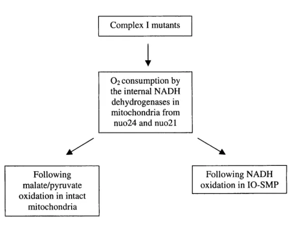
2. RESULTS AND DISCUSSION
2.1.1. Characterisation of the internal NADH oxidation by nuo24 38
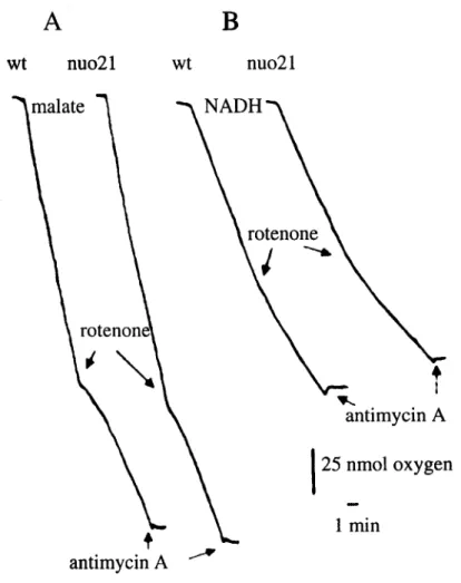
2.1.2. Characterisation of the internal NADH oxidation by nuo21 40
2.2. Identification, mapping and inactivation of the gene gncoding a
putative rotenone-insensitive NAD(P)H dehydrogenase 44
2.2.1. Gene characterisation 45
2.2.1.1. Structural analysis of the NM1C2 product 45
2.2.1.2. Chromosomal location of the gene encoding NDE1 53
2.2.2. Inactivation of the ndel gene 54
2.2.3 Analysis of crosses involving the ndel mutant 57
2.3. Characterisation of NAD(P)H oxidation in Neurospora mitochondria 59
2.3.1. Localisation of the NDE1 protein 60
2.3.1.1.Localisation of NDE1 in the inner mitochondrial membrane 60
2.3.1.2. NDE1 faces the intermembrane space 63
2.3.2. Phenotype of the nde mutant 67
2.3.3. Characterisation of exogenous NAD(P)H oxidation by
Neurospora mitochondria - pH and calcium effects 69
2.3.4. Kinetic properties of the external rotenone-insensitive
NAD(P)H dehydrogenases 74
2.3.5. Physiogical role of NDE1 76
2.3.6. Final considerations 79
3. CONCLUDING REMARKS 82
CHAPTER III - EXPERIMENTAL PROCEDURES
1. Chemicals 84 2. Media used 84
3. Strains of E. coli and N. crassa 86
4. Growth of Neurospora strains 87 5. Isolation of plasmid DNA 87
5.1. Small-scale preparation of plasmid DNA for analytical purposes 87 5.2. Large-scale preparation of plasmid DNA for preparative purposes 87
6. Enzymes used in cloning 88 6.1. Digestion of DNA with restriction enzymes 88
6.2. Dephosphorylation of DNA 88
6.6. Labelling a DNA probe 89 7. Gel electrophoresis of DNA 89
7.1. Agarose gel electrophoresis 89 7.2. DNA sequencing gels 90 7.3. Isolation of DNA bands from agarose gels 90
8. Isolation of genomic DNA from N. crassa 90
9. Analysis of genomic DNA by Southern blotting 91
9.1. Mapping of the ndel gene from N. crassa 91
10. Transformation of Neurospora spheroplasts 91
10.1. Preparation of spheroplasts 91 10.2. Transformation of spheroplasts 92 11. Disruption of the ndel gene by repeat-induced point mutations 92
12. Isolation of mitochondria from N. crassa 92
12.1. Preparation of mitochondria 92 12.2. Purification of mitochondria 92 12.3. Preparation of IO-SMP 93 13. SDS-polyacrylamide gel electrophoresis 93
14. Western blotting 93 14.1. Semi-dry 93 14.2. Wet-blot 93 15. Immunodecoration of proteins 93
16. Expression and purification of NDEl-fusion protein and preparation of
antibodies 94 16.1. Expression of NDE1 94
16.2. Purification of NDE1 94 16.3. Rabbit immunisation 94 17. In vitro translation of precursors in a rabbit reticulocyte system 94
18. Import of NDE1 and F i p precursors into Neurospora and yeast
mitochondria 95 18.1. Neurospora mitochondria 95
18.2. Yeast mitochondria 95 19. Protein determination 95 20. Enzyme assays 96
20.1. Cytochrome c oxidase 96
20.2. Malate dehydrogenase 96 20.3. Adenylate kinase 96 21. Oxygen electrode measurements 96
22. Digitonin fractionation of mitochondria 97 23. Alkaline extraction of mitochondrial proteins 97
24. Programs used in data processing 97
List of publications resulting from the doctorate work
This thesis is based on the following publications, from which tables and
figures were used with the permission of the editors.
I Melo, A. M. P., Duarte, M., and Videira A. (1999) "Primary structure and
characterisation of a 64 kDa NADH dehydrogenase from the inner membrane
of Neurospora crassa mitochondria" Biochim. Biophys. Acta 1412, 282-287.
II Almeida, T., Duarte, M., Melo, A. M. P., and Videira A. (1999) "The 24 kDa
iron-sulfur subunit of complex I is required for enzyme activity" Eur. J. Biochem. 265, 86-92.
III Ferreirinha, F., Duarte, M., Melo, A. M. P., and Videira A. ( 1999) "Effects of
disrupting a 21 kDa subunit of complex I from Neurospora crassa" Biochem.
J. 342,551-554.
IV Melo A. M. P., Duarte, M., Moller, I. M., Prokisch, H., Dolan, P., Pinto, L.,
Nelson, M., and Videira, A. (2001) "The external calcium-dependent
NADPH dehydrogenase from Neurospora crassa mitochondria" J. Biol.
Abbreviations
ACP acyl carrier protein
APS ammonium persulfate
ADP adenosine diphosphate
ATP adenosine triphosphate
ATPase adenosine triphosphatase
bp base pairs
BSA bovine serum albumin
cAMP Cyclic adenosine monophosphate
CCHL Cytochrome c heme lyase
CHAPS 3-[(3-cholamidopropyl)-dimethylammonium]-1 -propanesulfonate
dATP deoxyadenosine triphosphate
dCTP deoxycytosine triphosphate
dGTP deoxyguanosine triphosphate
dTTP deoxythymidine triphosphate
ddATP dideoxyadenosine triphosphate
ddCTP dideoxycytosine triphosphate
ddGTP dideoxyguanosine triphosphate
ddTTP dideoxythymidine triphosphate
DMSO dimethyl sulphoxide
DNAse deoxyribonuclease
DNA deoxyribonucleic acid
DPI diphenyleneiodonium
DTT dithiothreitol
EDTA ethylenediamine tetraacetate
EGTA ethyleneglycol-bis-aminoethylether tetraacetate
FAD flavin adenine dinucleotide
FMN flavin mononucleotide
IPTG isopropyl (3-D-thiogalactopyranoside
kDa kilo Dalton
LB luria broth
MES 2-[N-Morpholino]ethanesulfonic acid
min minutes
MOPS N-Morpholinopropanesulfonic acid
MPP matrix processing peptidase
MRI molecular resonance imaging
NADH nicotinamide adenine dinucleotide, reduced form
NADPH nicotinamide adenine dinucleotide phosphate, reduced form
OD optical density
PCR polymerase chain reaction
PEG polyethyleneglycol
PMSF phenylmethylsulphonylfluoride
RNAse ribonuclease
RNAsin ribonuclease inhibitor
SDS sodium dodecyl sulphate
SDS-PAGE SDS-polyacrylamide gel electrophoresis
TEMED N,N,N',N'-tetramethylethylenediamine
TES 2-([2-hydroxy-l,l-bis(hydroxymethyl)-ethyl]amino)ethanesulfonic
Resumo
Em sistemas eucarióticos não fotossintéticos, a mitocôndria é o organelo
responsável pela síntese de ATP, uma molécula muito energética usada na
biossíntese e manutenção celulares, bem como na tomada de iões. É, também, na
mitocôndria que ocorre a síntese de intermediários para várias reacções
biossintéticas.
O NADH é o maior dador de electrões à cadeia transportadora de electrões
mitocondrial e, consequentemente, da sua oxidação resulta a maior parte da energia
produzida no organelo. Dois tipos de enzimas levam a cabo a transferência de
electrões do NADH para a ubiquinona: as que acoplam a transferência de electrões à
translocação de protões, através da membrana interna mitocondrial, e as que realizam
a mesma transferência sem transdução de energia. No primeiro caso, temos o
complexo I (NADH:ubiquinona oxidorredutase), sensível à rotenona. Trata-se de
uma enzima multimérica ancorada à membrana interna mitocondrial, com um
domínio matricial, presente na maioria dos sistemas eucarióticos. O segundo grupo
integra as NAD(P)H desidrogenases alternativas, resistentes à rotenona, que
contribuem para a formação do potencial de membrana apenas através dos
complexos III e IV. O fungo Neurospora crassa, o organismo escolhido para realizar
este projecto, contém os dois tipos de NAD(P)H desidrogenases nas suas
mitocôndrias.
O complexo I de dois mutantes da enzima foi caracterizado no que se refere à
sua actividade de NADH: ubiquinona oxidorredutase. Estes mutantes não têm as
subunidades de 24 e 21 kDa e são chamados de nuo24 e nuo21, respectivamente.
Determinou-se o consumo de oxigénio por mitocôndrias e partículas
submitocondriais invertidas dos mutantes do complexo I nuo24 e nuo21 a oxidar,
respectivamente, malato/piruvato e NADH. A oxidação sensível à rotenona
observada no nuo21 foi semelhante à do tipo selvagem, enquanto o nuo24 não
apresentou qualquer oxidação sensível ao inibidor. Assim, em contraste com a
subunidade de 21 kDa, a subunidade de 24 kDa é indispensável para a actividade do
complexo I. O facto de mitocôndrias do nuo24 apresentarem actividade NADH:
oxidação do NADH matricial, para além do complexo I, em mitocôndrias de
Neurospora.
Um clone parcialmente sequenciado, codificante de uma hipotética NAD(P)H
desidrogenase, foi obtido do Fungal Genetics Stock Center e totalmente sequenciado
nas duas cadeias. O clone contém 2570 pares de bases e uma região codificante de
2019 pares de bases. Estes codificam uma proteína de 673 aminoácidos com uma
massa molecular aparente de 64 kDa, de acordo com electroforese em gel
desnaturante de poliacrilamida. Os primeiros 74 aminoácidos da cadeia polipeptídica
representam uma possível pré-sequência mitocondrial. A estrutura primária da
proteína de 64 kDa apresenta cerca de 35 % de identidade com NADH
desidrogenases, de outros organismos, também resistentes à rotenona. Uma
sequência de consenso para um domínio de ligação ao cálcio está presente na
estrutura primária da proteína de 64 kDa, o que sugere que o catião tem um papel na
regulação da actividade da enzima.
A região C-terminal da hipotética NAD(P)H desidrogenase foi expressa em
Escherichia coli, como proteína de fusão, purificada e utilizada para imunizar
coelhos, visando a produção de anticorpos policlonais. O gene ndel (que codifica a
proteína de 64 kDa ou NDE1) foi inactivado pelo processo de indução repetida de
mutações pontuais (RIP). Esferoplastos de Neurospora foram transformados com um
vector transportando uma cópia do cDNA do ndel, e a estirpe resultante foi cruzada
com a estirpe do tipo selvagem. Prepararam-se mitocôndrias da progenia do
cruzamento e os mutantes ndel foram seleccionados com anticorpos contra a NDE1.
Cruzamentos homozigóticos do mutante ndel originaram descendência, indicando
que a proteína não é necessária ao desenvolvimento sexual. Vários mutantes
deficientes em subunidades do complexo I foram cruzados com o mutante ndel,
observando-se mutantes duplos em todos os cruzamentos. Isto sugere que o produto
do gene ndel não está envolvido na oxidação do NADH matricial, ou, se este
envolvimento se verificar, existem outras vias para assegurar o processo em
mitocôndrias de N. crassa.
O subfraccionamento de mitocôndrias, com concentrações progressivas de
digitonina, localizou a NDE1 na membrana interna mitocondrial. Experiências
tratadas com proteases, mostraram que esta tem domínios expostos ao espaço
intermembranar. A região codificante da NDE1 foi usada num sistema de
tradução/transcrição para a síntese in vitro de um precursor. Este foi importado para
mitocôndrias de Neurospora e levedura, e processado na presença de potencial de
membrana. O precursor processado ficou acessível à tripsina em mitoplastos de
levedura, indicando que, tal como in vivo, a proteína processada e importada
apresenta domínios no espaço intermembranar.
Prepararam-se mitocôndrias e partículas submitocondriais invertidas das
estirpes do tipo selvagem e do mutante ndel e procedeu-se à caracterização da
oxidação de NADH e NADPH. Relativamente às partículas submitocondriais, não há
diferenças entre as duas estirpes. Este resultado sugere que as NAD(P)H
desidrogenases alternativas internas não foram afectas pela inactivação do ndel. A
comparação da oxidação de NADH por mitocôndrias das duas estirpes também
revelou características semelhantes. Pelo contrário, enquanto mitocôndrias do tipo
selvagem oxidam NADPH a pH fisiológico, mitocôndrias do mutante ndel não
apresentam esta actividade, o que indica que a NDE1 é uma NADPH desidrogenase
externa. Uma caracterização mais detalhada da actividade da NDE1 demonstrou que
a oxidação de NADPH, por esta enzima, varia em função do pH e é estimulada pela
presença de cálcio. Apesar da ausência de actividade da NDE1, mitocôndrias do
mutante ndel oxidam NADPH e NADH exógenos na zona acídica e ao longo da,
escala de pH, respectivamente. Este facto evidencia a existência de uma segunda
NAD(P)H desidrogenase resistente à rotenona, a NDE2. A NDE1 e a NDE2
contribuem para o "turnover" do NAD(P)H citosólico, em células de N. crassa.
Como se pode inferir do presente trabalho, a mitocôndria de N. crassa tem,
pelo menos, três NAD(P)H desidrogenases insensíveis à rotenona. Até ao presente, o
gene que codifica a NDE1 foi o único a ser clonado, sequenciado e identificado.
Num futuro próximo, deverá ser possível identificar os outros dois genes,
inactivá-los e caracterizar a actividade das suas mitocôndrias, possibilitando a clarificação do
papel fisiológico das NAD(P)H desidrogenases resistentes à rotenona. A produção de
uma estirpe de Neurospora sem NAD(P)H desidrogenases resistentes à rotenona
deficientes em subunidades desta enzima. Deste modo, poder-se-á estabelecer a
Resume
Chez les eucaryotes qui n'utilisent pas la photosynthèse, la mitochondrie est
1'organelle responsable de la synthèse d'ATP, une molécule très énergétique,
utilisées dans la biosynthèse, l'entretien des cellules et l'acquisition d'ions. La
mitochondrie est aussi le lieu de synthèse d'intermédiaires des différents reactions
biosynthétiques.
La NADH est le principal donneur d'électrons dans la chaîne mitochondriale
de transfert d'électrons. Il existe deux types d'enzymes qui permettent le transfert
d'électrons du NADH à l'ubiquinone: les enzymes qui couplent le transfert
d'électrons à la translocation de protons à travers la membrane interne de la
mitochondrie; et les enzymes qui permettent le même transfert mais sans
transduction d'énergie. Dans le premier type d'enzymes, il y a le complexe I
(NADH:ubiquinone oxydoréductase) qui est sensible au rotenone. C'est une enzyme
multimerique ancrée à la membrane interne mitochondriale, avec un domaine
matriciel, qui existe chez la majorité d'eucaryotes. Chez le second groupe se trouvent
les NAD(P)H déshydrogénases résistant au rotenone. Dans ce cas, seulement les
complexes III et IV contribuent pour la formation du potentiel de la membrane. Le
champignon Neurospora crassa a servi de modèle d'études afin de réaliser ce travail,
étant donné que les deux types de NAD(P)H déshydrogénases sont présentes dans cet
organisme.
Deux mutants ont servi pour la caractérisation de l'activité de
NADH:ubiquinone oxydoréductase du complexe I. Ces mutants sont dépourvus des
sous-unités de 24 et 21 kDa du complexe I et sont désignés nuo24 et nuo21,
respectivement. La consommation d'oxygène par les mitochondries et particules
sous-mitochondriales inversées chez les mutants nuo24 et nuo21, oxydant
respectivement le malate/piruvate e la NADH, a été comparée avec la souche
sauvage. L'oxydation sensible au rotenone observée dans nuo21 était comparable à
celle de la souche sauvage, alors que l'oxydation dans nuo24 n'a montré aucune
sensibilité à l'inhibiteur. Au contraire de la sous-unité de 21 kDa, la sous-unité de 24
Grâce au Fungal Genetics Stock Center, il nous a été possible d'obtenir un
clone de cDNA (NM1C2) partiellement séquence, en codant une hypothétique
NAD(P)H déshydrogénase. Les deux chaînes de ce clone ont été entièrement
séquencées. La région codante, de 2019 pbs, code pour une protéine de 673
aminoacides et de poids moléculaire d'environ 64 kDa selon son électrophorèse sur
gel dénaturant de polyacrilamide. Les premiers 74 aminoacides de la chaîne
polipeptidique représentent une possible pré-séquence mitochondriale. L'analyse de
sa structure primaire a révélé environ 35% d'identité avec la NADH déshydrogénase
d'autres organismes, et une séquence consensus d'un domaine de liaison au calcium
en suggérant un rôle du calcium dans la regulation de l'activité de cet enzyme.
L'expression de la région C-terminale de la possible NAD(P)H
déshydrogénase a été induite chez Escherichia coli, et la protéine de fusion a été
purifiée en vue de production d'anticorps polyclonaux chez le lapin. Le gêne ndel
(qui codifie la protéine de 64 kDa ou NDE1) a été inactivé au moyen d'induction
répétée de mutations ponctuelles (RIP). Un vecteur, en transportant une copie du
cDNA de ndel, a transformé les spheroplastes de Neurospora, la souche résultante a
été croisée avec une souche sauvage. Mitochondries de la descendence du croisement
ont été préparées et analisées, par Western blot en utilisant des anticorps anti-NDEl,
pour la sélection des mutants ndel. Les croisements homozygotiques du mutant ndel
ont produit une descendance, ce qui indique que la protéine n'est pas nécessaire pour
le développement sexuel. Différents mutants des sous-unités du complexe I ont été
croisés avec ndel, ayant donné tous des doubles mutants. Ce qui sugére que la
protéine de 64-kDa ne s'occupe pas de la oxidation du NADH matriciel, ou qu'il y a
d'autres routes pour faire cette oxidation dans les mitochondries de Neurospora.
Le sous-fractionnement de mitochondries, avec des concentrations progressives
de digitonine, a permit de localiser NDE1 dans la membrane interne mitochondriale;
le traitement des fractions résultantes avec des proteases a montré que cette protéine
possède des domaines exposés à l'espace intermembranaire. La région codant de
NDE1 a été utilisée dans la synthèse in vitro d'un précurseur qui a été importé vers
les mitochondries de Neurospora et levure, et ce processus a été effectué en présence
la trypsine dans les mitoplastes de levure, en indiquant que , comme in vivo, la
protéine processé et importé présente des domaines à l'espace intermembranaire.
Des mitochondries et particules sous-mitochondriales inversées des souches
sauvage et ndel ont été préparées et l'oxydation de NADH et de NADPH a été
caractérisée. En ce qui concerne les particules sous-mitochondriales, il n'y a pas de
différence quant à l'oxydation des deux substrats par les deux souches, suggérant que
les NAD(P)H déshydrgénases alternatives internes n'ont pas été affectées par
l'inactivation de NDE1. L'analyse comparative de l'oxydation de NADH par les
mitochondries des deux souches a révélé aussi un comportement similaire.
Cependant, alors que les mitochondries de type sauvage oxydent la NADPH à pH
physiologique, les mitochondries de ndel n'ont pas montré cette activité, ce que
indique que la NDE1 est une NADPH déshydrogénase externe. Une caractérisation
plus poussée de l'activité de NDE1 a démontré que l'oxydation de NADPH par cette
enzyme varie en fonction du pH et est stimulée en présence du calcium. Malgré
l'absence d'activité de NDE1, les mitochondries de ndel oxydent NADPH et NADH
exogènes, au niveau de la région acidique et le long de l'échelle de pH,
respectivement. Ce fait montre l'existence d'une deuxième NAD(P)H
déshydrogénase alternative, la NDE2. La NDE1 et la NDE2 contribuent pour le
«turnover» de la NAD(P)H cytosolique, dans les cellules de N. crassa.
Ce travail montre que la mitochondrie de N. crassa possède au moins trois
NAD(P)H déshydrogénases insensibles au rotenone. Jusqu'à présent, le gêne codant
pour la NDE1 a été le premier à être clone, séquence et identifié. Dans un futur très
proche, il devrait être possible d'identifier et puis inactiver les deux autres gênes afin
de caractériser l'activité de leurs mitochondries. Ceci aiderait à éclaircir le rôle
physiologique des NAD(P)H déshydrogénases résistantes au rotenone. La production
d'une souche de Neurospora dépourvus des NAD(P)H déshydrogénases résistantes
au rotenone nous donnera des modelles pour l'étude du complex I sauvage et des
complexes I defficientes en sous-unités de cette enzyme. Ansi, sera possible
Summary
In non-photosynthetic eukaryotic systems the mitochondrion is the organelle
responsible for the synthesis of ATP, a very energetic molecule that is used in
cellular biosynthesis and maintenance as well as in ion uptake. It is also in the
mitochondrion that the synthesis of many intermediates for several biosynthetic
reactions takes place.
NADH is the major electron donor to the mitochondrial electron transport
chain. There are two distinct types of enzymes carrying out the electron transfer from
NADH to ubiquinone: those that couple the electron transfer to proton translocation
across the inner mitochondrial membrane, and the others that perform the transfer of
electrons without energy transduction. In the first group is the rotenone-sensitive
complex I, or NADH:ubiquinone oxidoreductase, a multimeric enzyme anchored to
the inner mitochondrial membrane, with a matrix domain, that is present in most
eukaryotic systems. The second group of enzymes comprises the alternative
NAD(P)H dehydrogenases, also known as rotenone-insensitive NAD(P)H
dehydrogenases. In this case, only complexes III and IV contribute to the generation
of a membrane potential. The fungus Neurospora crassa, the organism chosen to
develop this project, contains both types of NAD(P)H dehydrogenases in its
mitochondria.
Studies were performed in order to characterise the complex I from two
mutants with respect to its NADH:ubiquinone oxidoreductase activity. They lack the
subunits of 24 and 21 kDa and are called nuo24 and nuo21, respectively. The oxygen
uptake by mitochondria and IO-SMP oxidising malate/pyruvate and NADH,
respectively, from nuo24 and nuo21 was measured. The rotenone-sensitive oxidation
of substrates by nuo21 and wild type was similar, while nuo24 did not present any
rotenone-sensitive activity. Thus, in contrast to the 21-kDa subunit, the subunit of 24
kDa is required for complex I activity. The fact that nuo24 mitochondria can still
oxidise NADH dehydrogenase confirms the presence of other enzymes, beyond
complex I, responsible for the oxidation of matrix NADH in Neurospora
A partially sequenced clone, encoding a putative rotenone-insensitive
NAD(P)H dehydrogenase, was obtained from the Fungal Genetics Stock Center and
sequenced fully in both strands. It has 2570 bp and contains an open reading frame of
2019 bp. The encoded protein consists of 673 amino acid residues, with an apparent
molecular mass of 64 kDa, as determined by SDS-PAGE. The first 74 amino acid
residues of the polypeptide constitute a putative mitochondrial-targeting sequence.
When compared with rotenone-insensitive NADH dehydrogenases from other
organisms, the primary structure of the Neurospora protein displays around 35 %
identity. A consensus sequence for a calcium-binding domain is also present in the
primary structure of the 64-kDa protein, what suggested that this cation might have a
role in the regulation of the enzyme.
The C-terminal region of the putative NAD(P)H dehydrogenase was expressed
in Escherichia coli as a fusion protein, purified, and used to immunise rabbits in
order to produce polyclonal antibodies, ndel (the gene encoding the 64-kDa protein
or NDE1) was inactivated by repeat-induced point mutations. Neurospora
spheroplasts were transformed with a vector carrying a copy of the ndel cDNA and
the resulting strain was crossed with the wild type strain. Mitochondria from the
progeny were prepared and antibodies against NDE1 polypeptides were used to
select ndel mutants. The homozygous crosses of ndel mutant yielded progeny
indicating that the protein is not essential for sexual development. Several mutants
deficient in complex I subunits were crossed with the ndel mutant producing double
mutants. This suggested that either the ncfei-encoded protein is not involved in the
oxidation of matrix NADH or, if it is involved, there are other ways to carry out this
activity in the matrix of N. crassa mitochondria.
Subfractionation of mitochondria with increasing concentrations of digitonin
localised the protein to the inner membrane of the mitochondrion. Similar
experiments were performed, where the digitonin-solubilized fractions were
submitted to protease treatment, showing that the protein has domains facing the
intermembrane space. The open reading frame of NDE1 was used as template for in
vitro synthesis of a precursor that was imported into Neurospora and yeast
precursor was accessible to trypsin in yeast mitoplasts indicating that, as verified in
vivo, the processed and imported protein has intermembrane facing domains.
Mitochondria and IO-SMP from wild type and ndel mutant strains were
prepared and NADH and NADPH oxidation was characterised. The oxidation of
NADH and NADPH by IO-SMP from both wild type and ndel mutant strains shows
a similar behaviour, indicating that the internal alternative NAD(P)H dehydrogenases
were not affected by the inactivation of ndel. The pattern of NADH oxidation by
wild type mitochondria and mitochondria from the ndel mutant is similar. In
contrast, while wild type mitochondria oxidise NADPH at physiologic pH, in ndel
mutant mitochondria this activity is absent, showing that NDE1 is an external
NADPH dehydrogenase. Further characterisation of NDE1 activity showed that
NADPH oxidation by the enzyme varies as a function of the pH and is stimulated by
the presence of calcium. In spite of lacking NDE1 activity, ndel mutant
mitochondria can still oxidise exogenous NADH throughout the pH range and also
NADPH at acidic pH. This observation provided evidence for the presence of a
second alternative NAD(P)H dehydrogenase, NDE2. Both NDE1 and NDE2 can
contribute to the turnover of cytosolic NAD(P)H in N. crassa cells.
As deduced from the present work, N. crassa mitochondria contain at least
three rotenone-insensitive NAD(P)H dehydrogenases. So far, the gene encoding
NDE1 is the only gene cloned, sequenced and identified. In a near future it should be
possible to identify the other two genes and very interesting to inactivate them and
characterise the activity of their mitochondria. This information can be crucial to
illuminate the physiological role of the rotenone-insensitive NAD(P)H
dehydrogenases. The production of a Neurospora strain lacking the
rotenone-insensitive NAD(P)H dehydrogenases will provide models to study the wild type
complex I and the complex I deficient in different subunits. In this way it will be
possible to establish the importance of each complex I subunit in the activity of the
Chapter I - General Introduction
1. Neurospora, the model
Though presenting some peculiarities, fungi share the basic characteristics of
eukaryotic organisms. The narrow range of DNA concentration values among fungi
suggests a remarkably uniform mechanism regulating nuclear division as compared
with the otherwise great plasticity characterising the organisms from this kingdom.
This narrow range of values for DNA composition contrasts with the huge variability
in the composition of other materials. The variability for the same fungus with
different growth conditions, ages and stages of development is in fact larger than
differences between species (Griffin, 1994). Among eukaryotes, fungi are ideal
organisms to be used as models for studies of sub-cellular structure, because of their
ease of handling, simplicity, short life cycles and the considerable amount of genetic
information available on them.
The species Neurospora crassa is a multicellular eukaryote that belongs to the
kingdom Fungi, division Eumycota, class Ascomycetes, sub-class Euascomycetidae,
order Euascomy ce tales, family Euascomycetacea and genus Neurospora. The
mitochondrion of Neurospora was the model used in the present work. The fact that
the organism grows strictly as a haploid makes it convenient to study introduced
phenotypes, allowing an accurate characterisation of deleted or added genes.
1.1. The life cycle of N. crassa
The life cycle of N. crassa is typical of the Euascomycetidae (Fig. 1).
Ascospores germinate into multinucleate cells that form branched filaments
(hyphae). The hyphal system spreads out fast to form a haploid mycelium. Asexual
reproduction is accomplished by macroconidia and microconidia produced on
specialised aerial hyphae, called conidiophores. The sexual cycle is initiated when
Figure 1. Neurospora life cycle. Redrawn from Griffin (1994).
An ascogonium, a swollen coiled hypha with an apical branched trichogyne,
differentiates. The differentiated hypha is quickly enclosed by nearby hyphae that
grow closely around it, forming a protoperithecium with the trichogyne projecting
out. A spermatial element of the opposite mating-type (spermatia can be
microconidia, macroconidia or hyphae) fuses with trichogyne. The nucleus of the
spermatium enters and migrates along the trichogyne to the ascogonium, creating the
"dicaryotic" stage. Ascogenous hyphae grow from the fertilised ascogonium,
generating a basal hymeneal layer within the developing perithecium. Most of the
tissues of the perithecium arise from the monocaryotic mycelium that formed the
ascogonium. The spermatial and ascogonial nuclei migrate into growing ascogenous
hyphae, dividing as they go. As development proceeds, nuclei in the tips of the
ascogenous hyphae pair together and divide simultaneously, while the tip of the
hyphae grows back on it to form a crozier. Two septa form in the crozier. The tip cell
is uninucleate, the second cell is binucleate, and the third cell is uninucleate. The first
fuses with the third, re-establishing the binucleate condition and a short branch grows
ascus; its nuclei fuse, rising a zygote, which immediately proceeds to meiosis.
Pre-meiotic synthesis of DNA occurs prior to nuclear fusion, so that when karyogamy
takes place, the resulting diploid nucleus immediately undergoes meiosis. Thus the
diploid phase of Neurospora life cycle is very short and restricted to a single cell.
After meiosis, a single mitosis gives rise to an eight-nucleate ascus and each nucleus
is enclosed within a cell wall within the ascus, forming each ascospore. At maturity,
the ascus elongates up the perithecial canal, exposing its tips, and the ascospores are
ejected. Genetic studies have shown that the nuclei that fused in the ascus were of
two genotypes, one from the ascogonial parent and the other from the spermatial one,
thus allowing genetic analysis (Selker, 1990; Griffin, 1994).
1.2. The genome of N. crassa
The genome of N. crassa consists of seven chromosomes ranging in size from
4 to 11 Mbp, as estimated by physical and cytological measurements. Eight percent
of the 37 Mbp present in Neurospora genome are repetitive sequences, mainly
ribosomal RNA genes. Besides nuclear DNA, Neurospora also contains
mitochondrial DNA. It encodes subunits 6, 8 and 9 of ATPase (the latter is a
pseudogene, meaning that it does not encode a protein), subunits 1, 2 and 3 from
cytochrome c oxidase, apocytochrome b, seven subunits from complex I and various
tRNA, ribosomal proteins, rRNA (Griffin, 1994). Several unidentified open reading
frames are also present in the mtDNA of N. crassa (Davis, 2000).
1.3. Inactivation of Neurospora genes
A commonly used strategy for protein characterisation is the disruption of their
encoding genes and the analysis of the induced mutants. A peculiarity of Neurospora
is the phenomenon of repeat-induced point mutations (RIP) acting on nuclear DNA
(Selker and Garrett, 1988). DNA duplications in the genome are detected and
both regions of the duplications introducing multiple GC to AT transition mutations
in the DNA. Sequences mutated by RIP are typically methylated at most of the
remaining cytosine residues. Any duplicated region bigger than 500 bp is susceptible
to RIP'ing. Though timing and effects of this phenomenon are well established, little
is known about its mechanism (Waiters et al, 1999). The RIP can be used to
inactivate genes in N. crassa.
Inactivation of genes in Neurospora may also be achieved by homologous
recombination. This process is accomplished by introducing a genetic marker, for
instance a gene coding for resistance to hygromycin B, in the middle or in the place
of a target gene. Neurospora spheroplasts are transformed with the DNA construct,
which carries the genetic marker flanked by Neurospora sequences. After
transformation, the recombination of the flanking sequences with the homologous
sequences in the genome results in an exchange of the host gene with the DNA
construct. The DNA sequence of the target gene is thus altered (gene disruption) and
the correct protein can not be synthesised (Videira, 1998).
2. Mitochondria: a brief story
The mitochondrion (Fig. 2) of Neurospora is a small organelle (1-2 u,m), found
in eukaryotic cells, where ribosomes, RNA and DNA are present which makes it a
semiautonomous organelle. This organelle is abundant in eukaryotic tissues, easy to
purify and a rich source of vital enzymes. The general appearance, structure and
organisation of mitochondria are similar for fungi, animals or plants. They are
composed of two bilayered membranes, a smooth outer membrane surrounding a
highly invaginated inner membrane that contains the electron transport chain. The
invaginations of the inner membrane are known as cristae. Inside the inner
membrane is a very concentrated protein suspension, the matrix, where the
tricarboxylic acid cycle takes place. The space between the two mitochondrial
membranes is called the intermembrane space. Intact mitochondria are osmotically
active, which means that they take up water and swell when placed in a
Outer membrane
Intermembrane space
Inner membrane
Matrix Cristae
Figure 2. Three-dimensional drawing of a mitochondrion.
NAD(P)H
OMM
NAD(P)H NAD(P)+
NDOMM
NADH NAD; Cyt c . A ^ v A
C1{V
Y
02 |H20
NAD(P)H
<r
H+
i
ADP + Pi ATP
H+
Figure 3. N. crassa respiratory chain. The gray fill represents common features with the mammalian
respiratory chain. Alt ox, alternative oxidase; C I, complex I or NADH:ubiquinone oxidoreductase; C
II, complex II or succinate dehydrogenase; C III, complex III or cytochrome bc\\ C IV, complex IV or
cytochrome c oxidase; C V, complex V or ATP synthase; IMM, inner mitochondrial membrane; IMS,
intermembrane space; NDE, rotenone-insensitive NAD(P)H dehydrogenase from the outer surface of
the inner mitochondrial membrane; NDI, rotenone-insensitive NADH dehydrogenase from the inner
surface of the inner mitochondrial membrane; NDOMM, NADH dehydrogenase from the outer
freely into the matrix due to the inner membrane, which works as an osmotic barrier.
In contrast, the outer membrane is permeable to solutes with molecular masses
smaller than 10 kDa (Douce, 1985; Taiz and Zeiger, 1991).
In non-photosynthetic eukaryotes, mitochondria are the organelles in charge of
producing most of the energy for cellular metabolism by a process called oxidative
phosphorylation. Electrons from the oxidation of substrates like NADH, NADPH
and FADH2 are passed along the electron transport chain to oxygen (Fig. 3). This
transfer is coupled to proton pumping across the inner membrane and when the
protons pass back, through the ATP synthase, ATP is synthesised (Hatefi, 1985).
Beta-oxidation of fatty acids and regeneration of intermediates for cellular
biosynthesis through the tricarboxylic acid cycle also occur in mitochondria.
Another very important task involving mitochondria is programmed cell death,
or apoptosis. Though a lot of mystery still surrounds the issue, it is recognised that
mitochondria play a central role in the regulation of this process. The organelles can
trigger cell death by disrupting electron transport and energy metabolism, releasing
or activating apoptosis-mediating proteins, or altering the cellular
reduction-oxidation potential (Green and Reed, 1998).
2.1. Description of respiratory chains
There are five inner-membrane complexes in the respiratory chain of
mammalian mitochondria (Fig. 3). Complex I, or NADH:ubiquinone oxidoreductase,
is the largest complex of the respiratory chain, and is responsible for the transfer of
electrons from NADH to ubiquinone. Complex II, or succinate dehydrogenase, is a
component of the tricarboxylic acid cycle. The electrons from succinate oxidation are
transferred to ubiquinone. Complex II contains FAD, several Fe-S clusters, and a
b-type cytochrome. Cytochrome bc\, complex III, transfers electrons from the reduced ubiquinone, ubiquinol, to cytochrome c through an iron-sulphur center, two 6-type
cytochromes, and a membrane-bound cytochrome c\. Complex IV, or cytochrome c
oxidase, receives electrons from the water-soluble hemoprotein cytochrome c,
cooper- containing active site, where they are used to reduce oxygen to two
molecules of water. Complexes I, III and IV couple proton translocation across the
inner membrane to the oxidation of their substrates, thus building an electrochemical
proton gradient. This is used as a proton-motive force by ATP synthase (complex V)
in the synthesis of ATP. The protons are driven back to the matrix simultaneously
with ATP synthesis, collapsing the electrochemical gradient. Complex V is a
functionally reversible enzyme which also hydrolyses ATP producing ADP (Saraste,
1999). The general mechanistic principle of oxidative and photosynthetic
phosphorylation - the chemiosmotic theory - explaining the coupling between
respiration and ATP synthesis was proposed by Peter Mitchell (Mitchell and Moyle,
1965). This theory awarded him the Nobel Prize in chemistry, in 1978.
In addition to these inner-membrane complexes, there is an NADH-cytochrome
bs reductase in the outer membrane of mammalian mitochondria that does not pump
protons. This enzyme directs electrons to cytochrome c, which will transfer them to complex IV (Bernardi and Azzone, 1981).
Fungal and plant mitochondria present slightly different respiratory chains than
those of animals. The former contain additional proteins that oxidise NAD(P)H in a
rotenone-resistant manner (Yagi, 1991). Moreover, an alternative way to drive
electrons from ubiquinol to water have been described in these respiratory chains, the
alternative oxidase (Li et al, 1996; Mcintosh, 1994). None of these proteins is
involved in pumping protons with the concomitant production of ATP. However, the
alternative NAD(P)H dehydrogenases give electrons to ubiquinone, contributing to
proton-pumping at complexes III and IV (Yagi, 1991; Vanlerberghe and Mcintosh,
1997).
2.2. NADH: The super fuel
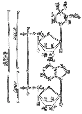
NADH (Fig. 4) is the major electron donor to the electron transport chain of
mitochondria and many bacteria, playing a crucial role in the synthesis of ATP
through oxidative phosphorylation. In eukaryotes, respiratory dissimilation of sugars
Figure 4. Structural representation of NAD(P)H. AMP is coupled to a nicotinamide-containing
nucleotide (NMN) through a pyrophosphate bond. The two non-equivalent hydrogens, labelled a and
P, in NADH are in position 4 of the nicotinamide ring. NADPH differs from NADH by having a
phosphate group instead of a hydroxyl group in position 2 of the ribose in AMP.
NADPH (Fig. 4) is also produced in the cytosol via the pentose phosphate pathway
(Overkamp et al, 2000). The generated NAD(P)H can be reoxidised by
mitochondria. In plants (Roberts et al, 1995) and fungi (Weiss et al, 1970) this
oxidation can be carried out by external NAD(P)H dehydrogenases. In the case of
mammalian mitochondria, with the exception of ct-glycerol phosphate
dehydrogenase from the outer surface of the inner mitochondrial membrane,
NAD(P)H needs to be transported, by redox shuttle mechanisms, into the matrix,
where it will be oxidised by complex I, (Dawson, 1979), since the inner
mitochondrial membrane is impermeable to NAD(P)H (von Jagow et al, 1970).
plants (Krorner and Heldt, 1991) and yeast (Overkamp et al, 2000), despite the
presence of external NAD(P)H dehydrogenases.
NADH is also produced in the matrix by pyruvate dehydrogenase, and by three
enzymes in the Krebs cycle: isocitrate dehydrogenase, alfa-ketoglutarate
dehydrogenase and malate dehydrogenase (Stryer, 1995). The oxidation of the matrix
substrate can occur via the complex I or the internal rotenone-insensitive NAD(P)H
dehydrogenases. Usually, though with different affinity, NADH-oxidising enzymes
can also oxidise NADPH, a compound with very similar structure to NADH with the
exception of a phosphate group replacing the hydroxyl in position 2 in the ribose of
AMP (Fig. 4) (Finel, 1998). At alkaline pH, this oxidation might be prevented by
electrostatic repulsion between the phosphate group of NADPH and the
phospholipids of the membrane (Moller et al, 1982).
3. Two types of NAD(P)H dehydrogenases
The existence of distinct NADH dehydrogenases was first reported by Bragg
and Hou (1967) who partially purified two NADH:menadione reductases from
Escherichia coli with distinct properties.
3.1. Complex I
The NADH:ubiquinone oxidoreductase, or complex I, is a well-characterised
enzyme that catalyses the transfer of electrons from NADH to ubiquinone, through a
number of protein-bound prosthetic groups, coupling this transfer to energy
transduction (Hatefi, 1985).
Electron microscopy of single complex I molecules revealed this enzyme as an
L-shaped structure with two major domains, a hydrophobic arm imbedded in the
inner membrane, and a peripheral arm protruding into the matrix. A thin collar,
missing in the N. crassa complex I, separates the two arms in E. coli (Guenebaut et
pathways have been shown for the peripheral and the membrane arms of Neurospora
complex I. In chloramphenicol-treated cells only a smaller form of the complex is
present (Friedrich et al, 1989). Pulse-labelling experiments have shown that some
complex I subunits are found in intermediate complexes that resemble the membrane
arm (Tushen et al, 1990) and in Mn deficient growth conditions, the peripheral arm
was not formed but the membrane arm could be detected (Schmidt et al, 1992).
Complex I has a dual genetic origin: the majority of its protein subunits is
nuclear-encoded, synthesised in the cytoplasm and imported into mitochondria,
where they are assembled with a few mitochondrially encoded polypeptides (Walker,
1992). The number of subunits that constitute the enzyme varies from 14, in bacteria,
which constitute the so-called minimal functional unit, to 43 in the bovine complex I.
The Neurospora homologue of the minimal functional unit contains seven
mitochondrially encoded subunits, ND1, ND2, ND3, ND4, ND4L, ND5 and ND6,
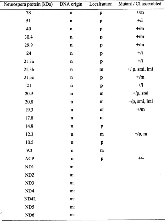
and seven nuclear-encoded subunits. This last group includes the 78, 51,49, 30.4, 24,
21.3c, and 19.3-kDa subunits. The 51-kDa subunit binds NADH, the FMN and a
tetranuclear iron-sulphur cluster. The subunits of 78, 24, 21.3c and 19.3-kDa also
bind iron-sulphur clusters (Videira, 1998). In addition to the minimal functional unit,
21 other subunits are estimated to be present in Neurospora complex I, one of which
is an acyl-carrier protein (Schulte et al, 1994).
Gene disruption experiments in Neurospora were carried out using the two
approaches described above: the replacement of the endogenous gene with defective
copies by homologous recombination and the method of RIP. Both methods have
been successful and already produced 15 mutants in nuclear encoded subunits: 11 in
subunits of the peripheral arm, and 4 in subunits of the membrane arm. The
inactivation of these genes gave mutants where the peripheral and/or the membrane
arm was detected independently (Table 1) confirming previous observations
(Videira, 1998). All mutants are viable in the vegetative state, although with
decreased growth rates, meaning that complex I is not essential for the survival of N.
crassa. In these mutants it is likely that the alternative NADH dehydrogenases
replace complex I, performing the turnover of NADH and contributing to the
production of ATP although with lower yield. Nevertheless, complex I is required for
Several natural and synthetic substances have been shown to be specific
inhibitors of complex I activity. These compounds fall into two groups, those that
prevent NADH oxidation and those that prevent reduction of ubiquinone (Esposti,
1998). In the first group, there is the competitive inhibitor ADP-ribose that, due to its
Table 1. Characteristics of subunits of complex I from Neurospora. i, intact; m, membrane arm; mt, mitochondrial; n, nuclear; p, peripheral arm; smi, small membrane intermediate; lmi, large
membrane intermediate; +, viable mutant. Adapted from Videira et al, 1998.
Neurospora protein (kDa) DNA origin Localisation Mutant I CI assembled
78 n P +/m
51 n P +/i
49 n P +/m
30.4 n P +/m
29.9 n P +/m
24 n P t-/i
21.3a n P +/i
21.3b n m +/ p, smi, lmi
21.3c n P +/m
21 n P +/i
20.9 n m +/p, smi
20.8 n m +/p, smi, lmi
19.3 n cf +/m
17.8 n m
14.8 n P
12.3 n m +/p, m
10.5 n P
9.3 n m
ACP n P
+/-ND1 mt
ND2 mt
ND3 mt
ND4 mt
ND4L mt
ND5 mt
analogous structure, can bind to the NADH binding site (Zharova and Vinogradov,
1997) and diphenyleneiodonium (DPI) which binds to the FMNH2, preventing its
oxidation (Majander et al, 1994). Rotenone, and capsaicin and piericidin A are in the
second group, and each one is representative of a different type of inhibition,
non-competitive, competitive and antagonist of the quinol, respectively (Esposti, 1998).
3.2. Rotenone-insensitive NAD(P)H dehydrogenases
Type II NADH dehydrogenases are nuclear-encoded polypeptides without an
energy-transducing site (Yagi, 1991). Unlike complex I, they are usually composed
of a single polypeptide, though the Sulpholobus NDH-2 (Yagi, 1991) and the 43 kDa
internal NAD(P)H dehydrogenase from red beetroot mitochondria (Menz and Day,
1996) were reported to be homodimers. These proteins lack FMN and iron-sulphur
clusters as co-factors, but contain a non-covalently bound FAD instead. They are
resistant to the complex I specific inhibitors rotenone and piericidin A, and no
general specific inhibitor has been described though a few compounds can prevent
their activity (Yagi et al, 1993).
3.2.1. Rotenone-insensitive NAD(P)H oxidase activities in different organisms
The respiratory chain of E. coli, beyond the energy-transducing NDH-1 (the
complex I equivalent in this organism), displays a membrane-bound
rotenone-insensitive NADH dehydrogenase (NDH-2) with the catalytic site facing the cytosol.
In 1987, Matsushita and others, comparing the deamino-NADH:ubiquinone 1
reductase and the NADH:ubiquinone 1 reductase activities, verified that the former
activity showed more sensitivity to piericidin A than the latter. Furthermore, the
membranes exhibited two apparent Kms for NADH, but only one for
deamino-NADH. They produced strains, whose inside-out membrane vesicles were deficient
in deamino-NADH:ubiquinone 1 reductase activity but could oxidise NADH
they proposed the existence of two species of NADH dehydrogenases. One of these
was able to oxidise deamino-NADH and NADH and its turnover lead to the
production of a proton gradient at a site between the primary dehydrogenase and
ubiquinone. The other enzyme oxidises exclusively NADH and does not generate a
proton gradient before ubiquinone. Spiro et al. (1989, 1990) suggested that oxygen
positively regulates the expression of NDH-2 in E. coli but not of NDH1.
In the cyanobacterium Synechocistys sp. strain PCC 6803 there are three open
reading frames coding for type II NADH dehydrogenases, ndbA, ndbB and ndbC.
Their primary structures display sequence identities with other NADH
dehydrogenases that do not exceed 30 %. NAD(P)H and FAD binding motifs are
conserved in all sequences. The genes have been cloned and deletion mutants
produced which lead only to small changes in the respiratory activity. An expression
construct of ndbB complemented an E. coli strain lacking NDH-1 and NDH-2
(Howittétfa/., 1999).
The obligate aerobic yeast Yarrowia lipolytica has a type II NAD(P)H
dehydrogenase on the outer surface of the inner mitochondrial membrane. Deletion
mutants of the enzyme were fully viable. Total inhibition of NADH oxidation in
mitochondria solubilized with CHAPS was achieved with piericidin A, indicating
that complex I activity was the sole NADH oxidation activity left in those strains.
The orientation of the alternative NADH dehydrogenase was assessed measuring
NADH:5-nonylubiquinone oxidoreductase activity before and after permeabilisation
of the inner mitochondrial membrane. In the presence of piericidin A,
NADH:5-nonylubiquinone oxidoreductase activity was not affected by permeabilisation,
showing that the active site of the enzyme faced the intermembrane space (Kerscher
etal., 1999).
The respiratory chain of the facultative aerobic yeast Saccharomyces cerevisiae
lacks complex I. The oxidation of NAD(P)H from the cytosol and from the matrix is
carried out by two external and one internal NAD(P)H dehydrogenases, respectively,
de Vries and Grivell (1988) described the purification of a presumptive external
NADH dehydrogenase, in mitochondria from S. cerevisiae. The protein consisted of
a single subunit with molecular mass of 53 kDa that contained a FAD and was
the nuclear gene coding for one of these NAD(P)H dehydrogenases. A null mutant
was constructed and the oxidation of several substrates by mitochondria from wild
type and mutant strains was measured. The oxidation of external NADH was not
affected in mutant mitochondria, while the oxidation of substrates generating internal
NADH was severely decreased (lactate) or missing (pyruvate/malate and ethanol).
This study showed that the inactivated enzyme was the internal NADH
dehydrogenase. Latter, Small and McAlister-Henn (1998), and Luttik et al, (1998)
have identified two other genes coding for mitochondrial NADH dehydrogenases,
NDE1 and NDE2, oxidising NADH from the cytosol. Both genes were deleted and
the NADH oxidation was followed in mitochondria from wild type and nde deletion
mutants. Compared to wild type mitochondria, exogenous NADH oxidation was
drastically reduced in one of the mutants, albeit the other displayed no difference.
However, in mitochondria from the double mutant, oxidation of external NADH was
completely absent. In conclusion, the external location of NDE1 and NDE2 was
confirmed.
In Neurospora mitochondria, two rotenone-insensitive NAD(P)H
dehydrogenases have been reported, one on each side of the inner membrane. Weiss
et al, (1970) observed oxygen consumption after addition of rotenone to
mitochondria respiring pyruvate/malate. This result indicated the presence of a
rotenone-resistant NADH dehydrogenase facing the matrix of Neurospora
mitochondria. Weiss et al, (1970) and Moller et al, (1982) showed that Neurospora
electron transport chain was able to oxidise external NADH and NADPH in a
rotenone-insensitive manner as well.
Rotenone-insensitive NAD(P)H dehydrogenases have also been described in
the electron transport chain of plant mitochondria. In plants, several efforts have
been done to purify them. There are reports of the purification of a 42-kDa (Luethy
et al, 1991), a 26-kDa (Rasmusson et al, 1993) and a 43-kDa (Menz and Day, 1996)
NAD(P)H dehydrogenases from red beetroot mitochondria. In red beetroot
mitochondria, the purification of a 58-kDa protein was associated to external
NAD(P)H oxidation activity (Luethy et al, 1995). The purification of a 32-kDa
external NADH dehydrogenase from maize mitochondria has been described
Studies of NADH and NADPH oxidation by intact mitochondria from Arum maculatum and potato tubers (Roberts et al, 1995), and by IO-SMP from potato tubers and Jerusalem artichoke (Moller and Palmer, 1982; Rasmusson and Moller,
1991b; Melo et al, 1996) lead to the conclusion that there are four distinct enzymes,
two on each side of the inner membrane. Rasmusson et al, (1999) described two
different cDNAs, from potato, homologous to genes encoding rotenone-insensitive
NADH dehydrogenases in yeast and bacteria. The encoded proteins have
approximate molecular masses of 55 and 65 kDa and are located in the inner and in
the outer surfaces of the inner mitochondrial membrane, respectively. The latter
protein could be homologous to the 58-kDa protein isolated from red beetroot.
3.2.2. Effects of cations on rotenone-insensitive NAD(P)H oxidase activities
In the matrix of mammalian mitochondria, the physiological concentration of
free calcium is estimated to be between 0.05 and 5 pM. A few matrix NAD+
-reducing enzymes are stimulated by calcium, namely pyruvate dehydrogenase and
the tricarboxylic acid cycle enzymes 2-oxoglutarate dehydrogenase and NAD+
-isocitrate dehydrogenase (McCormack et al, 1990).
Oxidation of matrix NADPH by potato tuber mitochondria is also enhanced by
the presence of calcium. The internal NADH oxidation is not really affected by
additions of external calcium (Rasmusson and Moller, 1991a, Melo et al, 1996). The presence of a calcium-dependent NADPH dehydrogenase on the matrix side of the
inner mitochondrial membrane potato tuber mitochondria, linked to the electron
transport chain, opens the possibility of a regulation of the redox potential of
NADPH pool, in the matrix, by calcium (Rasmusson and Moller, 1991a).
The stimulation of exogenous NADH oxidation by cations through screening
of the negative charges on the surface of the inner membrane was reported in N.
crassa mitochondria (Moller et al, 1982). Calcium-sensitivity has frequently been associated with these enzymes. Cytosolic NADH oxidation by plant mitochondria is
totally dependent on calcium (Coleman and Palmer, 1971; Moller et al, 1981).
same behaviour for external NADPH oxidation. The same authors proposed that
cation concentration, in general, and the concentration of calcium, in particular, can
regulate exogenous NAD(P)H oxidation. The primary structure of the 65 kDa
NAD(P)H dehydrogenase from potato mitochondria (Rasmusson et al, 1999)
contains an EF-hand motif, suggesting that this protein might bind calcium. This is in
sharp contrast with the primary structure of most alternative NAD(P)H
dehydrogenases such as the NDH-2 from E. coli (Young et al, 1981), NDI1 (de
Vries et al, 1992), NDE1 and NDE2 (McAlister-Henn et al, 1998; Luttik et al,
1998) from S. cerevisiae, which all lack calcium-binding domains.
3.2.3. The external NADH dehydrogenase from rat heart mitochondria - a
mysterious exception
There are several articles describing the oxidation of exogenous NADH by an
external NADH dehydrogenase on the outer surface of the inner membrane of rat
heart mitochondria (Nolh and Shõnheit, 1996, Oliveira et al, 2000). Nevertheless,
this issue is the center of an intense debate, and consensus is far from being achieved.
Studies have been performed regarding the possible physiological role of such
an enzyme and its behaviour in different pathological processes. The external NADH
dehydrogenase has been associated with pathological conditions related to the
production of oxygen free radicals, released during metabolic events under
conditions of ischemia/reperfusion. Similar association was found in cardioselective
toxicity of adrianmycin, an anticancer drug accepting the electrons from the external
NADH dehydrogenase (Nohl, 1998). It was also suggested that this NADH
dehydrogenase could be responsible for the oxidation of exogenous NADH when
there is an excessive accumulation of that substrate in the cytosol (Nolh and
Shõnheit, 1996).
Recently, Oliveira et al, (2000) reported a specific inhibitor to the external
NADH dehydrogenase, carvedilol, that does not affect the oxygen uptake resulting
from complex I activity. In contrast to glutamate/malate or succinate, the oxidation
membrane potential. Nonetheless, this activity is inhibited by antimycin A,
potassium cyanide, sodium azide and mixothiazol. Surprisingly, exogenous NADH
oxidation by rat heart mitochondria is also inhibited by rotenone.
4. Human pathologies associated with NADH oxidation
In humans, complex I is solely responsible for the oxidation of matrix NADH
and shuttle mechanisms exist for the oxidation of cytosolic NADH. Mutations in
mtDNA genes, for instance complex I genes, can be responsible for drastic
phenotypes (Wallace, 1992). An example of a pathology caused by point mutations
in mtDNA is the Leber's hereditary optic neuropathy (LHON) (Schapira, 1998).
Complex I deficiency and oxidative damage have been identified in the substantia
nigra (Schapira, 1996) and platelets (Swerdlow et al, 1996; Gu et al, 1998) of
patients with Parkinson's disease. Platelets from these patients have been used in
genome transplantation experiments with cells lacking mtDNA (p cells). This
involves fusion of the platelets (mtDNA but no nucleus) from a patient with
Parkinson's disease and complex I deficiency with p° cells (nucleus but no mtDNA).
In these experiments, the complex I defect was transferred with the patients mtDNA
to the resulting fused cells, indicating that it was due to a defect in the mtDNA (Gu et
al, 1998). The defects in mtDNA may be solely responsible for the mitochondrial
malfunctioning in Parkinson's disease but it is not known if that malfunctioning is
enough to cause the disease, or if other genetic or environmental factors are involved
(A. H. V. Schapira (2000) personal communication).
Nuclear gene mutations have also been associated with human pathologies. For
instance, a mutation in the nuclear gene coding for the 18-kDa subunit of complex I
was identified in a patient with encephalomyopathy (van den Heuvel et al, 1998).
Other mutations in nuclear genes from complex I subunits have been described for
patients with Leigh's syndrome. These involved two iron-sulphur proteins: the
subunits encoded by NDUFS8 and NDUFS7 (homologous to the 21.3c-kDa and the
19.3-kDa subunits of the N. crassa complex I, respectively), possibly involved in the
1999). Three children have been reported with mutations in the gene NDUFV1
encoding the NADH binding subunit, homologous to the 51-kDa subunit of the N.
crassa complex I. Two of them developed vomiting, hypotonia, myoclonic epilepsy
and psychomotor delay at 5 months and died at 14 and 17 months, respectively. The
third child MRI presented with myoclonic epilepsy at 6 months and progressed with
psychomotor delay and spasticity. At 12 months psychomotor development stopped
and macrocephaly was observed. Still alive at 10 years old, MRI showed macrocytic
leukodistrophy. In all cases, the patients were either homozygous or compound
hétérozygotes for the respective mutations and inheritance was autosomal recessive
(Schuelkeefa/., 1999).
Complex I-defects associated with human pathologies failed to be healed by
chemotherapies (Chrazanowska-Lightowlers et al, 1995). The capacity of type II
NADH dehydrogenases to carry out the turnover of NADH may be more important
to health than their inability to pump protons. Therefore, a possible approach to
overcome complex I-defects is to introduce in patient cells a type II NADH
dehydrogenase to restore the function of oxidising NADH in their mitochondria
(Kitajima-Ihara and Yagi, 1998).
The internal NADH dehydrogenase from S. cerevisiae (NDI1) was expressed
in E. coli (Kitajima-Ihara and Yagi, 1998) and in complex I-deficient Chinese
hamster cells (Seo et al, 1998), whereby functioned as a member of the respiratory
chain in the host cells. NDI1 was able to restore the NADH oxidase activity in the
latter case. Human kidney cells were also transfected by the gene encoding NDI1.
The transfected enzyme was successfully transcribed and translated to produce a
functional enzyme linked to the electron transport chain of the host cell mitochondria
(Seo etal, 1999).
5. Final comments
Our understanding of the structure, mechanisms and physiological roles of
NAD(P)H:ubiquonone oxidoreductases is far from clear, particularly with respect to
contribution to further characterise the oxidation of NAD(P)H by the respiratory
chain of mitochondria from the fungus N. crassa is described. This work paid special attention to genetics, biogenesis and physiology of the crucial enzymes responsible
for accomplishing this function in Neurospora. Contributing to the development of
models where the human mitochondrial disease condition can be simulated and
studied, allowing future progress concerning therapeutics, was also the target of our
Chapter II - Research Project
1. OBJECTIVES
Although much is already known about the NADH:ubiquinone oxidoreductase
(complex I) from Neurospora mitochondria, a full understanding of the structure and
function of several subunits of the enzyme has not been accomplished yet. In
Neurospora mitochondria, the presence of at least two rotenone-insensitive
non-proton-pumping NAD(P)H dehydrogenases has been reported. Nevertheless, the
genes encoding these proteins have not been identified and their structure was
unknown when this project started.
The overall purpose of the present work was to provide a better understanding
of the bioenergetical processes occurring in the mitochondrion, by improving our
knowledge of the NAD(P)H dehydrogenases of the Neurospora crassa organelle.
This project aimed to detect and characterise the different NAD(P)H
dehydrogenase activities in mitochondria from the wild type strain and from strains
deficient in complex I subunits. The isolation and sequencing of genes encoding
rotenone-insensitive NAD(P)H dehydrogenases was another goal. Envisaging a
detailed characterisation of the rotenone-insensitive NAD(P)H dehydrogenases, a
special attention was given to the induction and characterisation of mutants and also
of double mutants deficient in both the alternative NAD(P)H dehydrogenases and in
complex I subunits. The characterisation of the alternative NAD(P)H
dehydrogenases and the production of mutants deficient in these enzymes can
provide new models for the study of complex I and associated pathologies in