Dissecting the biophysical mechanisms underlying regeneration of complex organs in vertebrates
314
0
0
Texto
(2)
(3) Universidade de Lisboa Faculdade de Medicina de Lisboa. “Dissecting the Biophysical Mechanisms Underlying Regeneration of Complex Organs in Vertebrates” Joana Freire de Castiço Monteiro. Tese orientada pelo Professor Doutor António Alfredo Coelho Jacinto Tese co-orientada pelo Professor Doutor Joaquín Rodríguez-Léon e pelo Professor Doutor Domingos Manuel Pinto Henrique Doutoramento em Ciências Biomédicas Especialidade em Ciências Morfológicas. As opiniões expressas nesta publicação são da exclusiva responsabilidade do seu autor..
(4)
(5) A impressão desta dissertação foi aprovada pelo Conselho Científico da Faculdade de Medicina de Lisboa em reunião de 23 de Julho de 2014..
(6)
(7) Acknowledgements/ Agradecimentos First of all I want to thank to Ana Catarina Certal, who made this project possible. Thank you for all the teaching, trust and encouragement.. Joaquin Rodríguez-Léon, thank you for the prompt help, patience and encouragement. Your mentoring during the thesis writing was fundamental. You’ll always be the BOSS!. António Jacinto, Leonor Saúde, Rita Fior, thank you for the data discussion and. support. Moisés Mallo, you were always available to help and let me borrow your. “crew” for so many times: thanks for making me feel part of the group. Alan Shipley, thanks for the prompt SIET technical support, and thanks for caring.. To the “Organogenics” and to the “lab next door”, thank you for providing a great working environment, for listening to my frustrations and applauding my. achievements: Rita Aires, Rita Félix, Teresa Gomes, Raquel Tomás, Fernando Ferreira, Diana Chapela, Rui Castanhinha, Isabel Guerreiro, Sofia Pereira, Ana Nóvoa, Arnón. Juberg, Ana Casaca, Ana Cristina, Aibüke: what a great team!!! Rita Aires, thank you. for your help regarding the molecular biology techniques and for all the support. “Nandinho”, thank you for your collaboration regarding the chloride-specific flux. measurements. Teresa Gomes, my SIET-mate, thanks for the SIET-help, and thanks for listening to me so patiently!. It was a great pleasure to be part of the IGC/IMM community, that provided an. outstanding working environment and cooperation between all people. Particularly, I. would like to leave my appreciation to Lara Carvalho, Maysa Franco and Liliana Vale,. who took so great care of “my babies”; to Alessandro Ramos, for all the expertise. during SIET optimization; and to Mariana Simões, for the availability to discuss data. and protocols. Also, to Ana Homem, to the ladies from the cleaning room and the guys from the maintenance, especially Monteiro and Sr. Sousa, all of you made my life. Page | i.
(8) much easier for so many times. Also, this project was not possible without the FCT financial support.. E agora em bom português, uma palavra especial aos meus pais, Gabi e Tó: obrigada pela educação que me proporcionaram e que me trouxe até aqui! Esta tese também é. vossa. Tenho muito orgulho em ser vossa filha!. Às minhas irmãs, Sofia, Filipa e Inês, porque é tão bom ter irmãs com quem partilhar a vida!! Ao Pedro e à Leonor, que fazem de mim uma tia babadíssima. Ao Ricardo, por. nunca duvidar de que eu seria capaz. À avó Lili, pelo exemplo de aceitação e sentido. de humor para com a vida. Ao avô Castiço, pelo orgulho nas netas que lhe salta pelos olhos e pelo esforço para entender o que eu faço. Ao avô Mário, por ter respondido a. tantos “e se…?” sobre números e afins. À avó Julieta, agradeço os abraços sempre prontos, e a ajuda nestes últimos anos.. A todos os meus amigos, e em especial a Susana C, a Marta, a Raquel, o Ricardo S, o Ricardo L, o Mário, o Miguel, a Joana, a Paula, a Susana Z, o André, o Flacho, a Maria. João, obrigada por escutarem os meus entusiamos e frustrações ao longo deste projeto, mesmo quando não percebiam nada do que é que eu estava para ali a dizer! E pela amizade que não tem preço.. Page| ii.
(9) Table of contents Acknowledgments/ Agradecimentos ................................................................................................... i. Table of contents ........................................................................................................................................ iii List of figures ............................................................................................................................................ viii. List of tables ............................................................................................................................................... xii List of abbreviations .............................................................................................................................. xiii Resumo ......................................................................................................................................................... xv. Summary ................................................................................................................................................. xviii. Chapter I- Introduction .......................................................................... 1 1. Animal Regeneration ....................................................................................................... 1.1. Historical overview ....................................................................................................................... 1.2. Types of regeneration .................................................................................................................. 1.2.1. Cellular reprogramming ..................................................................................................... 1.3. Regeneration among Metazoa: distribution and evolution .......................................... 3 3 4 5 6. 2. Zebrafish regeneration ................................................................................................ 10 2.1. Structure of the caudal fin ............................................................................................................ 11 2.2. Stages of epimorphic regeneration in the zebrafish caudal fin .................................... 13 2.2.1. Wound healing ......................................................................................................................... 14 2.2.2. Blastema formation ................................................................................................................ 16 2.2.2.1. Cellular origin of the blastema ............................................................................... 18 2.2.3. Regenerative outgrowth ...................................................................................................... 19 2.2.4. Positional memory ................................................................................................................. 23. 3. Endogenous electrical signals of biological significance .................................... 25 3.1. Origin of endogenous electric currents and electric fields ............................................ 25 3.2. Endogenous electrical signals as morphogenetic signals ............................................... 28 3.2.1. Endogenous electric currents and electric fields during development ......... 28 3.2.2. Endogenous electric currents and electric fields during wound healing and regeneration ........................................................................................................................... 29 3.3. Bioelectrical signals control cell behaviour .......................................................................... 32 3.3.1. Bioelectrical control of cell behaviour during wound healing ............................ 32 3.3.2. Bioelectrical signals control cell behaviour required for regeneration ......... 34 3.3.2.1. Cell proliferation .......................................................................................................... 34 3.3.2.2. Cell differentiation/ de-differentiation .............................................................. 35 3.3.2.3. Apoptosis ........................................................................................................................ 37 3.3.2.4. Tissue patterning ......................................................................................................... 37 Page | iii.
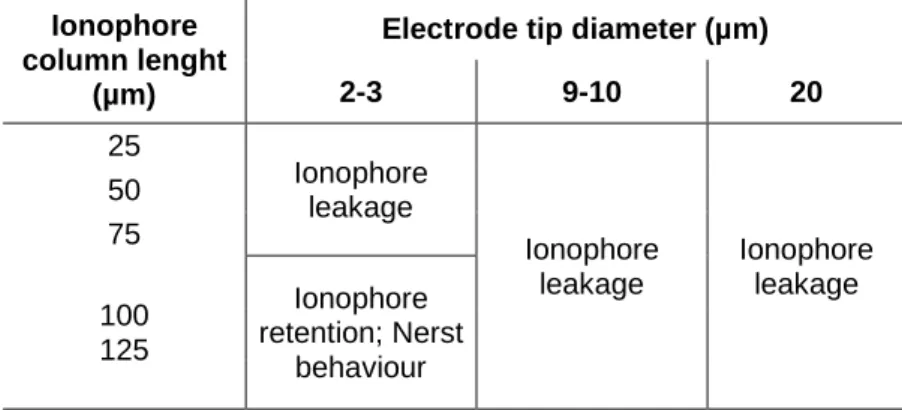
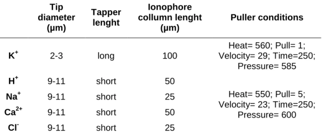
(10) 3.4. Chemical signals mediated by ion translocators affect cell behaviour ..................... 38 3.5. How do cells sense and transduce bioelectrical signals into cell behaviour .......... 39 4. Protons: chemical and electrical functions ............................................................... 43 4.1. Proton transport across membranes ....................................................................................... 44 4.2. V-ATPase: main proton (H+) pump in animal cells ............................................................ 46 4.2.1. Structure and subunit function ......................................................................................... 46 4.2.2. Mechanism of activity ........................................................................................................... 49 4.2.3. Regulation of activity ............................................................................................................. 50 4.2.4. V-ATPase functions ................................................................................................................ 51. 5. Tools for dissecting bioelectrical signals ................................................................... 54 5.1 Scanning Ion-selective Electrode Technique (SIET) .......................................................... 55. 6. AIMS OF THE THESIS .................................................................................................... 57. Chapter II – Materials and Methods .................................................. 59 1. Animal model ..................................................................................................................... 61 1.1. Zebrafish strains and husbandry ............................................................................................... 61 1.2. General procedures in adult zebrafish .................................................................................... 61 1.2.1. Anaesthesia and euthanasia ................................................................................................ 61 1.2.2. Caudal fin amputation and tissue collection ................................................................ 62 1.3. General procedures in zebrafish embryos and larvae ..................................................... 63 1.3.1. Embryo and larvae anaesthesia and euthanasia ........................................................ 63 1.3.2. Fin fold amputation ................................................................................................................. 63 1.3.3. Embryo and larvae collection, fixation and initial processing .............................. 63. 2. Scanning Ion-selective Electrode Technique (SIET) .............................................. 65 2.1. Construction of ion-selective electrodes (ISE) .................................................................... 65 2.1.1. Pulling conditions .................................................................................................................... 65 2.1.2. Silanization .................................................................................................................................. 65 2.1.3. Electrode filling ......................................................................................................................... 66 2.2. Completing the SIET system ........................................................................................................ 66 2.3. Calibration of the ISE ...................................................................................................................... 67 2.4. Recording solution and anaesthesia optimization ............................................................ 68 2.5. Recording chamber optimization .............................................................................................. 68 2.6. Artificial Source ................................................................................................................................. 68 2.7. Data acquisition and analysis ...................................................................................................... 69 3. Microarray ........................................................................................................................... 71. 4. Quantitative real-time PCR (qRT-PCR) ....................................................................... 72 5. Whole mount in situ hybridization .............................................................................. 73 5.1. Cloning of gene- specific DNA sequences .............................................................................. 73. Page| iv.
(11) 5.1.1. Isolation and recombination of DNA sequences for the genes of interest ...... 73 5.1.2. Transformation and clone selection ................................................................................ 75 5.2. Recombinant DNA amplification ............................................................................................... 75 5.3. Plasmid linearization and in vitro RNA probe transcription ......................................... 76 5.4. In situ hybridization protocols for caudal fin and whole embryos ............................. 77. 6. Whole mount immunohistochemistry ........................................................................ 78 6.1. Cell proliferation assay .................................................................................................................. 78. 7. Histological techniques .................................................................................................... 80 7.1. Cryopreservation and sectioning .............................................................................................. 80 7.2. Hematoxylin-Eosin staining ........................................................................................................ 80 8. In vivo manipulation techniques .................................................................................. 81 8.1. Microinjection .................................................................................................................................... 81 8.2. Electroporation ................................................................................................................................. 81 8.3. Dose-response curves for ion transporter inhibitors ...................................................... 81 8.4. Pharmacological inhibition of V-ATPase activity in regenerating caudal fins ....... 82 8.5. Morpholino-mediated knockdown of V-ATPase subunit in the caudal fin ............. 83 8.6. Fin fold regeneration ...................................................................................................................... 84. 9. Microscopy techniques ................................................................................................... 85 9.1 Scanning electron microscopy ..................................................................................................... 85 9.2. Microscopy and image analysis .................................................................................................. 85. Chapter III – Results ……………………….......…………….……………….. 87 Part 1. Optimization of the Scanning Ion-selective Electrode Technique … ……………………………………………………………………………………………………………... 91 1.1. Ion-selective electrode ................................................................................................................... 93 1.2. Recording media ............................................................................................................................... 93 1.3. Recording chamber ......................................................................................................................... 96 1.4. Artificial sources ............................................................................................................................... 97 Discussion .................................................................................................................................................... 98. Part 2. Ion-specific fluxes during adult zebrafish caudal fin regeneration .. …..................................................................................................................................... 101. 2.1. Spatial ion-specific flux profile ................................................................................................ 101 2.2. Temporal ion-specific flux profile ......................................................................................... 101 2.3. Ion-specific composition of electric currents during regeneration ......................... 104 Discussion ................................................................................................................................................. 105. Part 3. Molecular source of proton efflux during regeneration of the adult zebrafish caudal fin …………….…...……………………………………………….. 111. 3.1. V-ATPase is up-regulated during adult zebrafish caudal fin regeneration .......... 111 Page | v.
(12) 3.1.1. Transcriptomic analysis of the regenerating caudal fin ………………………..… 111 3.1.2. Analysis of V-ATPase expression in the regenerating caudal fin ……………… 117 3.1.2.1. qRT-PCR analysis of atp6v1e1b relative expression during regeneration ........................................................................................................................................... 117 3.1.2.2. Expression pattern of atp6v1e1b and atp6v1a during zebrafish embryonic development .................................................................................................. 118 3.1.2.3. Expression pattern of atp6v1e1b and atp6v1a during regeneration of the caudal fin ………………..………………..….………………………………………….... 120 3.1.2.4. Immunolocalization of Atp6v1a during caudal fin regenerating ….. 121 3.1.3. Analysis of Nhe7 expression in the regenerating caudal fin ............................... 124. 3.2. V-ATPase inhibition affects adult caudal fin regeneration .......................................... 125 3.2.1. Effect of atp6v1e1b knockout in larval fin fold regeneration ............................. 125 3.2.2. Pharmacological inhibition of the V-ATPase activity in the regenerating caudal fin …...…………………………………………......................………………………………………… 127 3.2.3. Morpholino-mediated knockdown of atp6v1e1b in the regenerating caudal fin ....................................................................................................................................................... 131 Discussion ................................................................................................................................................. 135. Part 4. Roles of V-ATPase and H+ efflux in the regenerative process ..... 145. 4.1. V-ATPase expression correlates with position-dependent regeneration rate and affects H+ efflux ...................................................................................................................................... 145 4.1.1. atp6v1e1b expression pattern during regeneration after proximal-distal (PD) amputation ............................................................................................................................. 146 4.1.2. Immunolocalization of Atp6v1a during regeneration after PD amputation .. 147 4.1.3. H+ flux pattern after PD amputation ........................................................................... 150 4.1.3.1. H+ flux pattern using sedation methods alternative to Tricaine .......... 151 4.1.4. H+ flux pattern after fin amputation at the caudal peduncle ............................ 153 4.1.5. H+ efflux is affected by V-ATPase subunit atp6v1e1b knockdown ................ 156 4.2. Histological comparison of the regenerating caudal fin after proximal and distal amputation …………………………………………………………………………………………….… 157 4.2.1. Hematoxylin-Eosin staining of regenerating fins after PD amputation …… 158. 4.2.2. Scanning electron microscopy analysis of the wound healing stage during caudal fin regeneration after proximal and distal amputation ........................ 160. 4.3. V-ATPase is required during regeneration in an amputation plane- dependent. manner ........................................................................................................................................................ 165. 4.4. V-ATPase affects molecular and cellular events during regeneration ................... 169 4.4.1. Blastema cell proliferation is reduced upon atp6v1e1b knockdown ............ 169 4.4.2. V-ATPase subunit knockdown affects fin innervation ........................................ 174 4.4.3. Comparison of regeneration markers expression in proximal and distal stumps .................................................................................................................................... 177. Page| vi.
(13) 4.4.4. V-ATPase is required for normal expression of aldh1a2 and mkp3 .............. 181 Discussion .................................................................................................................................................. 184. Chapter IV – General Discussion ……………….……………………….. 189 Chapter V – References ….…………………….…………………………….. 205 Chapter VI - Appendixes ……………………….……………………………. 247. Page | vii.
(14) List of figures Page Figure I.1 – Phylogenetic distribution of epimorphic regeneration among the Metazoa ..8. Figure I.2 – Zebrafish structures with regenerative ability .................................................. 10. Figure I.3 – Structure of the zebrafish caudal fin ...................................................................... 13. Figure I.4 – Stages of epimorphic regeneration ......................................................................... 14. Figure I.5 – Fin ray morphology and signalling during epimorphic regeneration ..... 22 Figure I.6 – Origin of endogenous electric signals .................................................................... 26. Figure I.7 – Membrane potential (V mem ) affects developments .......................................... 29. Figure I.8 – Electric field (EF) affects wound healing rate in rat cornea ......................... 30. Figure I.9 – Bioelectrical signals correlate with regenerative ability in amphibians ...... 32. Figure I.10 – Electric fields (EF) affect cell division and nerve sprouting during wound healing in rat cornea ............................................................................................................... 33. Figure I.11 – Membrane voltage (V mem ) correlates with proliferative potential and differentiation state of cells .................................................................................................................. 36 Figure I.12 – Membrane potential (V mem ) mediated by the H+,K+-ATPase regulates patterning during planarian regeneration .................................................................................... 38. Figure I.13 – Transduction of bioelectrical signals into cell behaviour .......................... 41 Figure I.14 – Structure of the V-ATPase ........................................................................................ 48. Figure I.15 – Scanning Ion-selective Electrode Technique (SIET) ..................................... 56 Figure II.1 – Planes of caudal fin amputation ............................................................................. 62. Figure II.2 – Plane of caudal peduncle amputation .................................................................. 63. Figure III.1 – Optimized ion-selective electrodes (ISE) .......................................................... 91 Figure III.2 – Caudal fin regeneration rate under different fish bathing solutions .... 92 Figure III.3 – Recording set-up for SIET application to adult zebrafish caudal fin .... 94. Figure III.4 – Artificial source for potassium (K+), sodium (Na+), protons (H+), calcium (Ca2+) and chloride (Cl-) ........................................................................................................ 95. Figure III.5 – Pattern of potassium (K+), sodium (Na+), calcium (Ca2+), chloride (Cl-) and proton (H+)-specific fluxes during the main regeneration stages of the adult zebrafish caudal fin ............................................................................................................................... 101 Figure III.6 – Contribution of individual ion-species to the total ionic flux during the first 6hpa, when wound healing is taking place ………………………….…………..….. 102. Figure III.7 – qRT-PCR for fgf20a at 24 hpa .............................................................................. 110 Page| viii.
(15) Figure III.8 – Dynamic expression of ion transport- related transcripts during regeneration ............................................................................................................................................. 114. Figure III.9 – qRT-PCR for atp6v1e1b during main stages of caudal fin regeneration . 116. Figure III.10 – atp6v1e1b is not expressed in the median fin fold of zebrafish embryos ................................................................................................................................................................................. 117. Figure III.11 – Expression of V-ATPase subunits atp6v1e1b and atp6v1a during zebrafish embryonic development ................................................................................................. 117. Figure III.12 – atp6v1e1b expression in zebrafish embryos: whole body view ........ 118. Figure III.13 – Expression of V-ATPase subunits atp6v1e1b and atp6v1a during caudal fin regeneration ....................................................................................................................... 119. Figure III.14 – Atp6v1a localizes specifically to the H+-ATPase rich cells in zebrafish embryos ...................................................................................................................................................... 120 Figure III.15 – Immunolocalization of Atp6v1a in the regenerating caudal fin ........ 121. Figure III.16 – Atp6v1a localizes to the blastema .................................................................. 121. Figure III.17 – Detailed view of Atp6v1a localization in the regenerating fin tissue ... 122. Figure III.18 – In situ hybridization for nhe7 during caudal fin regeneration ........... 123. Figure III.19 – Phenotypic characterization of V-ATPase- mutant zebrafish embryos and larvae ............................................................................................................................... 124. Figure III.20 – Larval fin fold regeneration in wild type and V-ATPase mutant zebrafish embryos/larvae .................................................................................................................. 125. Figure III.21 – Effects of Concanamycin A (concA) in the embryonic development of AB wild type zebrafish ......................................................................................................................... 127. Figure III.22 – DMSO and Concanamycin A (concA) dose-response curves for larval zebrafish mortality, survival and abnormal phenotype, at 72 hours post fertilization . 128. Figure III.23 – Inhibition of V-ATPase activity in the regenerating caudal fin using concA ........................................................................................................................................................... 128. Figure III.24 – Analysis of the specificity and toxicity of atp6v1e1b-specific fluorescein- tagged morpholinos (fluo-MO), in zebrafish embryos ................................. 131 Figure III.25 – atp6v1e1b knockdown during fin regeneration, using fluorescein – tagged morpholino (fluo-MO) .......................................................................................................... 132 Figure III.26 – Regeneration of zebrafish caudal fin after proximal-distal (PD) amputation ................................................................................................................................................ 143. Figure III.27 – atp6v1e1b is differently up-regulated during regeneration of caudal fin after PD amputation ....................................................................................................................... 144 Figure III.28 – Comparison of Atp6v1a domain in proximal versus distal regenerates ......................................................................................................................................................................... 146. Figure III.29 – Detailed view of Atp6v1a blastemal domain in proximal stumps and distal stumps at 24 hpa .............................................................................................................. 147 Page | ix.
(16) Figure III.30 – Detailed view of Atp6v1a blastemal domain in proximal stumps and distal stumps at 48 hpa ............................................................................................................... 148. Figure III.31 – H+ flux pattern during caudal fin regeneration after PD amputation ......................................................................................................................................................................... 149. Figure III.32 – Tricaine does not affect the detection of extracellular H+ efflux during regenenation of the caudal fin .......................................................................................................... 151. Figure III.33 – Analysis of caudal fin regeneration after amputation at the caudal peduncle .................................................................................................................................................... 153 Figure III.34 – atp6v1e1b knockdown using vivo- MO affects H+ efflux during caudal fin regeneration ...................................................................................................................... 155. Figure III.35 – Hematoxylin-Eosin (H-E) staining of regenerating caudal fins 6 and 24 hours after proximal and distal amputation ........................................................................ 157 Figure III.36 – Hematoxylin-Eosin (H-E) staining of regenerating caudal fins, 48 hours after proximal and distal amputation ................................................................... 157/ 158. Figure III.37 – Scanning electron microscopy imaging of the caudal fin immediately upon amputation at proximal and distal planes .......................................... 160. Figure III.38 – Scanning electron microscopy imaging of the caudal fin during the wound healing stage after amputation at proximal or distal planes ............................... 161. Figure III.39 – Scanning electron microscopy imaging of the caudal fin during the wound healing stage after amputation at proximal or distal planes – high magnification detail of images in Fig. III.38 ............................................................................. 162 Figure III.40 – Injection of control vivo-MO (cvivo-MO) did not affect fin regeneration 164. Figure III.41 – atp6v1e1b knockdown after proximal and distal fin amputation, using vivo-MO .......................................................................................................................................... 165. Figure III.42 – atp6v1e1b knockdown after proximal and distal fin amputation in atp6v1e1b hi577aTg/+ zebrafish, using vivo-MO ............................................................................ 166. Figure III.43 – Immunodetection of apoptotic cells in regenerating fins after atp6v1e1b knockdown ......................................................................................................................... 168. Figure III.44 –Quantification of proliferating cells in the blastema upon V-ATPase knockdown ............................................................................................................................................... 169 Figure III.45 – Cell proliferation in atp6v1e1b knocked down fins, 24h after proximal or distal amputation ............................................................................................................................. 170. Figure III.46 – Cell proliferation in atp6v1e1b knocked down fins, 48h after proximal or distal amputation ............................................................................................................................. 171. Figure III.47 – Caudal fin innervation 24 h after proximal or distal amputation and atp6v1e1b knockdown ......................................................................................................................... 173. Figure III.48 – Caudal fin innervation 48 h after proximal or distal amputation and atp6v1e1b knockdown ......................................................................................................................... 174 Page| x.
(17) Figure III.49 – Expression of aldh1a2 in the regenerating caudal fin after PD amputation ................................................................................................................................................ 177. Figure III.50 – Expression of wnt10a and wnt5b in the regenerating caudal fin after PD amputation ......................................................................................................................................... 177. Figure III.51 – Expression of msxb and mkp3 in the regenerating caudal fin after PD amputation ................................................................................................................................................ 178. Figure III.52 – Expression of osx and shh during caudal fin regeneration after PD amputation ................................................................................................................................................ 179. Figure III.53 – atp6v1e1b knockdown affects the expression of specific genes during regeneration .............................................................................................................................. 181 Figure IV.1 – V-ATPase H+ pumping activity is required during adult zebrafish caudal fin regeneration: Working model ………….……………………..………………………..….………. 200. Page | xi.
(18) List of tables Table II.1 – Ion-specific electrode filling solutions ................................................................... 66. Table II.2 – Ion-specific calibration solutions ............................................................................. 67 Table II.3 – Ion-specific artificial source solutions ................................................................... 69. Table II.4 – Sequence-specific primers for quantitative real-time PCR .......................... 72. Table II.5 – Sequence-specific primers for amplification of the genes of interest ...... 74 Table II.6 – Enzymes used to produce in vitro RNA probes from recombinant DNA .76. Table II.7 – Primary antibodies ......................................................................................................... 79. Table II.8 – Sequences of the morpholinos used ....................................................................... 84. Table III.1 – Optimization of ion-selective electrodes for H+, Na+, Ca2+ and Cl- - specific flux recordings using SIET .................................................................................................................... 90. Table III.2 – Optimization of K+-selective electrodes for flux recordings using SIET. ............................................................................................................................................................................ 90 Table III.3 – Characteristics of the optimized ion-selective electrodes. for SIET. application to adult zebrafish caudal fin ........................................................................................ 91. Table III.4 – Response of fish to different Tricaine concentrations (mM) ..................... 92 Table III.5 – Optimized recording medium for each ion species investigated with SIET ................................................................................................................................................................. 93. Table III.6 – Validation of the microarray ....................................................................... 110/111. Table III.7 – Differentially expressed transcripts during regeneration ........................ 111. Table III.8 – Differentially expressed ion transport- related transcripts during. regeneration ............................................................................................................................................ 114 Table III.9 – V-ATPase subunits differentially expressed in the microarray ... 114/115. Table III.10 – Response of fish to different BTS concentrations (mM) ......................... 151. Page| xii.
(19) List of abbreviations µV - microvolt Ab – antibody. AEC – apical epidermal cap. AER– apical ectodermal ridge. ASET - Automated Scanning Electrode Technique software ATP – adenosine triphosphate BEL – basal epithelial layer. BTS – N-benzyl-p-toluenesulfonamide Ca2+ – calcium. cfluo-MO – control fluorescein- tagged morpholino Cl- – chloride. ConcA – concanamycin A Cu2+ – copper. DB – distal blastema. DC – direct electric current. DMSO – Dimethyl sulfoxide. DNA – Deoxyribonucleic acid. Dpa – days post-amputation EC – electric current EF – electric field. Fluo-MO – fluorescein- tagged morpholino. FMUL – Faculdade de Medicina da Universidade de Lisboa H+ – proton. H 3 O+ – hydronium. H3P – Phospho-Histone-3. Hpa – hours post-amputation. IGC – Instituto Gulbenkian de Ciência ISE – ion-selective electrode K+ – potassium. MCS – multiple cloning site Page | xiii.
(20) MO – morpholino mV – miliVolts Na+ – sodium. O/N – overnight. PB – proximal blastema. PBS – phosphate buffered saline PBT – PBS with 0.1% Tween-20. PCR – polymerase chain reaction PD – proximal-distal. PFA – paraformaldehyde pHi – intracellular pH PZ – patterning zone. RNA – ribonucleic acid. RT – room temperature. RT-PCR – real time polymerase chain reaction. SIET – Scanning Ion-selective Electrode Tecnique T°C – temperature Celsius. V – electric potential difference, voltage V mem – membrane potential WE – wound epidermis. WPI – World Precision Instruments WT – Wild type. ZIRC – Zebrafish International Resource Center Zn2+ – zinc. Page| xiv.
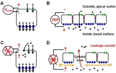
(21) Resumo O corpo humano não consegue regenerar após a perda ou dano severo de um órgão. ou secção do corpo. Pelo contrário, outros metazoários têm essa capacidade,. alimentando a esperança de que mecanismos de regeneração similares possam ser induzidos no Homem se formos capazes de fornecer os sinais apropriados. Nesse sentido, ao longo de vários séculos, cientistas de todo o mundo têm-se dedicado ao. estudo dos mecanismos de regeneração em diferentes organismos. Atualmente encontra-se disponível uma vasta biblioteca de informação relevante, mas ainda há. muitos aspetos da regeneração que precisam de ser explicados.. O teleósteo Danio rerio (peixe-zebra) é capaz de regenerar vários órgãos internos e também as barbatanas. Estas últimas possuem uma estrutura simples, de fácil acesso. e exercem uma função não vital, constituindo por isso um excelente modelo para o. estudo da regeneração em vertebrados adultos. Após a amputação, uma nova barbatana é formada no espaço de aproximadamente duas semanas, através de um. processo chamado regeneração epimórfica, que inclui três fases principais: fecho de. ferida (0-12 horas pós-amputação - hpa), formação do blastema (12-48 hpa), e crescimento regenerativo (48 hpa até cerca de 2 semanas). O blastema é a estrutura. fundamental para a regeneração epimórfica, contendo a informação morfogenética. necessária para formar e modelar os tecidos perdidos. A regeneração é regulada pela. ação concertada de diversas vias moleculares de sinalização, incluindo Wnt (canónico e não-canónico), Fgf, Shh, Bmp, Activin-βA, Notch e Ácido Retinoico.. Paralelamente às tradicionais vias moleculares de sinalização, a importância de um outro grupo, o dos canais e transportadores iónicos, é cada vez mais evidente. A sua. ação coordenada resulta na acumulação de iões, e por isso carga elétrica, através da. membrana celular. As propriedades elétricas das células e organismos, incluindo o potencial de membrana, correntes e campos elétricos endógenos, têm origem nessa. segregação de cargas. Entre os anos 60 e 80 (séc. XX), estes fenómenos bioelétricos. foram alvo de intensa investigação, e foi demonstrado que eles não só têm um papel ativo na regeneração, como também são capazes de aumentar a capacidade. regenerativa de espécies que normalmente não regeneram. Já nos anos mais recentes, têm sido desenvolvidas novas ferramentas para estudos de biologia celular e Page | xv.
(22) molecular bem como novas tecnologias de imagiologia, que permitem novas abordagens para investigar a natureza iónica dos sinais elétricos e identificar os seus mediadores moleculares.. Assim, esta tese pretende contribuir para a compreensão das bases fisiológica e molecular das correntes iónicas endógenas presentes durante o mecanismo de. regeneração epimórfica em animais vertebrados adultos. Além disso, esta tese visa. também o estudo da interação dos sinais bioelétricos com as tradicionais vias de. sinalização molecular envolvidas na regeneração.. A fim de identificar a composição iónica das correntes elétricas endógenas durante a regeneração da barbatana caudal do peixe-zebra, recorremos a uma técnica. designada “Scanning Ion-selective Electrode Technique” (SIET), que permite medir. isoladamente os fluxos de cada espécie iónica de interesse. Após a otimização e. adaptação da técnica ao nosso modelo animal, confirmámos a existência de um perfil. dinâmico de fluxos iónicos durante a regeneração. Fluxos de sódio, potássio, cloreto e cálcio parecem contribuir para o potencial de ferida, que é uma corrente elétrica com. carater universal, que se estabelece sempre após o ferimento de qualquer tecido e. dura até à ferida fechar. Por outro lado, detetámos o estabelecimento de um efluxo de. protões (H+) em fases posteriores ao fecho de ferida, o que sugere uma função específica durante a regeneração.. Para identificar a fonte molecular do efluxo de H+ detetado, combinámos a abordagem biofísica com técnicas de biologia molecular. Dessa forma, conseguimos demonstrar. que a V-ATPase, que é a principal bomba de protões em células animais, contribui para o efluxo de H+. Descobrimos que tanto a expressão da V-ATPase como o efluxo. de H+ variam de intensidade de acordo com o plano de amputação ao longo do eixo. proximal-distal da barbatana, e estabelecemos uma correlação entre este fenómeno e. a cinética de regeneração, que também depende do plano de amputação. Mais,. demonstrámos que a inibição da atividade da V-ATPase diminui significativamente a regeneração e que os tecidos do toco dependem mais da atividade da V-ATPase. quando a amputação é proximal do que após uma amputação distal. Estes resultados. sugerem um papel para a V-ATPase na regeneração associado à posição de amputação. Page| xvi.
(23) Foram também investigados os mecanismos através dos quais a atividade desta. bomba de H+ se articula com as tradicionais vias moleculares de sinalização, de forma a comandar o comportamento celular e dar origem aos tecidos perdidos.. Demonstrámos que a V-ATPase é necessária para a expressão de pelo menos dois. importantes genes, aldh1a2 e mkp3, para a proliferação celular no blastema e para a. inervação normal da barbatana.. Tanto quanto conseguimos apurar, esta é o primeiro trabalho relativo ao papel da V-. ATPase durante a regeneração em vertebrados adultos.. Page | xvii.
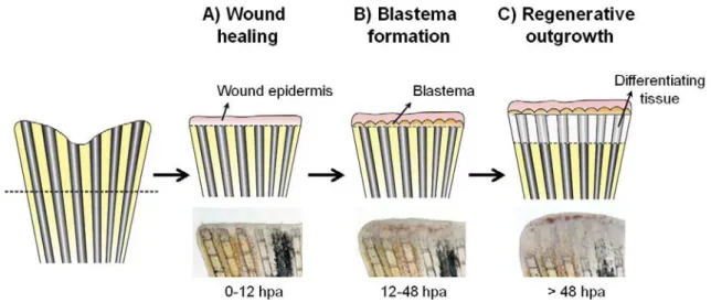
(24) Summary Humans are unable to regenerate after severe organ loss or amputation of body parts.. Notwithstanding, other metazoans have such a capacity, giving grounds for hope that similar regeneration mechanisms can be induced in humans if the correct signals are provided. This has driven scientists to investigate regeneration for centuries.. Nowadays, even though a great amount of data is available, there are still many aspects of regeneration that still need eliciting.. The teleost Danio rerio (zebrafish) is able to regenerate several internal organs and the fins. The latter constitutes a great model to study adult vertebrate regeneration. due to its simple structure, easy access and non-vital function. Upon amputation, a new fin is produced roughly within two weeks through a process called epimorphic. regeneration, including three main stages: wound healing (0-12 hours post amputation - hpa), blastema formation (12-48 hpa), and regenerative outgrowth (48. hpa to 2 weeks). Importantly, the blastema is the crucial structure for epimorphic. regeneration, containing the morphogenetic information required to give rise and repattern all the missing tissues. Regeneration is regulated by the orchestrated action of several signalling pathways activated after injury, including Wnt (canonical and noncanonical), Fgf, Shh, Bmp, Activin-βA, Notch and Retinoic acid.. Alongside classical signalling pathways, the relevance of ion channels and transporters for regeneration is becoming increasingly evident. Their coordinated activity results in the differential accumulation of ions, thus electric charge, across. cells membranes. The electrical properties of cells and organisms, including. membrane potential, endogenous electric currents and electric fields, arise from this charge segregation. In the 1960’s-1980’s, these bioelectrical phenomena were. extensively investigated, and it was demonstrated that they are crucial for wound healing and regeneration. It was demonstrated that electrical cues not only have an. active role governing regeneration, but they can also augment regenerative ability in species that normally do not regenerate. In the recent years, as new tools become. available regarding cellular and molecular biology and imaging technology, scientists are beginning to unveil the ionic nature of electrical signals and the molecular players. that generate them. In that regard, this thesis intended to be a contribution to the. Page| xviii.
(25) understanding of the physiological and molecular basis of endogenous ionic currents during adult vertebrate regeneration, and their interaction with canonical molecular pathways involved in the regeneration.. We used the scanning ion-selective electrode technique (SIET) to identify the ion nature of electric currents during regeneration in an adult vertebrate model, the. caudal fin of zebrafish (Danio rerio). After extensive optimization and adaptation of the technique to the working model, we were able to show that there is a dynamic profile of ion-specific fluxes during regeneration. Sodium, potassium, chloride and. calcium-specific fluxes seem to contribute to the injury potential, an electrical current that establishes upon wounding of any tissue and lasts until the wound is closed. On. the other hand, we found that a proton (H+) outward current (efflux) is specifically set. during caudal fin regeneration in stages later that the wound healing, arguing in favour of a regeneration-specific function.. To identify the molecular source of the regeneration-associated H+ efflux, we. combined biophysical and molecular approaches. This way, we were able to demonstrate that the V-ATPase, which is the main H+ pump in animal cells,. contributes to the relevant H+ efflux. Interestingly, we found that the onset and intensity of both V-ATPase expression and H+ efflux vary with the amputation plane. along the PD axis in a way that correlates with the regeneration kinetics. Specifically,. we could demonstrate that inhibition of V-ATPase activity impairs regeneration and. that proximal stumps have a stronger dependence on V-ATPase activity compared to. distally amputated fins. These findings suggest a role for V-ATPase in position-. dependent regeneration.. Another important question addressed regards how the activity of V-ATPase H+ pump. articulates with molecular signalling pathways to affect cell behaviour and give rise to the missing tissues. We show that V-ATPase is required for aldh1a2 and mkp3 expression, blastema cell proliferation and normal fin innervation.. Overall, our findings show that V-ATPase contributes to a regeneration-specific proton efflux, and is required for position-dependent regeneration by interfering with the expression of genes and cell behaviours crucial for regeneration success.. To the best of our knowledge, this is the first report on the role of V-ATPase during adult vertebrate regeneration.. Page | xix.
(26) Page| xx.
(27) Chapter I - Introduction. Chapter I Introduction. Page | 1.
(28) Chapter I - Introduction. Page | 2.
(29) Chapter I - Introduction. 1. Animal Regeneration 1.1. Historical overview The ability to replace lost body parts has always been part of the human imaginary. Ancient records go back to the Greek mythology, which described powerful creatures like the Hydra, a multi-headed monster that regrew two heads every time one was cut. off, and the snake or dragon Uroborus that ate its own tail as it continuously regenerated (Goss 1991).. The first reference to regeneration in a known organism belongs to Aristotle (384-. 322 BC), who mentioned lizard tail regeneration, but it wasn’t until the 18th century. that regeneration became a field of scientific interest. Following the description of limb regeneration in crayfish by René de Réaumur (1712) and the finding that hydra. regenerates after being cut into two or more pieces by Abraham Trembley (1740),. many other authors investigated the regenerative potential of both invertebrate (for. example annelids by Charles Bonnet in 1745, planarians by P.S. Pallas in 1770s and Charles Darwin in 1839, among others) and vertebrate species (for example amphibians by Spallanzani in 1769, and Todd in 1823, fish by Broussonet in 1786). In. 1901, Thomas H. Morgan critically revised the findings done on the field of regeneration until then, instituting a standard scientific terminology and data that. continues to inform regeneration studies today (Sunderland 2010). Later, with the. significant improvement of histological techniques in the beginning of the 20th. century, and the advent of genetic and molecular tools in the late 20th and early 21th. centuries, regeneration research progressed from gross observations to detailed histological descriptions, to molecular studies identifying the cellular and molecular networks that underlie regeneration in a variety of model systems (Dinsmore et al 1991, Carlson 2007, Tanaka and Galliot 2008). Although the understanding of the. regenerative machinery is still far from complete, the available data already allowed. the development of several stem cell based therapies that are now on clinical trial and aim the improvement of different human health conditions, such as visual disorders,. Page | 3.
(30) Chapter I - Introduction. cardiac disfuncion and diabetes (Chhabra et al 2013, Koudstaal et al 2013, Ramsden et al 2013).. 1.2. Types of regeneration The term “regeneration” has been used for centuries to refer to “the natural. restoration of the lost parts of an organism” (Morgan 1901). It is generally accepted to distinguish two main types of regeneration based on the presence or absence of injury or disease as the triggering factor:. • “Physiological regeneration”, defined as the regular replacement of worn-out or short-lived structures in order to maintain the integrity of the multicelular body.. Some examples are the replacement of the skin, hair, bone, gut epithelium and blood cells, and the shedding of crustaceous exoskeleton and snakes skin.. • “Reparative regeneration”, defined as the replacement of cells, tissues or body. sections upon injury or disease. This process is not common to all organisms, and though it may represent survival upon trauma, it is not essential for life under normal circumstances.. (Morgan 1901, Carlson 2007, Stoick-Cooper et al 2007a, Kawakami 2010, Poss 2010) Reparative regeneration has been particularly subjected to investigation and discussion due to its relevance for regenerative medicine. The diversity of. mechanisms that ensure the re-establishment of the shape and/or function of the lost part is huge, and placing them into defined categories has not been easy or consensual (Morgan 1901, Sánchez Alvarado 2000, Reddien and Sánchez Alvarado. 2004, Agata et al 2007, Stoick-Cooper et al 2007a). Nevertheless, it is possible to. distinguish different types of reparative regeneration.. “Morphallaxis” is the reconstitution of the lost structure, in both shape and function,. by remodelling of the pre-existing tissues without need for cell proliferation. The resulting organism is smaller than the original. Then, cell proliferation may occur to bring the structures to the original size (Morgan, 1901, Galliot and Chera 2010). A. typical example of this process is apical head regeneration in the cnidarian Hydra (Park et al 1970, Holstein et al 1991, Chera et al 2009). Page | 4.
(31) Chapter I - Introduction. Oppositte to morphallaxis, the term “epimorphosis” was originally used by Morgan. (1901) to refer to the reparative regeneration strategies that rely on cell. proliferation. However, as regeneration research progressed, it became clear that there are distinct modes of regeneration that depend on cell proliferation. Nowadays,. epimorphosis or “epimorphic regeneration” refers only to regeneration via formation. of a blastema, which is a population of undifferentiated progenitor cells that contains. intrinsic morphogenetic information required to re-pattern the regenerating structure to its original tridimensional polarity, form and function (Mescher 1996,. Stoick-Cooper et al 2007a, Tanaka and Reddien 2011). Classical examples of this. mechanism are the regeneration of transected planarians and appendages in amphibians and fish (tail and limb, and fins, respectively), but the process occurs in many other organisms (Sánchez Alvarado and Tsonis 2006). Epimorphosis is many. times seen as the bona fide or perfect regeneration process, because new structures. are formed de novo and are very similar to the lost parts in both shape and function. Consequently, this type of regeneration has been the most studied.. Other proliferation-dependent but blastema-independent types of regeneration are: “hypertrophy” or “compensatory growth”, defined as the increase in mass of the. remainder part of an organ such as the mammalian liver and kidneys to compensate the lost or non-functional portion (Carlson 2007, Stoick-Cooper et al 2007a); and “tissue regeneration”, characterized by the repair of local and limited damage to an. organ predominantly via restoration of only one cell type (for example skeletal muscle, bone).. 1.2.1. Cellular reprogramming The advances made so far in understanding natural regeneration processes have allowed the development of artificial or induced regeneration strategies that improve regeneration in humans. Two main differences are often invoked to justify the lack of. adult human regenerative ability compared to regenerating species: the shortage of natural stem cells from normal tissues, and the inability of differentiated somatic cells to proliferate and/or switch identity into the missing cell types. To overcome this, scientists have found ways in which a somatic cell can be stably transformed into a. Page | 5.
(32) Chapter I - Introduction. distinct cell type by forced expression of lineage-determining factors, a process generally called cellular reprogramming (Vierbuchen and Wernig 2012). This can be achieved by several experimental approaches, from which two stand out for their. remarkable clinical value. The first is nuclear transfer from a differentiated somatic cell into an enucleated oocyte (Gurdon 1962), that results in the production of viable blastocysts that can be grown to adulthood or induced to differentiate into specific somatic cell lines of medical interest (Gurdon and Simonsson 2003). The second is the over-expression of a small number of specific transciption factors, namely Oct4, Sox2,. Klf4, c-Myc, Nanog and Lin28. Different combinations of at least 3 of these genes. (Oct4, Sox2 and KLF4) are sufficient to convert a somatic cell type directly into another (Weintraub et al 1989, Gurdon and Melton 2008, Zhou et al 2008, Yamanaka. and Blau 2010) and to reprogram somatic cells such as fibroblasts, hepatocytes and gastric epithelial cells into induced pluripotent stem cells (iPS cells) (Takahashi and Yamanaka 2006, Takahashi et al 2007, Aoi et al 2008, Chakraborty et al 2014).. Cellular reprogramming will eventually be used to generate cells for tissue repair or replacement while avoiding the ethical issues inherent in the use of human ES cells and the need for immunosuppression since the cells would be derived from each. pacient. Furthermore, it enables the culture of defective cells, allowing disease modelling and the screening for therapeutic drugs (Takahashi and Yamanaka 2006,. Gurdon and Melton 2008). But for now much remains to be learned about the molecular basis of both transcriptional and epigenetic machinery involved in cellular reprogramming in different contexts (normal and disease) before it is fully understood and its potential can be safely harnessed (Yamanaka and Blau 2010). A. major contribute in that regard will be the deep understanding epimorphic. regeneration, since it is a natural mechanism in which some cells naturally switch lineage identity and proliferate in a controlled and limited manner.. 1.3. Regeneration among Metazoa: distribution and evolution In Metazoa, the ability to undergo reparative regeneration is widespread among. phyla with great evolutionary distances (Fig. I.1). Among invertebrates, sponges,. hydra, star fish, annelid worms and planarians can all rebuild a complete organism Page | 6.
(33) Chapter I - Introduction. from a fragment of the original one (Bely 2006, Hernroth et al 2010, Wulff 2010, Galliot. 2012, Elliot and Sánchez Alvarado 2013). Decapode crustaceans and nymphs of. hemimetabolous insects such as cockroaches and crickets regenerate entire limbs. (Nakamura et al 2008, Das and Durica 2013). Among Mollusca, examples of regeneration include the rebuilt of excised mantle in bivalves (Gustaf et al 2009) and. the central nervous system in gastropods (Matsu and Ito 2011), regeneration of the. cornea in octopus (Dingerkus and Santoro 1981) and the tentacular arms in squid (Aldrich and Aldrich 1968).. Some basal Chordata, like the colonial ascidians can regenerate functional adults from minute vasculature fragments (Rinkevich et al 2007), and adult amphioxus rebuild. anterior and posterior structures, including neural tube, notochord, fin, muscle, intestine and tail (Somorjai et al 2011).. Among higher vertebrates, there are also species with impressive regeneration after trauma. Urodele amphibians such as newts and axolotls can replace lost appendages. (Tweedell 2010), lens, retina (Barbosa-Sabanero et al 2012), several internal organs. such as the heart (Cano-Martínez et al 2010) and the liver (Michalopoulos and DeFrances 1997), and the central nervous system (Zukor et al 2011), throughout. their life cycle. Teleost fish have also great regenerative ability (chapter I: section 2). However, in the majority of higher vertebrates, regeneration is very limited. Anuran. amphibians (frogs and toads) can regenerate the tail and limbs, but only during larval stages; reptiles can only regrow the tail (McLean and Vickaryous 2011); and birds appear to be nearly or entirely unable to regenerate any structure (Bely 2012). As for. mammals, including humans, they can repair damage to skeletal muscle (Chargé and Rudnicki 2004) and peripheral nervous system (Bosse 2012) and can recover from. damage to internal organs such as the liver (Michalopoulos and DeFrances 1997), kidney (Angelotti et al 2012), pancreas (Shu et al 2012) and intestine (Tsonis 2000).. Newborn mice, monkeys and even human children can regenerate the distal tip of the. digits (Illingworth 1974, Singer et al 1987, Han et al 2008). Nevertheless, humans cannot recover from serious damage or loss of organs or body sections.. Page | 7.
(34) Chapter I - Introduction. Almost all metazoan phyla include species with regenerative ability (Fig. I.1), but the. ability to regenerate and the type of regeneration employed vary a lot, even between. closely related species. For example, urodele and anuran amphibians have opposite. regenerative capacity during adulthood. Given this ambiguous phylogenetic distribution, it is difficult to understand how regeneration originated and evolved to the present days (Sánchez Alvarado 2000, Carlson 2007).. Figure I.1 – Phylogenetic distribution of epimorphic regeneration among the Metazoa. Modified from Galliot and Chera 2010.. One hypothesis is that regeneration is a homologous trait, an attribute of the common. ancestral of all metazoans that was lost in some groups as a neutral or negative trait,. in a variety of contexts. The other main view is that regeneration is an analogous trait that arose independently in each group, and then converged in animals sharing the same evolutionary context (Sánchez Alvarado 2000, Brockes and Kumar 2008). Even. though the former theory is generally more accepted, there are phylogenetic and molecular data pointing in either direction, and therefore a consensus is still far from. reaching (Goss 1969, Bely and Sikes 2010, Somorjai et al 2011, Bely 2012 against Anderson et al 2008, Khalturin et al 2009, Garza-Garcia et al 2010). Adding to this Page | 8.
(35) Chapter I - Introduction. evolutionary puzzle, regeneration is often seen as the counterpart of development, which is highly conserved within metazoans, in agreement with a common early. ancestral (Wolpert 1994, Sánchez Alvarado 2000, Brockes et al 2001, Khalturin et al. 2009, Shubin et al 2009). Indeed, both processes share many cellular and molecular. mechanisms and their end result is almost indistinguishable, as extensively. highlighted for vertebrate limb development and regeneration (Imokawa and. Yoshizato 1997, Simon et al 1997, Sánchez Alvarado 2000). Notwithstanding,. regeneration involves some mechanisms that are not employed in the deeply conserved embryonic development, such as the dependence on the injury signal and on nerve supply (Brockes and Kumar 2008).. Taken all the above, it is difficult to predict which animals hold regenerative. mechanisms closer to humans. Consequently, it is unclear which species should be. further investigated in order to find new strategies to improve human health conditions resulting from injury, aging and disease. Nevertheless, understanding the. mechanisms that are involved in regeneration in diverse model systems is potentially advantageous for biomedicine. For instance, understanding why particular. regenerative processes take place in some animals but not in human tissues could provide new pathways to stimulating human regeneration, especially if endogenous human pathways are unavailable (Sánchez Alvarado and Tsonis 2006).. The most common regeneration model organisms are hydra, planarians, urodeles (salamanders, newts and axolots), Xenopus and zebrafish. They represent some of the. first organisms in which regeneration was described and they all have extreme regenerative ability, including epimorphosis (Morgan 1901, Slack 2003).. Page | 9.
(36) Chapter I - Introduction. 2. Zebrafish regeneration The ability of zebrafish to regenerate its fins was first scientifically described in 1786 by Broussonet. Over the years, it was showed that besides the appendages, zebrafish. can undergo epimorphic regeneration of the heart muscle (Poss et al 2002), maxillary. barbels (LeClair and Topczewski 2010) and retina (Cameron 2000). They can also regenerate the optic nerve (Bernhardt et al 1996), liver (Sadler et al 2007), pancreas. (Moss et al 2009), spinal cord (Becker et al 1997), sensory hair cells (Lopez-Schier and Hudspeth 2006) and scales (Sire et al 2000) (Fig. I.2). Many other teleosts can. also regenerate some external and internal organs (Shao et al 2009, Watanabe et al 2009, Sîrbulescu and Zupanc 2011), but this is not a property common to all teleosts (Geraudie and Singer 1977).. Figure I.2 – Zebrafish structures with regenerative ability.. In the last two decades the zebrafish has emerged as a standard model for. regeneration studies, much because it combines a remarkable regenerative ability with several practical advantages, including: simple and low cost husbandry and maintenance requirements; small size and good social behaviour that allows raising and maintaining large numbers of fish in restricted space; high fecundity; short. generation time; and rapid organism development. Besides, zebrafish and humans share several functional organs and orthologous genes (Brittijin et al 2009).. Moreover, the zebrafish has long been a standard model organism to study animal Page | 10.
(37) Chapter I - Introduction. development (Eisen 1996), and more recently it also became a model of excellence for immunology and behaviour studies (Oliveira et al 2011, Renshaw and Trede 2012, Medina and Royo 2013). As a consequence, the amount of available tools and. protocols for molecular and genetic manipulation and characterization is much greater compared to other fish and also compared to the regeneration models previously used, such as amphibians (Slack 2003). Those include complete genome. sequence (Ensembl project, Zv9 2014), mutagenesis screens (Jonhson and Weston 1995, Amsterdam et al 1999, Poss et al 2002, Mathew et al 2007), gene knockdown. and transgenesis techniques (Ivics et al 1993, Tawk et al 2002, Bayliss et al 2006,. Thummel et al 2006, Moens et al 2008, Ishida et al 2010, Hans et al 2011, Dahlem et al. 2012), microarrays (Ton et al 2002, Schesbesta et al 2006), antibodies, expressed sequence tag (EST) (Baxendale et al 2009).. Among the zebrafish structures with regenerative ability, the caudal fin is a. particularly attractive model to study epimorphic regeneration in adult vertebrates. due to the easier access and non-vital function relative to other epimorphic regenerating structures, like the heart. Also, it has a simple anatomical structure and limited cell types compared, for example, to the classically studied urodele limb (Stoick-Cooper et al 2007a).. 2.1. Structure of the caudal fin In gross mode, the caudal fin is a fan-like frame of dermal exoskeletal elements called fin rays, which are arranged longitudinally and separated by inter-ray connective. tissue. The whole structure forms two symmetrical lobes, dorsal and ventral, and is covered with a scaleless epithelium (Fig. I.3A).. Detailed structure of the caudal fin is represented on Fig. I.3B. Each bone ray, or. lepidotrichium, is comprised of two concave and symmetrical segmented hemirays which, in cross section, appear like a parenthesis (Becerra et al 1983). The hemirays. are made of acellular bone that mineralizes directly from a collagenous matrix secreted by scleroblasts - skeletogenic cells equivalent to mammalian osteoblasts. (Santamaria et al. 1992, Hall 2005) that surround each hemiray as a monolayer Page | 11.
(38) Chapter I - Introduction. (Nechiporuk and Keating 2002). The fin grows distally from its base by sequential addition of pairs of hemiray segments connected end-to-end by segment joints of. dense fibrous connective tissue (Haas et al 1962, Becerra et al 1983). Once formed,. the segments can become increasingly thicker but cannot elongate (Géraudie et al 1995, Azevedo et al 2011). At some point the rays bifurcate by splitting along the proximal-distal axis, with exception of the more dorsal and ventral ones (Becerra et al. 1983, Géraudie et al 1995). Both the length of the segments and the local of. bifurcation are carefully instructed by genetic signals (Murciano et al 2002, Murciano et al 2007). The rays encircle the intra-ray mesenchyme and also blood vessels,. nerves, and pigment cells, which are also present in the inter-ray mesenchyme that connects the lepidotrichia (Poss et al 2003). All of these structures are covered by a. thin but multilayered epithelium, the skin, which has a characteristic basal cell layer at its bottom.. The fin exoskeleton meets the rest of the body at the fin base, in the proximal end of the lepidotrichia. This region is covered with scales and contains striated muscle that. connects the lepidotrichia exoskeleton to the hypural bones of the fish endoskeleton. (cartilaginous origin) (Géraudie et al 1995, Poss et al 2003). At the opposite, distal tip of the fin, each ray ends in a double palisade of small, rigid and fusiform spicules called actinotrichia. They are composed of hyperpolymerised elastoidin (collagen-like. protein) and have been reported to play skeletal and morphogenetic roles (Santamaria and Becerra 1991, Böckelmann and Bechara 2009).. Two main axes are present in the fin: the proximal-distal axis, which separates the regions closer to the fin base (proximal) from those nearer the fin tip (distal); and the. dorsal-ventral axis, which divides the fin into an upper, dorsal half and a similar bottom, ventral half (Fig. I.3A).. Page | 12.
(39) Chapter I - Introduction. Figure I.3 – Structure of the zebrafish caudal fin. (A) Overview of caudal fin and main fin axes: dorsal-ventral and proximal-distal. (B) Detail of the fin exoskeleton. The fin exoskeleton is mainly composed of bone rays or lepidotrichia separated by inter-ray connective tissue. Each bone ray is made of pairs of hemiray segments connected end-to-end by segment joints of connective tissue. Most rays bifurcate at least once. Schematic view in B (bottom) modified from Akimenko et al 2003.. 2.2. Stages of epimorphic regeneration in the zebrafish caudal fin Regardless the amount of times the caudal fin exoskeleton is amputated, a new fin is always produced (Azevedo et al 2011) as a result of epimorphic regeneration. The new structure is not a perfect copy of the original fin but it is very similar in both. shape and function (Azevedo et al 2012). Interestingly, the rate of regeneration is temperature sensitive; at 33° C a new fin is rebuilt in about 2 weeks, but the process will take longer under lower water temperature (Jonhson and Weston 1995).. Epimorphic regeneration includes always three main and partially overlapping stages that will be further described in the following subsections: wound healing, blastema. formation and maturation, and regenerative outgrowth (Fig. I.4). These stages are common to all epimorphic regeneration events.. Page | 13.
Imagem


+7


Documentos relacionados