UNIVERSIDADE DA BEIRA INTERIOR
Ciências da Saúde
O efeito dos extractos de cereja em células
humanas do cancro da próstata:
Do cultivo à clínica?
Gonçalo Emanuel Duarte Rosas da Silva
Dissertação para obtenção do Grau de Mestre em
Ciências Biomédicas
(2º ciclo de estudos)
Orientador: Prof. ª Doutora Ana Paula Duarte
Co-orientador: Prof. ª Doutora Sílvia Socorro
Dedicatória
Existe um número quase embaraçoso de pessoas sem o apoio das quais eu não teria moral, nem financiamento, nem tampouco a paciência e força de vontade necessárias para escrever qualquer tipo de dissertação.
Como suspeito que o poder de uma dedicatória seria diluído ao ponto desta não valer nada ao ser dividida por duas ou mais pessoas, não sendo eu crente na homeopatia, dedico-a a quem a ler.
Agradecimentos
Queria começar por agradecer à minha orientadora, a Professora Doutora Ana Paula Duarte. Acredito que ser directora do CICS não dá pouco trabalho, especialmente com um orientando como eu. Mesmo assim, sempre se mostrou disponível, empenhada, paciente, e bem-disposta. Em todas as facetas desta travessia, em todos os aspectos, não me faltou nada.
À Professora Doutora Sílvia Socorro, que guiou a maior parte da segunda metade desta tese, e que foi absolutamente incansável. Obrigado pela paciência, e desculpe pelas horas de sono perdidas.
Ao CICS, à Câmara Municipal da Covilhã e à Cerfundão, sem os quais a elaboração deste trabalho teria sido impossível.
Aos meus colegas de Trabalho. À Doutora Cátia Vaz, que contribuiu quase tanto como eu para esta dissertação, que trabalhou com afinco nos bons, maus, e péssimos dias, e que devia ganhar algum tipo de medalha por ter sido a pessoa que mais tempo passou comigo no laboratório. À Doutora Sara Correia, cujo toque mágico nas minhas placas de MTT me melhorarou os resultados, e cuja perpétua boa disposição me facilitou os piores dias. À Mestre Marília Figueira e ao Mestre Henrique Cardoso, que (felizmente) não mudaram nada ao longo destes sete anos. À Mestre Margarida Grilo, que partilha a minha paixão pela literatura, embora não propriamente pelos mesmos livros. Ao Doutor Luís Rato, a quem nunca lhe faltou a boa disposição, por muito atarefado que estivesse. Ao Doutor Ricardo Marques, obrigado especialmente pelas dezenas de vezes que utilizei a tua mini-espátula (sem a tua autorização). À Mestre Ana Silva, o sol radiante do grupo. À Anaísa Coelho, que sempre sorri e nunca dorme. Ao Luís “Santo” Espínola, graças a quem nunca me faltaram anticorpos feitos. A todos os investigadores e investigadoras do CICS; vocês são a razão pela qual este centro de investigação é a máquina bem-oleada que è, e foi uma honra ter feito parte dela convosco. Aos meus maravilhosos pais, Ilídio e Maria de Fátima, e ao resto da minha querida família; primos, tios e avós, presentes ou em paz, que sempre disseram que não se importavam com o que eu fizesse da minha vida logo que o fizesse bem, e fosse feliz a fazê-lo. Não tenho dedos, mãos, braços e pernas suficientes para apontar os nossos defeitos, mas não mudava nenhum deles. Adoro-vos.
Aos meus amigos no Porto (e alguns fora dele), que me fazem parecer monótono e calmo em comparação.
E por último, mas que isso não lhes faça pensar que é por isso que valem menos, à Irmandade; Joana Rodrigues, Joana Coelho, Inês Mateus, Ivo Duarte, Rita Simões e Ana Luísa (longe mas perto). Nas palavras imortais do Marcel Proust, “Devemos agradecer às pessoas que nos fazem felizes... São elas os jardineiros encantadores que fazem as nossas almas florescerem”. Que o meu canteiro nunca esteja longe dos vossos regadores.
Nenhuma combinação de palavras, pelo menos uma elaborada por mim, consegue fazer justiça ao apreço que tenho por vocês todos. Independentemente do final desta viagem, por me ajudarem, estimarem e (especialmente) aturarem, têm um cantinho especial reservado no céu e na terra.
“We do not have to visit a madhouse to find disordered minds; our planet is the mental institution of the universe.” -Johann Wolfgang von Goethe
“Do not wait to strike till the iron is hot; but make it hot by striking.” -W.B. Yeats
“Fear not for the future, weep not for the past.” -Percy Bysshe Shelley
Resumo
A proliferação descontrolada das células da próstata é uma condição observada normalmente na população masculina à medida que esta envelhece, e está associada com o desenvolvimento do cancro da próstata. Assim, estratégias que visem diminuir a proliferação dos tecidos prostáticos durante o seu envelhecimento deverão diminuir a prevalêcia desta condição médica. As cerejas são ricas em compostos fenólicos como as antocianinas, com efeitos antioxidantes e anti-proliferativos bem estudados em células cancerígenas como as MCF-7 (cancro da mama) e HCT116 (cancro colo-rectal). O estudo elaborado para esta dissertação pretendeu avaliar o efeito de extractos da cereja Saco, uma variedade local da região do Fundão, na viabilidade, apoptose, e metabolismo glicolítico das células não-neoplásicas do epitélio da próstata PNT1A, nas células não-neoplásicas androgeno-dependentes LNCap, e nas células neoplásicas androgeno-independentes PC3. As cerejas foram descaroçadas, liofilizadas, e a sua extracção efectuada recorrendo a ultrassons e utilizando metanol acidificado com 0,1% HCl como solvente. O extracto obtido foi então dissolvido em meio de cultura celular a várias concentrações (0, 2, 20, 200 e 2000 µg/mL) e utilizado para tratar as células não-neoplásicas e neoplásicas durante 48, 72 e 96 horas. A viabilidade celular nas diferentes condições experimentais foi determinada por ensaios MTT. Globalmente, a viabilidade celular mais baixa foi observada às 72 horas e com 20 µg/mL de extracto de cereja, e estas condições foram utilizadas para avaliar os efeitos dos extractos de cereja na proliferação,apoptose e metabolismo glicolítico das células PNT1A, LNCaP e PC3. A análise à expressão génica e a actividade dos reguladores-alvo destes processos biológicos foram efectuadas através da metodologia de Western Blots e kits de análises bioquimicas, respectivamente. O consumo de glucose e a produção de lactato foram ambos medidos por análise espectofotométrica. Os resultados obtidos demonstraram que a administração dos extractos metanólicos de cereja aumentou a apoptose das células LNCaP, um efeito não-confirmado nas células não-neoplásicas PNT1A. Curiosamente, as células PC3 mostraram uma diminuição da actividade apoptótica na presença dos extractos de cereja, apesar da proliferação diminuída. Quanto ao metabolismo glicolítico, foi observado que as células PNT1A tratadas com extractos de cereja apresentaram uma diminuição parcial deste, enquanto que as células LNCaP demonstraram uma supressão da metabolização da glucose em resposta aos extractos de cereja, com consumo de glucose e produção de lactato ambos diminuídos, acompanhados por alterações da expressão/actividade de transportadores e enzimas glicolíticas. Analogamente aos resultados da apoptose, as células PC3 apresentaram um comportamento distinto das LNCaP relativamente ao metabolismo glicolítico; os extractos de cereja induziram um aumento da glicólise e da produção de lactato. Os presentes resultados indicam que os extractos metanólicos de cereja podem ter efeitos benéficos nas células do cancro da próstata, tanto neoplásicas como não-neoplásicas, diminuindo a sua
proliferação e controlando as vias apoptóticas e glicolíticas, produzindo efeitos mais visíveis no caso das células sensíveis à ação dos androgénios, LNCaP.
Palavras-chave
Resumo Alargado
A proliferação descontrolada das células da próstata é uma condição observada normalmente na população masculina à medida que esta envelhece, estando associada com o desenvolvimento do cancro da próstata. Este último é uma das causas de morte oncológica mais frequente nos indivíduos do sexo masculino, com mais de um milhão de mortes e 300.000 novos casos anuais. O envelhecimento geral da população, entre vários outros factores de risco, também contribui para um aumento anual dos ritmos de incidência desta doença.
Assim, estratégias preventivas, que visem diminuir a proliferação dos tecidos prostáticos durante o seu envelhecimento, evitando a progressão de condições como a hiperplasia prostática benigna e posteriormente cancro da próstata, deverão diminuir a prevalêcia desta condição médica de difícil tratamento.
As cerejas são ricas em compostos fenólicos como as antocianinas, com efeitos antioxidantes e anti-proliferativos bem estudados em células cancerígenas como as MCF-7 (cancro da mama) e HCT116 (cancro colo-rectal), além de vários outros benefícios associados à prevenção e atenuamento dos efeitos do cancro e a outras patologias diversas. São também frutos populares, produzidos e consumidos um pouco por todo o mundo.
O estudo elaborado para esta dissertação pretendeu avaliar o efeito de extractos da cereja
Saco, uma variedade bastante consumida e local da região do Fundão, na viabilidade,
apoptose, e metabolismo glicolítico das células não-neoplásicas do epitélio da próstata PNT1A, nas células neoplásicas androgeno-dependentes LNCap, e nas células neoplásicas androgeno-independentes, e comparativamente mais agressivas, PC3.
As cerejas foram descaroçadas, liofilizadas, e a sua extracção foi efectuada recorrendo a ultrassons e usando metanol acidificado com 0,1% HCl como solvente. O extracto obtido foi então dissolvido em meio de cultura celular a várias concentrações (0, 2, 20, 200 e 2000 µg/mL) e utilizado para tratar as células não-neoplásicas e neoplásicas durante 48, 72 e 96 horas. A viabilidade celular nas diferentes condições experimentais foi determinada por ensaios MTT. A viabilidade celular variou consideravelmente entre as linhas celulares, mesmo para as mesmas concentrações e os mesmos periodos de tempo, mas globalmente, os extratos diminuíram a viabilidade celular, sendo esta consistentemente mais baixa em todas as linhas às 72 horas e com 20 µg/mL de extracto de cereja. Estas condições foram então utilizadas para avaliar os efeitos dos extractos de cereja na proliferação, apoptose e metabolismo glicolítico das células PNT1A, LNCaP e PC3. A análise da xpressão génica foi efectuada através da metodologia de Western Blot normalizando a quantidade de proteína com a marcação da β-actina. A atividade dos reguladores chave da apoptose e metabolismo foi avaliada por
ensaios bioquímicos usando kits específicos. O consumo de glucose e a produção de lactato foram medidos por análise espectrofotométrica .
Os resultados obtidos demonstraram que a administração dos extractos metanólicos de cereja aumentou a apoptose das células LNCaP pela via intrínseca, com um aumento no rácio BAX/Bcl-2 e uma maior expressão das caspases 9 e 3. O efeito nas células não-neoplásicas PNT1A foram menos concretos, com um aumento da expressão do gene supressor tumoral p53 mas sem alteração na expressão das caspases-9 e 3. A caspase-8, envolvida na via extrínseca, não se apresentou alterada em respostas aos extratos de cerejas em nenhuma das linhas. Curiosamente, as células PC3 mostraram uma diminuição da actividade apoptótica na presença dos extractos de cereja, com uma diminuição da expressão das caspases 9 e 3, apesar da proliferação diminuída.
Quanto ao metabolismo glicolítico, foi observado que as células PNT1A tratadas com extratos de cereja apresentaram uma diminuição parcial deste, evidente pela diminuição da expressão dos GLUTs 1 e 3 e das enzimas PFK-1 e LDH, apesar de um aumento da produção de lactato. As células LNCaP demonstraram uma supressão da metabolização da glicose em resposta aos extratos de cereja, com uma diminuição da expressão do GLUT3 e MCT4, e com o consumo de glucose e produção de lactato ambos diminuídos. Analogamente aos resultados da apoptose, as células PC3 apresentaram um comportamento distinto das LNCaP relativamente ao metabolismo glicolítico; os extractos de cereja induziram um aumento da glicólise e da produção de lactato, apesar de uma diminuição da expressão da PFK-1 e da actividade da LDH. Os presentes resultados indicam que os extractos metanólicos de cereja podem ter efeitos benéficos nas células do cancro da próstata, tanto neoplásicas como não-neoplásicas, diminuindo a sua proliferação e controlando as vias apoptóticas e glicolíticas, produzindo efeitos mais visíveis no caso das células LNCaP sensíveis à ação dos androgénios.
Abstract
The uncontrolled proliferation of prostate cells is a condition commonly observed in the ageing male population, which is associated with the development of prostate cancer. Thus, strategies to counteract the proliferative feature of aging prostatic tissues should prove beneficial by diminishing the widespread prevalence of this condition. Sweet cherries are rich in phenolic compounds such as anthocyanins, with widely studied anti-proliferative and antioxidant effects in different types of cancer cells such as MCF-7 (human breast) and HCT116 (human colorectal). The present study aimed to evaluate the effect of crude extracts of Saco sweet cherry, a local cultivar from the Fundão region (Portugal), on the viability, apoptosis and glycolytic metabolism of non-neoplastic PNT1A prostate epithelial cells, and androgen-sensitive LNCaP cells and androgen-insensitive PC3 prostate cancer cells. Sweet cherries were seeded, freeze-dried, and underwent ultrasound-assisted extraction with methanol acidified with HCl 0.1%. The extract was then dissolved in cell culture medium at several concentrations (0, 2, 20, 200 and 2000 µg/mL) and used to treat non-neoplastic and neoplastic prostate cells for 48, 72 and 96 hours. Cell viability at different experimental conditions was determined by MTT assay. Overall, the lowest viability was observed at 72 hours of culture with 20 µg/mL of cherry extract, and these conditions were used to evaluate the effect of cherry extracts on the cell proliferation, apoptosis and glycolytic metabolism of PNT1A, LNCaP and PC3 cells. Gene expression analysis and activity of target regulators of the aforementioned biological processes was assessed by means of Western blot and biochemical assays, respectively. Glucose consumption and lactate production were measured spectrophotometrically. The obtained results demonstrated that the administration of methanolic cherry extracts increased the apoptotic rate of LNCaP cells, an effect not confirmed in the non-neoplastic PNT1A cells. Curiously, PC3 cells had an overall down-regulation of apoptotic activity in the presence of cherry extracts, in spite of its decreased proliferation. Concerning metabolism, it was found that treated PNT1A cells had a partial down-regulation of glycolytic metabolism, while LNCaP cells markedly displayed a suppression of this process with decreased glucose consumption and lactate production, which was accompanied by altered expression/activity of glycolytic transporters and enzymes. Analogously with the results of apoptosis, PC3 cells had a distinct behavior relatively to LNCaP in what concerns glycolytic metabolism; cherry extracts induced an up-regulation of glycolysis and lactate production. The present findings indicate that cherry methanolic extracts may have beneficial effects on prostate cells, both neoplastic and non-neoplastic, diminishing proliferation and controlling the apoptotic and glycolytic pathways, producing more visible effects in the case of the androgen-sensitive LNCaP cells.
Keywords
Index
Chapter 1 - Introduction ... 1
1.1 - The anatomy and physiology of prostate ... 1
1.2 - Prostate cancer: the overall picture ... 2
1.2.1 - The development of prostate cancer ... 4
1.2.2 - Apoptosis of prostate cancer cells... 6
1.2.3 - Glycolytic metabolism of prostate cancer cells ... 8
1.3 - Cherries ... 10
1.3.1 - Chemical composition and biological activities ... 11
1.4 - Classification and properties of phenolic compounds ... 12
1.4.1 - The particular case of anthocyanins ... 15
1.4.1.1 - Anthocyanin extraction... 17
1.4.1.2 - Anthocyanins as anticancer molecules ... 19
Chapter 2 - Aim ... 25
Chapter 3 - Materials and methods ... 27
3.1 - Cell lines ... 27
3.2 - Cherry Extracts ... 27
3.2.1 - Extraction procedure ... 27
3.3 - MTT assays and stimuli ... 28
3.3.1 - Cell proliferation assays (MTT) ... 28
3.3.2 - Protein extraction ... 29 3.4 - Western blots ... 29 3.5 – Biochemical assays ... 30 3.6 - Statistical analysis ... 32 Chapter 4 - Results ... 33 4.1 – MTT assays ... 33 4.2 – Apoptotic pathways. ... 35 4.3 – Glucose metabolism ... 42 Chapter 5 - Discussion ... 51
Chapter 6 - Conclusions and future prospects ... 57
Chapter 7 - References ... 59
List of Figures
Figure 1 - Sagittal view showing the male reproductive structures ... 1
Figure 2 - The intrinsic and extrinsic apoptotic pathways. ... 7
Figure 3 - Glycolysis and the two main possible pathways of pyruvate. ... 10
Figure 4 - General flavonoid structure. ... 14
Figure 5 - Basic anthocyanin structure. ... 15
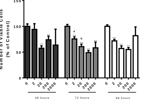
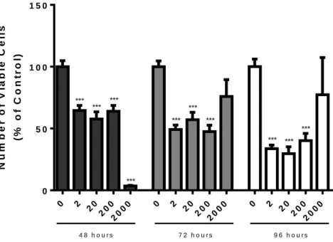
Figure 6 - Number of viable non-neoplastic human prostate epithelial PNT1A cells after exposure to different concentrations of saco sweet cherry extracts (2, 20, 200, and 2000 μg/mL) for 48, 72 and 96 hours determined by the MTT assay ... 33
Figure 7 - Number of viable human prostate cancer LNCaP cells after exposure to different concentrations of saco sweet cherry extracts (2, 20, 200, and 2000 μg/mL) for 48, 72 and 96 hours determined by the MTT assay. ... 34
Figure 8 - Number of viable human prostate cancer PC3 cells after exposure to different concentrations of saco sweet cherry extracts (2, 20, 200, and 2000 μg/mL) for 48, 72 and 96 hours determined by the MTT assay. ... 35
Figure 9 - Expression of apoptosis regulators (A-H) in the human prostate epithelial PNT1A cells after exposure to 20 μg/mL of saco (July crop) cherry for 72 hours, determined by Western blot analysis after normalization with β-actin. ... 38
Figure 10 - Activity of Caspase-3 in the human prostate PNT1A epithelial cells after exposure to 20 μg/mL of saco (July crop) cherry for 72 hours, determined by a specific assay kit ... 38
Figure 11 - Expression of apoptosis regulators (A-E) in the human prostate cancer LNCaP cells after exposure to 20 μg/mL of saco (July crop) cherry for 72 hours, determined by Western blot analysis after normalization with β-actin. ... 39
Figure 12 - Activity of Caspase-3 in the human prostate cancer LNCaP cells after exposure to 20 μg/mL of saco (July crop) cherry for 72 hours, determined by a specific assay kit... 40
Figure 13 - Expression of apoptosis regulators (A-E) in the human prostate cancer PC3 cells after exposure to 20 μg/mL of saco (July crop) cherry for 72 hours, determined by Western blot analysis after normalization with β-actin ... 41
Figure 14 - Activity of Caspase-3 in the human prostate cancer PC3 cells after exposure to 20 μg/mL of saco (July crop) cherry for 72 hours, determined by a specific assay kit. ... 42
Figure 15 - Glucose consumption (A) and lactate export (B) in human prostate PNT1A epithelial cells after exposure to 20 μg/mL of saco (July crop) cherry for 72 hours, determined by spectrophotometric assays. ... 43
Figure 16 - Expression of metabolism-associated proteins (A-F) in human prostate PNT1A epithelial cells after exposure to 20 μg/mL of saco (July crop) cherry for 72 hours, determined by Western blot analysis after normalization with β-actin. ... 44
Figure 17 - LDH enzymatic activity in human prostate PNT1A epithelial cells after exposure to 20 μg/mL of saco (July crop) cherry for 72 hours, determined by spectrophotometric assays 44 Figure 18 - Glucose consumption (A) and lactate export (B) in human prostate cancer LNCaP cells after exposure to 20 μg/mL of saco (July crop) cherry for 72 hours, determined by spectrophotometric assays ... 45
Figure 19 - Expression of metabolism-associated proteins (A-E) in human prostate cancer LNCaP cells after exposure to 20 μg/mL of saco (July crop) cherry for 72 hours, determined by Western blot analysis after normalization with β-actin ... 46
Figure 20 - LDH enzymatic activity in human prostate cancer LNCaP cells after exposure to 20 μg/mL of saco (July crop) cherry for 72 hours, determined by spectrophotometric assays. ... 46
Figure 21 - Glucose consumption (A) and Lactate export (B) in human prostate cancer PC3 cells after exposure to 20 μg/mL of saco (July crop) cherry for 72 hours, determined by spectrophotometric assays ... 47
Figure 22 - Expression of metabolism-associated proteins (A-F) in human prostate cancer PC3 cells after exposure to 20 μg/mL of saco (July crop) cherry for 72 hours, determined by Western blot analysis after normalization with β-actin. ... 48
Figure 23 - LDH enzymatic activity in human prostate cancer PC3 cells after exposure to 20 μg/mL of saco (July crop) cherry for 72 hours, determined by spectrophotometric assays. ... 49
List of Tables
Table 1 - The Harborne and Simmonds model………13 Table 2 - Ethanol content in the different cherry solutions………28 Table 3 - Synthesis of the effect of cherry extracts in proliferation, apoptosis and glycolytic metabolism of non-neoplastic (PNT1A) and neoplastic (LNCaP and PC3) cells…56
Acronym List
1,3BPG 1,3-Bisphosphoglyceric Acid
2PG 2-Phosphoglycerate
3PG 3-Phosphoglyceric acid
Akt Protein Kinase B
ALT Alanine Transaminase
ASCT2 ASC amino-acid Transporter 2
ATP Adenosine Triphosphate
BAK Bcl-2 Homologous Antagonist Killer
BAX Apoptosis Regulator BAX
Bcl-2 B-cell Lymphoma 2
BMI Body Mass Index
BPH Benign Prostatic Hyperplasia
C3G Cyanidin-3-Glucoside
DHAP Dihydroxyacetone Phosphate
DHT Dihydrotestosterone
DNA Deoxyribonucleic Acid
DTT Dithiothreitol
ECL Electrogenerated Chemiluminescence
EDTA Ethylenediaminetetraacetic Acid ErbB2 Receptor tyrosine-protein kinase erbB-2
F-6-P Fructose 6-Phosphate
FADD Fas-Associated Protein with Death Domain
FasL FAS Ligand
FasR FAS Receptor
GADP Glyceraldehyde 3-Phosphate
GAPDH Glyceraldehyde 3-Phosphate Dehydrogenase
GLS Glutaminase
GLUT Glucose Transporter
HCl Hydrochloric Acid
Il-6 Interleukin 6
LDH Lactate Dehydrogenase
LNCaP Lymph Node Carcinoma of the Prostate MAPK Mitogen-activated Protein Kinase MCP-1 Monocyte Chemoattractant Protein-1
MCT Monocarboxylate Transporter
MTT 3-(4,5-Dimethylthiazol-2-Yl)-2,5-Diphenyltetrazolium Bromide NADH Nicotinamide Adenine Dinucleotide (reduced)
NF-kB Nuclear Factor Kappa-light-chain-enhancer of Activated B Cells NKX-3.1 Homeobox protein Nkx-3.1
P53 Tumour Suppressor p53
PBS Phosphate-buffered Saline solution PCNA Proliferating Cell Nuclear Antigen
PFK-1 Phosphofructokinase 1
PGK Phosphoglycerate Kinase
PGM Phosphoglycerate mutase
PI3K Phosphoinositide 3-kinase
PIN Prostatic Intraepithelial Neoplasia PMSF Phenylmethylsulfonyl Fluoride
PPO Polyphenol Oxidase
PSA Prostate-specific Antigen
PTEN Phosphatase and Tensin Homolog
PUMA p53 Upregulated Modulator of Apoptosis PVDF Polyvinylidene Fluoride
RIPA buffer Radioimmunoprecipitation Assay buffer
ROS Reactive Oxygen Species
RPMI Roswell Park Memorial Institute medium
SDS Sodium Dodecyl Sulfate
T2D Type-2 Diabetes
TNF Tumor Necrosis Factor
TRAIL TNF-related Apoptosis-inducing Ligand
Chapter 1 - Introduction
1.1 The anatomy and physiology of prostate
The prostate is an accessory exocrine gland of the male reproductive tract. In a healthy human male prostate has a walnut shape and size. It is located dorsally in regards to the pubic symphysis, anterior to the rectum (being detectable via a rectal examination), and immediately below the urinary bladder. Prostate gland surrounds a portion of the urethra known as the prostatic urethra, and merges it with the two ejaculatory ducts (Fig. 1) [1, 2].
Figure 1 - Sagittal view showing the male reproductive structures. Modified from Seeley’s Essentials
of Anatomy and Physiology, p.532 [3].
Structurally, prostate can be described as a capsule of fibrous consistency, enveloping smooth muscle cells and a variety of nerves, with a system of veins that provides the entire organ with the necessary nutrients. It can be divided into anterior and apical regions, encompassing the anterior fibromuscular stroma (the anterior part of the prostatic capsule), and the posterior and lateral regions [4].
According to the model proposed by John E. McNeal in 1968, the prostate can be separated into 3 functionally and histologically-different separate zones: i) a central zone, which also comprises the area that envelops the ejaculatory ducts; ii) a transitional zone, enveloping the proximal portion of the prostatic urethra; and iii) a larger peripheral zone that occupies the bulk of the organ and surrounds the aforementioned central zone, contains the urethral ducts and is conical in shape [1, 4–6]. Although not unanimously, this model is generally accepted. Interestingly, the vast majority of prostate cancer cases (75-85%) seems to originate in the peripheral zone, suggesting this area of the prostate is naturally more susceptible to the mutations that might originate the disease [7]. Lastly, there is a “fourth” region that is
constituted of fibromuscular stroma and not glandular tissue, adjacent to the bladder and enveloping part of the urethra [5, 8].
In a developmental perspective, the prostate is extremely small in children (roughly 1.2 mL in volume) and ellipsoidal in shape, and its growth starts occurring at puberty reaching the full size in adult. The mass of the prostate is roughly stabilized until the male enters the 30s, when the organ has a tendency to once again start gaining mass, resulting in a condition known as benign prostatic hyperplasia (BPH). This condition affects a sizeable amount (as much as 50%) of the male population aged 51-60 and may cause adverse effects such as several urinary deregulations. BPH is predicted to become a larger issue as the world population has been ageing on average, especially since this growth of the prostate could in many cases evolve into prostate cancer [9–12].
At the cellular level, the prostatic epithelium has four main constituents: i) secretory epithelial cells (mostly luminal cells); ii) stem cells; iii) basal cells and iv) neuroendocrine cells, which promote the growth of the organ. The extra-cellular matrix is non-cellular in nature and contains connective tissue [4, 13].
A correct function of all these components ensure proper prostatic gland activity, which is important to semen constitution and sperm motility. The aforementioned secretory cells produce, in volume, what amounts to roughly 30% of the complete seminal fluid, and are responsible both for maintaining the alkaline pH that protects the sperm from the acidic environment of the female reproductive system, and for providing nutrients and several additional factors such as calcium, zinc, spermine, profibrinolysin. The prostatic fluid also contains clotting enzymes that ensure their good functioning and survival in male and female reproductive systems. [4, 14–16].
Also important is the fact that the prostate produces and releases the prostate-specific antigen (PSA), a protein that is normally found in seminal fluid, but whose concentrations in the bloodstream is used as an indicative of prostatic disease [17].
1.2 Prostate cancer: The overall picture
Prostate cancer is one of the most frequent forms of the oncological diseases, being the overall most frequent in men, and one of the leading causes of cancer death. Incidence is estimated to be over 1.1 million new cases and 300,000 prostate cancer-related deaths a year, and the fact that the average age at which the disease is detected is above 60 means that this prolonged but steady progression makes it a difficult condition to diagnose in a correct and timely fashion, which is troubling as the incidence of the disease is increasing as the population ages. Also notable is the fact that both incidence and mortality associated with prostate cancer have been shown to be significantly higher among males of African descendent, though this factor cannot be completely attributed to race alone [18]. The ratio
between the increases in incidence and mortality in men of African descent also seems to be disproportionate, with an increase in incidence of under 60% translating into an increase in mortality of 100-200% [19].
In the particular case of Portugal, the latest estimates indicate that by 2020 there will be an expected yearly occurrence of 8600 new cases and 1700 deaths due to prostate cancer. Yet, both in the case of Portugal and the world, mortality associated with prostate cancer seems to be steadily decreasing over time while incidence increases [18, 20].
Also of note is the fact that the study conducted on Portuguese prostate cancer statistics also showed that there is a significant difference in both incidence and mortality (with the former actually decreasing in areas to the north of the country annually) across separate regions of the country [20]. This geographic difference concurs with the global study, as these different regions also differ in their access to adequate medical care.
Prostate cancer is also a highly heterogeneous disease with several associated factors which aren’t completely understood. For example, unlike the cases for all other known forms of cancer, the risk of developing prostatic cancer significantly decreases in patients suffering from, or more genetically susceptible to, Type-2 diabetes (T2D) despite studies proclaiming that patients with T2D have an increased risk of prostate cancer mortality [21], [22, 23]. Nevertheless, the main risk factors for prostate cancer, aside from the aforementioned race/ethnicity factors and aging, are hereditary factors, which are considered to be the overall greatest determining factor in both the incidence and expected survivability of patients [24, 25]. Part of this risk is the aforementioned propensity for the prostate to grown with age and the development of BPH, which despite not being an indicative of the onset prostate cancer by itself, has been identified as a possible causal factor for carcinogenesis [26].
Aside from these set factors, there are also other possible risk factors, which have less to do with factors beyond the control of the patient and are more behavioral in nature. Perhaps the most obvious of these factors is the diet, both as a preventive behavior and as a risk factor, since excessive consumption of foods containing saturated fats and red meats seems to be associated with an increased risk of prostate cancer while a diet rich in flavonoids and other similar antioxidant compounds is associated with a decreased risk. Curiously, data regarding the association of obesity, and by extension sedentary routine, and high BMI and prostate cancer risk seems to be inconclusive and even conflicting [27, 28]. However, it is clearly accepted that an obesity condition is associated with poor prognosis and reduced survival times [29].
Sexual behavior and exposure to sexually transmitted diseases also have been implicated as risk factors for prostate cancer [30]. Other risk factors rise from environmental exposure,
such as an increase in prostate cancer risk was associated with frequent and routine contact with pesticides, possibly in conjunction with other agricultural occupations [31]. Lastly, chronic inflammation, which can originate from a variety of factors such as disease and exposure to noxious environmental agents, is estimated to be the cause of roughly 20% of all cancer cases, among them prostate cancer [32]. The mechanism through which inflammation drives carcinogenesis is related with an increase in the differentiation from basal to luminal cells, suggesting that the prostate cancer cases augmented by inflammation are basal in nature [32, 33].
Due to its asymptomatic nature, early-detection of prostate cancer is essential. There are two main methods of screening, a physical examination known as a digital rectal examination, and a biochemical examination of the serum content of PSA [34, 35]. It is important to note that neither of these methods of examination can effectively compensate for the other, and that there is a possibility that the PSA test can produce a false positives [36]. For this reason, new tests, such as the 4kscore blood examination that tests four biomarkers instead of just one [37] are being tested. Nevertheless, all these approaches produce preliminary results, with more advanced and indispensable examinations such as a transrectal ultrasound and, a biopsy, being needed to ascertain the presence of cancer tissue with certainty [38].
Finally, there is a wide variety of possible therapies for the treatment of prostate cancer that are dependent on the disease’s stage of progression. If the tumour is small, localized and deemed low-risk, a monitoring strategy called “Watchful Waiting”, is selectively employed today [39]. General pre-metastatic options include androgen-deprivation therapy, radical prostatectomy, prostate brachytherapy and external beam radiation therapy [40]. Prostate cancer in more advanced, metastatic stages is usually treated with first and second-line hormonal therapy, radiotherapy and chemotherapy [41]. Still, the reliability of these therapies is not overly high, and thus the delineation of innovative therapies is always desirable.
1.2.1 The development of prostate cancer
Prostate cancer generally develops in the basal epithelial compartment of the prostate, more specifically in the transit-amplifying population, a group of slow turnover stem cells. These cells express androgen receptors, and respond to androgen stimuli differentiating into luminal cells [42].
Developmentally, prostate cancer progresses slowly through a variety of developmental stages, beginning with mutations and aberrations at genetic level, that include punctual mutations and alternative splicing events, which are considered as one of the reasons for the disease’s wide array of possible phenotypes. Neoplastic focus progress to small and then large scale tissue invasions and alterations [43, 44]. Genomic lesions are also very frequent in prostate cancer cases [45]. As the disease slowly advances, the tissues and channels become
less and less organized, become more heterogenic and lose their original structures. Aided by extravasation and neovascularization, tumour cells spread out to the surrounding organs and bones and metastasis occurs [43].
At tissue/cellular level, the first event linked to carcinogenesis is the epithelium atrophy into a structure called a proliferative inflammatory atrophy. Aggravations of this stage will result in the abnormal proliferation of the luminal epithelial cells of the prostate, in a condition knowns as prostatic intraepithelial neoplasia (PIN lesion), which is a recognized precursor of prostate carcinoma [46, 47]. High-grade forms of this type of neoplasia have even been shown to share genetic and molecular markers with prostate cancer [47].
Progression of the disease from this stage on, like in the case of all forms of cancer, is not completely comprehended, but there are several pathways and mechanisms that are known to be altered as the cancer evolves and adapts. For example, phosphoinositide-3-kinase (PI3K), an enzyme associated with cell growth and proliferation, is activated in prostate cancer [48]. This is accompanied by a down/regulation and deletion of the important tumour suppressor phosphatase and tensin homolog (PTEN) [48, 49]. Other contributing factors to the uncontrolled initial growth of prostate cancer include the activation/upregulation of mitogen-activated protein kinases (MAPKs), also associated with cell survival, and mutation/deletion of other important tumour suppressors such as NKX-3.1. [50, 51]. The case of NKX-3.1 is particularly noteworthy since this particular suppressor is known to be an indirect down-regulator of PSA [51]. As cancer cells mutate, they start to release a variety of important growth factors that contribute to the perpetuation their growth [43].
The aforementioned inflammatory process associated with several cases of prostate cancer also aids development by the recruitment and migration of leukocytes and macrophages which release reactive oxygen species (ROS) around the area of the inflammation. This will in turn contribute to further DNA damage and mutations, and this will eventually even result in the loss of essential functions in the androgen receptors of prostate cells [52]. Another essential step in the progression and evolution of prostate cancer is its interaction with the stroma/connective tissue, with the latter providing the cancer with a variety of factors that promote growth and invasiveness [53].
Lastly, perhaps one of prostate cancer’s most defining features is its dependence on essential hormonal regulators, androgens. These steroid hormones exert their actions through the mediation of androgen receptors, and control the development of the healthy prostate from youth [54]. Testosterone, the most abundant androgen, is irreversibly metabolized into the more powerful 5 α-dihydrotestosterone (DHT) by the activity of 5α-reductase. Indeed, it is DHT binding to the androgen receptors that promotes a steady development of prostate gland, and controls cell growth and apoptosis [54].
Prostate cancer is, at initial phases, maintained by androgens and androgen receptors, being considered one of the most hormone-sensitive cancers. This makes the early stages of prostate cancer ideal targets for hormone therapy, as they respond very well to it in general [55].However, prostate cancer cases inevitably reach a hormone-resistant stage if the disease progresses far enough [56].
Indeed, given enough time to mutate, prostate cancer will eventually develop a set of complex sensitizations, reaching a stage where androgen receptor activity will subsist (ensuring continuous development) even in case of castration. This stage is known as hormone-refractory prostate cancer, and it marks a stage where hormone therapy loses almost all its effectiveness [57]. It is achieved by a great amplification of both the expression and sensitivity of androgen receptors, meaning they will continue to function, and promoting proliferation, even at extremely low levels of androgen. In fact, activity can be maintained with androgen levels reduced by as 80% [58]. Cells at this stage can even up-regulate their own levels of 5α-reductase in order to maintain high intratissue levels of DHT so they can function more effectively [59]. In some cases, the androgen receptors themselves mutate in order to allow their stimulation by compounds other than androgens, such as estrogens, and even allow for activation by completely separate methods, such as activation through phosphorylation by the aforementioned MAPKs [60, 61]. Co-activators that would impede cancer progression also seem to be able to be downregulated [62].
Finally, evidence also indicates that, in some malignancies, resistant forms of hormone-refractory prostate cancer can originate from mutated, continuously-renewing stem cells [63].
1.2.2 Apoptosis of prostate cancer cells
In normal, healthy conditions, cell tissue balance is heavily-regulated by apoptotic cell death mechanisms. Apoptosis can be triggered by two pathways, the intrinsic and extrinsic (Fig. 2), i.e., dependent and independent on the release of cytochrome-c from the mitochondria. However, the activation of apoptosis executioner caspase-3 is an end-point in both of these pathways [64].
Essentially, and in simplified terms that better fit the scope of this dissertation, the intrinsic mechanism is initiated through the accumulation of tumour-suppressor p53 in the cell in response to DNA damage and other stress factors (in a mechanism that is also associated with the process of aging) [65]. This will, in turn, up-regulate the expression of other apoptosis regulators, such as BAX, B-cell lymphoma 2 (Bcl-2) homologous antagonist killer (BAK) and PUMA [65, 66]. These apoptosis regulators, especially BAX and BAK, will change their conformation, oligomerize, and be attracted to the mitochondria inducing the creation apoptotic pores in mitochondrial membrane. This will release one of the designated main “killing factors” of the cell, the enzyme cytochrome-c, alongside with another pro-apoptotic
protein, Smac/DIABLO, which will aid in caspase activation [67]. The release of cytochrome-c will form a large quaternary protein structure called the apoptosome, resulting in the activation of caspase-9 via cleavage of pro-caspase-9. The activation of initiator caspase-9 will in turn result in the cleavage and activation of the final apoptotic effector, caspase-3 [68–70].
The extrinsic, or mitochondria-independent apoptotic pathway is initiated at cell membrane. The tumor necrosis receptor family member, FAS receptor (FasR), is activated through trimerization induced by binding of the transmembrane protein Fas-ligand (FasL) [71]. FasR will then, through its intracellular “death domain”, proceed with the FADD (Fas-associated protein with death domain)-mediated recruitment and activation of initiator caspase-8, which will then cleave and activate procaspase-3 into caspase-3 [72, 73]. Caspase-3 also feeds back into caspase-8 activity in a positive feedback loop [73].
Interestingly, these two pathways are not completely independent of each-other with, for example, caspase-8 mediated release of cytochrome-c being achieved via the cleaved death ligand tBID [74]. A simplified model of both pathways is observable in Figure 2.
Figure 2- The intrinsic and extrinsic apoptotic pathways. Intrinsic: p53 activation up-regulates Bax,
which triggers caspases 9 and 3, resulting in apoptosis. Extrinsic: external death stimuli trigger caspase 8 and 3, resulting in apoptosis.
Cancer cells, display a characteristic resistance to apoptosis and augmented survival rates, which occurs in consequence of the down-regulated expression of these pro-apoptotic factors, usually also associated with the augmented expression of the anti-apoptotic.
Moreover, these features become more pronounced with successive mutations and increasing cancer aggressiveness.
p53 is down-regulated in several forms of prostate cancer, and this pattern is present in roughly 40% of prostate cancer cases [75]. Most noticeably, the aggressive hormone-refractory prostate cancer cell type PC3 is known not to express any quantifiable levels of p53 [76]. BAX expression is also downregulated, though in this case it is directly downregulated by another protein, Bcl-2, an anti-apoptotic protein located in the membrane of the mitochondria and a promoter of cell survival (Fig. 2). This protein is not commonly found in measurable amounts in healthy prostatic tissue, and is only found in very high levels in several cases of prostate cancer (in particular in cases of hormone-refractory prostate cancer), with the ratio of expressed BAX/Bcl-2 being considered a direct indicator of caspase-3 activity and apoptotic cell death [77–79].
Prostate cancer cells also mutate in order to curb the activation of the extrinsic apoptotic pathway, becoming more insensitive to Fas-mediated apoptosis. This kind of apoptosis is notoriously difficult to induce in these cells, with attempts being made with virus infection and FasL agonists [80, 81]. Interestingly, when activation of this pathway is successful, the resulting cytotoxicity causes very high rates of apoptosis, which suggests that the mutations that prostate cancer undergoes do not have a great impact on the effectiveness of this pathway despite hampering their activation [80, 81].
1.2.3 Glycolytic metabolism of prostate cancer cells
Prostate cancer cells, as other cancer cell types, are known by its ability to modify and adapt several metabolic pathways in order to fulfil their energy needs. This includes quantifiable alterations to the metabolism of glucose, glutamine, and lipids [82–85].
In a healthy cell, the glycolytic process is tightly regulated and consists of three stages, the first of which involves glucose uptake from the extracellular space. The importation of glucose across the plasma membrane occurs via the glucose transporters (GLUTs) family of membrane proteins. GLUT1 and GLUT3 isoforms in particular are present across all mammalian cells and are responsible for a considerable amount of the basal glucose uptake [86]. Once inside the cell, this glucose is converted into pyruvate through a chain of reactions – glycolysis (Fig. 3). Firstly, glucose is phosphorylated into glucose-6-phosphate (consuming one molecule of ATP) through the action of the enzyme hexokinase. Glucose-6-phosphate is then converted, in a reversible reaction, into fructose-6-phosphate (F-6-P) through the action of the enzyme phosphoglucose isomerase (PGI). F-6-P in turn is phosphorylated into fructose 1.6-biphosphatase through the action of the enzyme phosphofructokinase-1 (PFK-1). This glycolytic step is of particular importance on the entire process, especially for studies analysing the regulation and good functioning of glycolysis, as PFK-1 is considered to be the main rate-limiting enzyme for the entire glycolytic process [86, 87].
The second stage involves the cleavage of the six-carbon fructose in the fructose 1.6-biphosphatase molecule in order to generate two separate molecules. This is achieved by the enzyme aldolase, and it generates D-glyceraldehyde 3-phosphate (GADP) and dihydroxyacetone phosphate (DHAP). The enzyme triose phosphate isomerase will then convert the DHAP molecule into a second GADP molecule, thus meaning the final glycolytic stage will occur twice [86].
The third and final glycolytic step is characterized by its generation of ATP, known as its “payoff” phase. One GADP molecule will be oxidised by the enzyme glyceraldehyde phosphate dehydrogenase (GAPDH) into D-1,3-bisphosphoglycerate (1,3BPG). One of the phosphate groups from 1,3BPG will then be transferred to an ADP molecule by the enzyme phosphoglycerate kinase (PGK) in order to generate the process’ first molecule of ATP and one molecule of 3-phosphoglycerate (3PG) [86].
The 3PG molecule is isomerized by the enzyme phosphoglycerate mutase (PGM) into 2-phosphoglycerate (2PG). This molecule will then be dehydrated into phosphophenolpyruvate which will, in the last step of the glycolytic process, be converted to pyruvate together with and one final ATP molecule by the action of the enzyme pyruvate kinase [86]. This pyruvate can then be used for several other metabolic pathways, such as the citric acid cycle, which is the common endpoint of this pathway for cells undergoing aerobic respiration, and in normal cellular function this is the pathway given priority to due to its higher ATP yield. The other main possible pathway is the anaerobic pathway, which has a lesser ATP yield and normally occurs in conditions of cellular stress and/or hypoxia. Finally, there are also alternative pathways to pyruvate such as the alanine cycle, which recycles pyruvate back into glucose by shuttling it to the liver [86, 88].
In the anaerobic pathway, pyruvate is converted to lactate by the enzyme lactate dehydrogenase, and is then exported from the cell by monocarboxylate transporters (MCTs), also known as “lactate shuttles”. The MCT4 is the MCTs family member being directly linked to lactate export [89].
It should also be noted that these metabolic pathways are not isolated, and interact with other metabolic systems. For example, glutaminase (GLS), a key enzyme in glutaminolysis that converts glutamine to glutamate, positively regulates glucose uptake [90]. A simplified model is observable in Figure 3.
Figure 3 - Glycolysis and the two main possible pathways of pyruvate.
Cancer cells, as is the case of prostate cancer cells, undergo a heavy re-structuring of glycolytic metabolism. The most immediately-noticeable of which is the prioritization of the anaerobic pathway over the transportation of pyruvate to the mitochondria for the citric acid cycle. This happens even in the presence of oxygen, in a phenomenon known as the Warburg Effect [91]. One of the main known reasons for this process being the ability for cancer to use this lactate as fuel to further augment its proliferative capabilities instead of discarding it completely like healthy cells do [92].
As expected from this, more aggressive, androgen-independent strains of prostate cancer produce more lactate when compared to their androgen-dependent counterparts with lesser proliferative capabilities. This is accompanied by increased activity of the enzyme LDH and the transporter MCT4, in order to accommodate this higher lactate production [82].
Not only the prioritization of pathways, but the glycolytic metabolism as a whole seems to be upregulated in prostate cancer cells, with increased expression of the GLUTs and the limiting glycolytic enzyme PFK-1 [93, 94].
1.3 Cherries
Cherries are the generally red-to-yellow drupe fruits of trees of the genus Prunus [95]. They both flower and grow in small bunches and grow in the temperate climates of America, Asia, and especially Europe, though they are grown all over the world in some measure. Despite there being more cherry types than the following overall, the vast majority of edible cherry cultivars are split into sweet cherries (Prunus avium) and sour cherries (Prunus cerasus).
These two species of cherries do not normally cross-pollinate, though hybrids have been artificially produced [96].
They are a popular fruit consumed all over the world, relatively expensive due to their fragility, and they are very nourishing fruits whose nutritional value is very noticeably impacted, both positively and negatively by the environment they grow in [97, 98].
1.3.1 Chemical composition and biological activities
On a very basic level, cherries can already be recognized as very nutritious (and nutritiously-versatile) fruits, containing a wide array of vitamins, minerals and other dietetically-relevant compounds [97].
They are rich in anthocyanins, a group of pigments that will be described further on, and in hydroxycinnamates, a class of phenolic acids with several biological properties of interest, most notably as strong antioxidants in their own right [97, 99–101].
Other components contained in cherry extracts are, inter alia, carotenoids, which are also pigments with well-established antioxidant potential; melatonin, a hormone widely studied for its positive, therapeutic effects on human sleep patterns, duration and quality across a wide range of ages, as well as other regulatory and immunological benefits. Finally, vitamin C has several modulatory functions in the immune system, associated to building up resistance to several pathogens, though claims of its preventive and therapeutic properties pertaining the common cold are heavily contested and appear to be exaggerated [97, 102–106]. The particular antioxidant properties of vitamin C also made it a focus for dermatological and bone loss studies [107, 108]. Barbados cherries, also known as acerola cherries, in particular have very large quantities of vitamin C [109].
Other components in sweet cherry extracts include fiber and potassium [97]. Both are essential nutrients with beneficial effects on blood pressure and cardiac health, and fiber in particular is very important to the maintenance of a healthy gastrointestinal function [110– 112].
Finally, cherries also contain perillyl alcohol, a monoterpene that has been repeatedly noted as a possible chemopreventive and chemotherapeutic agent for a wide variety of different cancers [113–116].
The crude extract has also proven beneficial for the treatment of diabetes, though the degree of effectiveness of these extracts could vary significantly between cherry cultivars due to the fact that different cultivars can contain their active compounds, such as polyphenols, including anthocyanins, in different proportions [117–119].
As an aside, the proportional polyphenolic content and subsequent antioxidant potential in extracts obtained from cherry stems is actually higher than in the extracts obtained from the fruit. However, in the study performed by Bastos et al (2015), only the fruits showed potential anticancer activity the fruits seemed to have more specific anticancer activity , possibly due to the presence of anthocyanins in the fruit and lack thereof in the stem [120]. The stems of certain varieties of cherry, however, are being studied for other purposes, such is the case with the common Prunus avium cherry, whose powdered stem has confirmed diuretic effects [121].
Lastly, the stems of a local Portuguese type cultivar of sour cherry called the “Ginja” cherry (Prunus cerasus austera), usually used for traditional liqueur infusions, was proved to have antimicrobial effects against both Gram-positive and Gram-negative strains of bacteria [122].
1.4 Classification and properties of phenolic compounds
Phenolic compounds are a set of organic chemicals, classified as aromatic secondary plant metabolites, and they have one characterizing feature among all of them, a hydroxyl group bonded directly to an aromatic hydrocarbon ring [123]. They are the largest group of phytochemicals, and are associated with an array of health benefits [124].
Actually enumerating the possible phenolic compounds would be impossible, as they are a very complex and numerous family of chemicals with over 8000 basic phenolic structures identified to this day [125, 126]. The number of different identified phenolic compounds at this stage is so vast that some have been identified and classified as exclusive to certain plants, such as sesamol (sesame seeds) and raspberry ketone [127, 128].
Their extremely-high variance in complexity also made it challenging to arrange them in a linear classification. Harborne and Simmonds suggested a classification of these phenols into groups in 1964, based on the number of carbons in the molecule, and although several more have been discovered in the interim years, the general classification has remained largely unchanged. Table 1 presents a phenolic compound classification according to the Harborne and Simmonds model [129, 130].
Table 1: The Harborne and Simmonds model, as seen in “Phenolic Compound Biochemistry” [130].
Basic Structure Class(es) C6 Simple phenolics
C6-C1 Phenolic acids and related compounds
C6-C2 Acetophenones and phenylacetic acids
C6-C3 Cinnamic acids, cinnamyl aldehydes, cinnamyl alcohols,
coumarins, isocoumarins, chromones
C15 Chalcones, aurones, dihydrochalcones, flavans, flavones,
flavanones, flavanones, flavanolols, anthocyanidins, anthocyanins C30 Biflavonyls
C6-C1-C6; C6-C2-C6 Benzophenones, xanthones, stilbenes
C6, C10, C14 Quinones
C18 Betacyanins
Ligands, neoligands Dimers or oligomers Lignin Polymers
Tannins Oligomers or polymers Phlobaphenes Polymers
Lastly, it should be noted that phenols are a very wide class of chemical compounds with several subgroups, with some proving particularly effective at specific tasks and even boasting comparatively unique properties. Quercetin, which is abundant in cherries, prevents thrombus formation by inhibiting platelet aggregation and selectively lowers blood pressure in hypertensive subjects in addition to boating anti-inflammatory and antioxidant properties similar to those previously-discussed, as well as unique anticancer properties due to its inhibition of mast cell secretion [97, 131–133].
Most interestingly of all, several phenolic compounds have been found to have positive synergies among themselves, meaning that the “extended family” of phenolic compounds benefits greatly from being studied as a whole [134–137].
Out of all the enumerated phenolic compound sub-groups, the largest and possibly the most thoroughly-studied, with over 4000 identified structures is flavonoids, with a three-ringed structure usually with a 15-carbons C6-C3-C6 backbone. The basic flavonoid structure is shown
Figure 4 - General flavonoid structure. With two phenyl rings (A, B) and a heterocyclic ring (C) [139].
Much of the available scientific literature seems to indicate that the general consensus on flavonoids and general phenolic compounds is that they contain a wealth of anticancer properties, preventive and therapeutic, observed across a variety of organs and tissues, and acting through several mechanisms, though trends are not always completely clear and more advanced and complex studies are still needed [140–144]. Despite the doubts raised by some of these studies, the anticancer effects of flavonoid compounds is established to the point of there being at least one commercially-available, “pharmaceutical-grade” green tea catechin concentrate with the confirmed capacity to induce both anoikis (programmed cell death) and necroptosis (programmed necrosis) in cancer cells [145].
This trend is likewise extended to other diseases such as cerebral amyloidosis and other neurodegenerative diseases, cardiovascular conditions, ulcer treatments and general antioxidant and anti-inflammatory capabilities [146–150]. A study of general flavonoid, including anthocyanin intake also demonstrated their usefulness in long-term weight maintenance [151].
In spite of these similarities with their parent groups, there are some noticeable differences between flavonoids and other phenolic compounds at large, aside from the preventive and therapeutic parameters that vary even between the elements of smaller sub-groups such as anthocyanins. One of these is the range of stabilities of these compounds when compared to the relatively unstable nature of, for example, anthocyanins by themselves, especially when isolated and purified. A greater number of certain groups (hydroxyl) will decrease stability while others (methoxyl) will increase it. This is verifiable in anthocyanins as well, but the higher structural variability within their enclosing class will result in a greater variability in stability [152, 153].
The use of flavonoids is also, according to present research, comparatively light on side effects due to their moderately low toxicity, though concerns when taking very large daily doses, especially in combination with other drugs that may result in toxic interactions, still apply, and will have an impact in all their future applications [154]. This makes them very desirable elements in treatments such as chemotherapy, where they are gaining relevance as P-glycoprotein inhibitors despite the aforementioned possible contraindications [155].
1.4.1 The particular case of anthocyanins
Anthocyanins are glycosides of athocyanidins, and the most abundant subset of flavonoids, themselves a subset of the larger phenolic compound family, in fruits and vegetables, mostly known as a group of very powerful antioxidants [156]. They are a class of pigments found in several separate sections of a very wide array of plants, and generally found in high quantities in several fruits and vegetables, such as blueberries, blackcurrants, blackberries, eggplants, some peppers, and even in grains, like some varieties of colored rice. Cherries, both sweet and sour, have both been noted as very rich sources of anthocyanins [157, 158].They have been successfully extracted from all these sources, and put to varied uses in several industries, such as in food and pharmaceutical. They have several confirmed anticancer related properties, many of which are shared by other flavonoids, including anti-inflammatory, free-radical-scavenging properties, as well as the induction of apoptosis and differentiation in cancer cells [157, 159–166].
Structurally, anthocyanins present the basic three-ring skeleton of flavonoids, and, most importantly, a positively-charged oxygen atom in their C ring, which confers them unique characteristics. They differ in the positioning/number of their hydroxyl/methoxyl/other active groups, the possible positions of which are marked as “Rs” on figure 5 [167].
Figure 5 - Basic anthocyanin structure [167].
Research seems to indicate that anthocyanins have distinct behavior in pharmacokinetics, concerning absorption and metabolism, when compared to other flavonoids. Anthocyanins are absorbed as glycosides, although a significant portion of them may be lost during digestion after consumption. This process is dependent on the anthocyanin’s type and degree of glycosylation. In some cases, this can result in low retention and subsequent urinary excretion of entire anthocyanins [153, 168, 169]. Despite this,, recent studies indicate that the stability of anthocyanins can be artificially ameliorated, and that the presumably poor bioavailability that results from this lack of stability might actually be naturally, improved [170, 171]. It should also be said that anthocyanins are only a fraction of the available antioxidant compounds in their plant extracts of origin. As an example, purple wheat is not only rich in anthocyanins, but it also contains noticeable amounts of the antioxidants secoisolariciresinol
diglucoside and melatonin, which add to its overall antioxidant capacity and nutritional value [172].
Regarding their chemical stability, anthocyanins are not very stable compounds by themselves under most conditions, and even within the plant cell, where they are synthetized in the cytoplasm, they need to be sequestered and accumulated in the vacuole (which has a very acidic pH) to be properly stabilized [173]. It is important to note, though, that the concentration and stability of these anthocyanins will differ dramatically depending on their source. For example, extracting anthocyanins in high quantities from sweet cherry cultivars is notoriously difficult, as they are considered less stable than their blackberry-borne counterparts in addition to the fact that cherries contain problematic quantities of Polyphenol Oxidase (PPO), a freeze-resistant enzyme that degrades phenolic compounds. This makes a swift albeit careful handling of these cherries very important in obtaining optimal amounts of anthocyanins, despite the fact that they are considered “rich” in phenolic compounds, including anthocyanins [157].
Conversely, strawberries appear to have an excellent amount of antioxidant activity and other nutritional factors even after being left to ripen post-harvest for relatively large periods of time, and other studies indicate that the anthocyanins themselves, along with other phenolic compounds, are generally unstable and hard to store regardless of their source. This does not invalidate the previously-discussed discrepancies in stability [174–176]. Both of these attributes are heavily influenced by the anthocyanin molecule’s unique structure [177]. This disparity in difficulty is not an immutable element, however, as in addition to the calibration and optimization processes inherent to these extractions, several new methodologies are being devised and tested in order to better extract, preserve and keep these compounds from more complicated sources, such as sweet cherries [157, 178, 179]. This is an essential factor to consider when evaluating the viability of cherries as antioxidant sources, and when considering the possible uses for these antioxidants. Cleaner, environmentally-friendly processes with stronger yields will always be desired [178]. The current standard methods are functional, but not ideal, as they yield small doses of anthocyanins, which are very low when compared to similar berries [157].
This dissertation’s focus on sweet cherries, as a source of anthocyanins and other phenols in spite of the aforementioned difficulties in extraction, is due to several reasons. Firstly, they are extremely popular fruits, enjoyed in large quantities all over the world, in some form, frequently processed, all year round despite them being seasonal drupes. Secondly, they are popular in the Cova da Beira region specifically, and their distribution and consumption is considered a part of the local economy. Lastly, they are (as previously-stated) an extremely versatile diet component, rich in several forms of antioxidants not limited to phenolic compounds, like (namely) melatonin.[97, 157].
While studying these compounds, one must also consider the fact that the amounts and proportionalities of these chemicals of interest within the cherries will vary significantly not only across different cultivars and geographical locations, but also according to a host of other factors, most of which can’t be controlled, and many can hardly be properly quantified or accounted for. Such factors include, but are not limited to, the amount of ultraviolet light the cherries are exposed to while growing, the storage conditions, the way the cherries are processed and even the aggressiveness of the meteorological conditions the cherries are subjected to while growing; for example, bruising and tearing may rend the fruit, resulting in infection and quality loss [97, 180]. This difference in the levels of active compounds within a certain source are not completely exclusive to cherries, however, as studies have demonstrated that anthocyanin content varies significantly between blueberry genotypes and cultivars, and specific strawberry cultivars [181–183].
Perhaps most importantly of all, thanks to the presence of certain enzymes like the aforementioned PPO that will act on the fruit while it’s still ripening, the ripeness of the cherries will severely impact their phenolic, antioxidant and nutritional contents. It can be said that virtually every stage of the cherry’s growth and extraction can have noticeable repercussions on its integrity [157, 184].
1.4.1.1 Anthocyanin extraction
As previously discussed, the methods used in the process of extraction will severely impact the amount of anthocyanins and other phenols present in the extract. This makes it necessary to optimize the extraction procedure to best fit the intended component proportions and integrity [157].
Accordingly, there is a wide array of parameters that must be accounted for and controlled in order to yield optimal results. Choosing a fruit’s (such as cherry or strawberry) cultivar over another will, as mentioned before, immediately influence both the amount and durability in storage of anthocyanins even before taking into account the entire extraction methodology [183, 185].
The extraction parameters that need to be controlled are varied, and different methodologies will introduce additional parameters, but among the most generalized are mass: solvent ratio, extraction time, type of solvent and extraction temperature [186, 187]. Amass solvent ratio of 1:20 is deemed appropriate, and extraction time will vary significantly between extraction techniques, taking 5 minutes in ultrasound extraction and 60 minutes in enzymatic) [157, 186, 188]; the best anthocyanin yields are generally obtained with an alcoholic based solvent, either ethanol or methanol, and water, possibly acidified a small amount, usually around 1%, of mild-to-strong acid. Studies featuring acetic, trifluoroacetic acid, HCl and gallic acid were observed [157, 161, 186, 188–191]. Data regarding temperature was also varied according to the used method, with some methods, namely accelerated
![Figure 1 - Sagittal view showing the male reproductive structures. Modified from Seeley’s Essentials of Anatomy and Physiology, p.532 [3]](https://thumb-eu.123doks.com/thumbv2/123dok_br/18203629.876215/23.892.195.772.337.703/figure-sagittal-reproductive-structures-modified-essentials-anatomy-physiology.webp)


![Table 1 : The Harborne and Simmonds model, as seen in “Phenolic Compound Biochemistry” [130]](https://thumb-eu.123doks.com/thumbv2/123dok_br/18203629.876215/35.892.157.777.123.552/table-harborne-simmonds-model-seen-phenolic-compound-biochemistry.webp)
![Figure 4 - General flavonoid structure. With two phenyl rings (A, B) and a heterocyclic ring (C) [139]](https://thumb-eu.123doks.com/thumbv2/123dok_br/18203629.876215/36.892.318.532.104.245/figure-general-flavonoid-structure-phenyl-rings-heterocyclic-ring.webp)
![Figure 5 - Basic anthocyanin structure [167].](https://thumb-eu.123doks.com/thumbv2/123dok_br/18203629.876215/37.892.347.584.600.782/figure-basic-anthocyanin-structure.webp)