2016
UNIVERSIDADE DE LISBOA
FACULDADE DE CIÊNCIAS
DEPARTAMENTO DE BIOLOGIA VEGETAL
Alternative Splicing and SR proteins in ABA-mediated
Stress Responses
Filipa Lara Fernandes Lopes
Mestrado em Biologia Molecular e Genética
Dissertação orientada por:
Doutora Paula Duque
Professor Doutor Pedro Fevereiro
I ACKNOWLEDGMENTS
To my supervisor, Paula Duque, first of all for giving me the opportunity to conduct this thesis in the Plant Molecular Biology (PMB) laboratory, which was a wonderful experience. Second, for all the support, motivation and knowledge transmitted during this period and for the precious advice and suggestions during the writing of this thesis. Additionally, Paula, I want you to know that you made me a better listener.
To my Professor, Pedro Fevereiro, for accepting to be my co-supervisor and for all shown availability and help during this thesis.
I would also like to thank all the PMB lab for the patience in all the endless questions I asked and for all the fun shared at work. Dale, Dora, Esther, Mafe and Tom, thanks for teaching me every time I needed. And thank you, Dale, for your valuable English editing.
Um especial obrigado ao Tom. Sei o quão trabalhosos foram para ti estes últimos meses. Peço desculpa por teres sido o escolhido para ajudares a orientar o meu trabalho e rever a minha tese, mas quero que saibas que foste um excelente professor e que estou de facto muito agradecida pela tua paciência e dedicação. P.S.: Espero que tenhas passado no exame de português!
I wish to also thank our lab neighbors, the Plant Stress Signaling group, for sharing knowledge, experience and lab material.
To Vera, our plant technician, for her excellent care of our plants and plant growth facilities. Verinha, minha Sensei, um especial obrigado pela tua paciência (em especial no que toca às armadilhas que arruinei e ao excesso de água). Mas acima de tudo, Vera, obrigada pelo teu maravilhoso sentido de humor diário, que sempre nos anima.
Por último e mais importante, gostaria de agradecer aos meus pais por todo o seu apoio e carinho. Sei que nunca foi fácil, mas mesmo assim quero que saibam que agradeço todas as oportunidades que me deram e espero não vos ter desiludido.
II ABSTRACT
Abscisic acid (ABA), the major plant stress hormone, plays a crucial role in the response to the two most pervasive causes of loss of crop productivity worldwide, drought and high salinity.
To adapt to an environment in constant change, plants, as sessile organisms, have evolved high degrees of developmental plasticity, which is ultimately regulated at the genome level. The exceptional versatility associated with gene regulation by alternative splicing is likely to play a prominent role in plant stress responses. By being alternatively spliced, a single gene can generate multiple transcripts, and eventually more than one polypeptide, upscaling the genome’s coding capacity, thus generating proteome diversity and functional complexity.
Serine/arginine-rich (SR) proteins are a conserved family of RNA-binding proteins that act as major modulators of alternative splicing. These RNA-binding proteins influence splice site selection in a concentration and phosphorylation-dependent manner and are known to be key players in mammalian alternative splicing. While their functional relevance in plants remains largely unknown, mounting evidence suggests a central role for these proteins in the response to stress, particularly by targeting the ABA pathway. Consistently, the expression of nearly half of the Arabidopsis thaliana SR genes is regulated by this phytohormone.
The work presented in this thesis sheds light on the functional significance of the Arabidopsis SR34 and SCL33, two uncharacterized ABA-responsive SR genes. Phenotypical analyses of the corresponding sr34-1 and scl33-1 mutants show that both of these genes play important roles in regulating ABA-related stress responses during early plant development. While the scl33-1 mutant does not appear to be impaired in stress responses, the loss of SCL33 function in the scl30a-1 mutant rescues the latter’s stress hypersensitive phenotype, suggesting that SCL33 and SCL30a have opposite functions in a same pathway controlling ABA-related stress responses. On the other hand, the sr34-1 mutant, in which SR34 expression is strongly downregulated, shows hypersensitivity to ABA, high salinity, and drought during seed germination, pointing to a role for this splicing factor as a negative regulator of ABA-mediated stress responses. Moreover, here we show that this gene generates at least three splice variants, for which the subcellular localization of the corresponding protein products was assessed by two different approaches. While in Nicotiana benthamiana transient assays all SR34 splice forms were localized in the nucleus, both a nuclear and cytoplasmic localization was detected in Arabidopsis transgenic root tissues.
Together, the results gathered during the execution of this work provide additional evidences for a key role of SR proteins in the regulation of plant stress responses.
III RESUMO ALARGADO
Condições ambientais de stress, tais como a seca, a elevada salinidade e temperaturas extremas, constituem as causas mais comuns de perda de produtividade agrícola a nível mundial. Estes tipos de stress resultam em stress osmótico, sendo caracterizados pela redução da água disponível que a planta consegue utilizar.
Não possuindo capacidade de locomoção, as plantas desenvolveram estratégias adaptativas únicas, quer a nível de desenvolvimento quer a nível fisiológico, que lhes permitem fazer face a um ambiente em constante mudança. As hormonas vegetais têm um papel crucial nesta resposta, atuando não só como reguladores do crescimento e desenvolvimento vegetal, mas também como mediadores da resposta ao stress. Dentro das diferentes fitohormonas, destaca-se o ácido abscísico (ABA). Em resposta ao stress osmótico é promovida na planta a síntese desta hormona, que será responsável pela ativação/repressão de diversas cascatas de sinalização, regulando numa última fase alterações na expressão génica de modo a tolerar ou combater o stress. Esta hormona é igualmente responsável pela regulação da maturação e do estado de dormência das sementes, atuando posteriormente no desenvolvimento do embrião e na sua germinação. Em condições de stress, o ABA reprime a germinação e, em estados mais tardios do desenvolvimento, é responsável pelo controlo da abertura estomática, prevenindo perdas excessivas de água por transpiração. O estudo das vias de sinalização dependentes desta hormona tem-se intensificado nos últimos anos, existindo cada vez mais evidências que apontam para uma ligação entre as respostas ao stress dependentes do ABA e o processamento do mARN através do mecanismo de splicing.
A maioria dos genes dos organismos eucariotas contém regiões codificantes, denominadas exões, interrompidas por regiões não-codificantes, os intrões. O processo pelo qual os intrões são removidos do mARN precursor (pré-mARN) e os exões ligados entre si denomina-se splicing. Múltiplas formas de mARN maduro podem ser obtidas a partir de um único pré-mARN através de um mecanismo conhecido por splicing alternativo. Este processo permite a produção de mais do que uma proteína a partir de um único gene, contribuindo assim decisivamente para a criação de diversidade transcritómica e proteómica. Por outro lado, este mecanismo proporciona uma forma rápida e versátil de regular a expressão génica, o que pode desempenhar um papel importante nos processos de adaptação das plantas. Nos últimos anos, o número de exemplos de splicing alternativo descrito em plantas tem aumentado exponencialmente, havendo evidências crescentes de que este mecanismo de regulação pós-transcricional desempenha um papel fundamental na resposta das plantas ao stress ambiental.
As proteínas SR, ricas em serina e arginina, constituem uma família altamente conservada de fatores de splicing, presentes tanto em metazoários como em plantas. Caracterizam-se estruturalmente por possuírem um ou dois domínios de reconhecimento do ARN (RRM) e um domínio rico em dipéptidos de serina e arginina (RS), responsável por interações proteína-proteína. Em
IV comparação com os animais, as plantas possuem um maior número de proteínas SR, em parte devido a duplicação génica intercromossómica. Não se sabe ainda se os genes parálogos resultantes destas duplicações possuem funções redundantes ou se são capazes de desempenhar papéis diferentes, tendo sido apenas demonstrado que alguns destes pares de parálogos possuem perfis de expressão diferentes durante o desenvolvimento vegetal. No genoma de Arabidopsis thaliana existem vinte genes codificando proteínas SR ou semelhantes, seis dos quais possuem um gene parálogo.
As proteínas SR são essenciais na regulação tanto do splicing constitutivo como no splicing alternativo, desempenhando um papel-chave nas etapas iniciais de montagem do spliceossoma, influenciando a escolha dos sítios de splicing em função da sua concentração e do seu nível de fosforilação. Os genes que codificam estas proteínas são eles próprios alvos de splicing alternativo, tornando possível aumentar em seis vezes a complexidade do seu transcritoma. Todavia, em Arabidopsis thaliana, a maioria destas variantes de splicing contém codões de terminação prematuros, codificando proteínas incapazes de realizar a sua função e que constituem alvos para degradação celular.
Existem poucos estudos disponíveis visando a caracterização de proteínas SR em plantas. No entanto, dados recentes apontam para estas proteínas como principais coordenadoras na resposta ao stress, tendo como alvo a via de sinalização do ABA. Um exemplo é o caso da proteína SCL30a, pertencente à subfamília SCL de proteínas SR de Arabidopsis thaliana. O laboratório de Biologia Molecular de Plantas, onde o trabalho desta tese foi desenvolvido, descobriu recentemente que um mutante para o gene SCL30a de Arabidopsis apresenta hipersensibilidade a condições de stress osmótico durante a germinação da semente, e que esta resposta é dependente da hormona ABA. Por outro lado, o mesmo laboratório reportou que o tratamento com ABA exógeno e/ou a alteração dos níveis de expressão de componentes essenciais à síntese ou sinalização desta hormona alteram significativamente a expressão de oito genes codificando proteínas SR em Arabidopsis.
O presente trabalho foi iniciado com o objetivo de compreender o papel biológico do splicing alternativo no crescimento e desenvolvimento das plantas, utilizando Arabidopsis thaliana como organismo modelo. Neste estudo, através da utilização de genética reversa e análises de localização subcelular, investigámos as funções in vivo de duas proteínas SR de Arabidopsis: a SR34, pertencente à subfamília SR, e a SCL33, um parálogo da SCL30a, ambas pertencentes à subfamília SCL. Os níveis de expressão destas dois genes SR, SR34 e SCL33, são regulados pelo ABA e, no presente trabalho, fornecem-se evidências de que de facto estes dois fatores de splicing desempenham um papel nas respostas ao stress mediadas por esta fitohormona.
Este estudo teve início com a caracterização funcional do gene SR34, que se descobriu gerar pelo menos três variantes de splicing alternativo. Um mutante com níveis de expressão deste gene fortemente reduzidos mostrou ser hipersensível durante a germinação da semente em condições de
V stress cuja resposta é mediada pela hormona ABA. Este resultado preliminar sugere que o factor de splicing SR34 constitui um regulador negativo da resposta ao stress durante a germinação. Adicionalmente, por análise da localização subcelular das três isoformas de splicing da SR34, concluímos que o splicing alternativo deste gene não influencia a localização subcelular da proteína que codifica. Quando expressas de forma transiente em N. benthamiana, todas as isoformas de splicing apresentaram uma estrita localização nuclear, substanciando o papel desta proteína no processo de splicing. Surpreendentemente, quando expressas de forma constitutiva em Arabidopsis thaliana, a sua localização não se limitou apenas ao núcleo celular, tendo sido observadas igualmente no citoplasma das células, sugerindo um papel mais generalizado para estas proteínas.
Por outro lado, este trabalho teve como objetivo caracterizar funcionalmente o gene SCL33 e compreender uma possível interação genética com o seu parálogo SCL30a. Por análise de um conjunto de mutantes onde a expressão destes genes se encontra afetada, demonstrámos que por si só o gene SCL33 não parece estar envolvido na resposta a condições de stress durante a germinação da semente ou durante o desenvolvimento vegetal precoce. No entanto, a utilização de um duplo mutante para estes genes permitiu verificar que, na ausência de ambos, o fenótipo descrito para o mutante do gene SCL30a é resgatado, sugerindo que as proteínas SCL30a e SCL33 atuam de forma antagónica na mesma via de sinalização de resposta a stress mediada pela hormona ABA.
Tanto para os genes SCL33 e SCl30a, como para o SR34, observou-se uma expressão generalizada dos seus transcritos em diferentes órgãos e ao longo do desenvolvimento vegetal. Estes resultados poderão assim sugerir um papel para estas proteínas para além da fase precoce do desenvolvimento. No entanto, ainda que globalmente expressos, este estudo demonstra que mutantes para estas proteínas não apresentam defeitos, comparativamente ao controlo, na regulação da abertura estomática.
Em suma, o trabalho aqui apresentado fornece uma importante contribuição para a elucidação do papel das proteínas SR, e consequentemente da relevância biológica do splicing alternativo, na resposta das plantas ao stress, durante a germinação da semente. Adicionalmente, aqui reportamos uma expressão generalizada para estas proteínas SR, sugerindo que estes fatores de splicing poderão regular variados processos celulares ao longo dos diferentes estágios de desenvolvimento vegetal, não estando a sua função limitada ao processo de germinação da semente ou a condições de stress.
Palavras-chave:
VI TABLE OF CONTENTS
ACKNOWLEDGMENTS _______________________________________________________________ I ABSTRACT _______________________________________________________________________ II RESUMO ALARGADO ______________________________________________________________ III TABLE OF CONTENTS ______________________________________________________________ VI INDEX OF FIGURES _______________________________________________________________ VIII INDEX OF TABLES ________________________________________________________________ VIII ABBREVIATIONS LIST ______________________________________________________________ IX
I. INTRODUCTION ___________________________________________________________ 1
1. Abiotic stress ___________________________________________________________________ 1 2. Pre-mRNA splicing in plants _______________________________________________________ 1 3. SR proteins ____________________________________________________________________ 3
II. MATERIALS AND METHODS _________________________________________________ 7
1. Plant materials and growth conditions ______________________________________________ 7 2. Isolation of T-DNA insertion mutants _______________________________________________ 7 3. RNA Extraction, cDNA Synthesis, and RT-PCR analyses _________________________________ 8 4. Quantitative real-time RT-PCR (RT-qPCR) ____________________________________________ 8 5. Phenotypical analyses ___________________________________________________________ 9 6. Generation of transgenic lines _____________________________________________________ 9 7. Microscopy ____________________________________________________________________ 9 8. Water loss assay _______________________________________________________________ 10
III. RESULTS _______________________________________________________________ 10
1. Functional analysis of the Arabidopsis SCL33 and SCL30a genes during early seedling
development ____________________________________________________________________ 10 1.1. Isolation of the Arabidopsis scl33-1, scl30a-1 and scl33-1 scl30a-1 mutants ___________________ 10 1.2. Functional characterization of the Arabidopsis scl33-1, scl30a-1 and scl33-1 scl30a-1 mutants ___ 11 1.2.1. Expression patterns of SCL33 and SCL30a during plant development _______________________ 11 1.2.2. Phenotypic characterization of the scl33-1, scl30a-1 and scl33-1 scl30a-1 mutants ___________ 12 2. Functional characterization of the Arabidopsis SR34 gene during early development ________ 17 2.1. Expression and splicing pattern of SR34 during plant development __________________________ 17 2.2. Isolation and phenotypic characterization of the sr34-1 mutant ____________________________ 18 2.3. Subcellular localization of the SR34 splice forms _________________________________________ 22
VII
IV. DISCUSSION ____________________________________________________________ 23
1. SCL33-SCL30a genetic relationships during ABA-related stresses ____________________ 24 2. Functional characterization of the SR34 gene ____________________________________ 26
V. CONCLUSIONS ___________________________________________________________ 28
VI. REFERENCES ____________________________________________________________ 30
VI. ANNEX ________________________________________________________________ 36
VIII INDEX OF FIGURES
Figure 1.1 Isolation and molecular characterization of the scl33-1, scl30a-1 and scl33-1 scl30a-1 mutants__________________________________________________________________________ 11 Figure 1.2 Expression profile of the SCL33 and SCL30a genes in different tissues and during early development______________________________________________________________________ 12 Figure 1.3 Germination rates of scl33-1, scl30a-1 and scl33-1 scl30a-1 mutant seeds_____________ 13 Figure 1.4 Germination rates of scl33-1, scl30a-1 and scl33-1 scl30a-1 mutant seeds under ABA-related stresses____________________________________________________________________ 14 Figure 1.5 Cotyledon greening and expansion rates of scl33-1, scl30a-1 and scl33-1 scl30a-1 mutant seeds_____________________________________________________________________________ 15 Figure 1.6 Part of the scl30a-1 population is incapable of proper development under control conditions_________________________________________________________________________ 15 Figure 1.7 SCL33 and SCL30a expression in the scl33-1, scl30a-1, and scl33-1 scl30-1 mutants______ 16
Figure 2.1 Expression profile of the SR34 gene ____________________________________________ 17 Figure 2.2 Schematic diagram of the Arabidopsis SR34 splice variants _________________________ 18 Figure 2.3 Isolation and molecular characterization of the sr34-1 mutant______________________ 19 Figure 2.4 Phenotypical analyses of the sr34-1 mutant_____________________________________ 20 Figure 2.5 Cotyledon greening and expansion rates of the sr34-1 mutant_______________________ 21 Figure 2.6 Leaf water loss rates of the sr34-1 mutant_______________________________________ 22 Figure 2.7 The Arabidopsis SR34 splice forms are localized in the nucleus of N. benthamiana cells___ 23
Figure 3. 1 Schematic model for the mode of action of SCL30a and SCL33_______________________ 25
Supplementary Figure 1 Schematic diagram of the full-length Arabidopsis SCL30a transcript_______ 37 Supplementary Figure 2 Schematic diagram of the Arabidopsis SCL33 splice variants_____________ 37 Supplementary Figure 3 Leaf water loss rates in SCL33 and SCL30a mutant lines_________________ 38 Supplementary Figure 4 The Arabidopsis SR34 splice forms are localized in both the nucleus and the cytoplasm of Arabidopsis thaliana root cells _____________________________________________ 39
INDEX OF TABLES
IX ABBREVIATIONS LIST ABA AS bp DNA eGFP ESE ESS hnRNP ISE ISS KO LB mRNA Pre-mRNA PTC NMD RNA RNP RRM RS snRNP SR protein TAIR UTR WT Abscisic acid Alternative splicing Base pair Deoxyribonucleic acid
Enhanced green fluorescent protein Exonic splicing enhancer
Exonic splicing silencer
Heterogeneous ribonucleoprotein Intronic splicing enhancer
Intronic splicing silencer Knockout
Left border Messenger RNA Precursor mRNA
Premature termination codon Nonsense-mediated decay Ribonucleic acid
Ribonucleoprotein RNA-recognition motif Arginine/serine-rich
Small nuclear ribonucleoprotein Serine/arginine-rich protein
The Arabidopsis Information Resource Untranslated region
1
I. INTRODUCTION
1. Abiotic stress
Abiotic stresses such as drought, high salinity and extreme temperatures represent the most pervasive causes of reduced crop productivity worldwide. Therefore, a major goal in plant science is to understand how plants respond to and withstand environmental stresses successfully. Abiotic stresses trigger many biochemical, molecular and physiological alterations as well as responses that influence various cellular and whole-plant processes [reviewed in 1]. Drought, high salinity, and
temperature stress lead to reduced water availability (also known as osmotic stress), characterized by a decreased turgor pressure and water loss. Osmotic stress promotes the synthesis of the plant stress hormone abscisic acid (ABA), which then triggers major changes in gene expression and adaptive physiological responses [reviewed in 2]. ABA coordinates several signal transduction pathways involved in the above-mentioned abiotic stresses, all of which are known to increase endogenous levels of the phytohormone. In seeds, ABA regulates embryo and seed development, dormancy establishment and the transition to germination [reviewed in 3, 4]. Under stress conditions, ABA blocks seed germination and, later during development, it regulates stomatal aperture, thus preventing excessive water loss from leaves [reviewed in 5, 6]. Moreover, genetic studies on Arabidopsis mutants exhibiting an abnormal response to abiotic stress or ABA revealed a clear link between mRNA processing/metabolism and ABA-related stress responses [reviewed in 7]. Importantly, several lines of evidence point to a major role for a posttranscriptional regulatory mechanism called alternative splicing (AS) during plant stress responses. Indeed, plant genes associated with various stresses are particularly prone to AS [8, 9], which in plants is also markedly affected by abiotic stress. An important link between AS and plant responses to ABA stresses has been provided recently with the discovery that HAB1, a type 2c phosphatase, known as a central regulator of the ABA signaling pathway, is alternatively spliced in response to ABA [10]. The ratio between the two generated transcripts is an important on-off switch in ABA signaling and plant responses to ABA.
2. Pre-mRNA splicing in plants
The majority of protein-coding genes in photosynthetic eukaryotic organisms are discontinuous, with coding sequences (exons) disrupted by noncoding regions (introns). Splicing is a fundamental posttranscriptional gene regulation process that removes the introns from the precursor messenger RNA (pre-mRNA) and joins the exons in order to obtain a mature mRNA that may later be translated into a protein [11, 12]. While the functional significance of pre-mRNA splicing in animal systems has been well-established, mounting evidence suggests a crucial role for this mechanism in plants as well,
2 where it acts during photosynthesis, defense responses, flowering, circadian clock control, as well as in hormone signaling, among other functions [13, 14].
The RNA cleavage and ligation reactions necessary for intron removal in protein-coding mRNAs (and long noncoding RNAs) occur in a multi-megadalton ribonucleoprotein (RNP) complex, called the spliceosome. Both the conformation and composition of the spliceosome are highly dynamic, allowing the splicing machinery to be both accurate and flexible. In plants as in animals, there are two types of spliceosomes. The more abundant type is called the U2-dependent spliceosome and performs splicing of U2-dependent introns, while the less abundant U12-dependent spliceosome is only present in a subset of eukaryotes and splices the rare U12-type class of introns. Both spliceosomes consist of a large number of non-small nuclear ribonucleoprotein (non-snRNP) and five small nuclear ribonucleoproteins (snRNPs), U1, U2, U5 and U4/U6 in the major spliceosome, and U11, U12, U5, U4atac/ U6atac in the minor spliceosome [reviewed in 15].
For accurate splicing of the pre-mRNA in higher organisms, the assembly of the spliceosome on introns in pre-mRNAs is also governed by different splicing signals. First, introns are delimited by short consensus sequences: the GU and AG, respectively at the 5’ and 3’ splice sites, which mark the border between exons and introns; the branch point, a sequence near the 3’ splice site that is defined by an adenosine residue; and the polypyrimide tract (Py), a stretch of pyrimidines downstream of the branch point site [16].
The use of the splice sites is also regulated by cis-regulatory sequences and trans-acting factors. The former include moderately conserved, short consensus sequences known as exonic splicing enhancers (ESEs), exonic splicing silencers (ESSs), intronic splicing enhancers (ISEs) and intronic splicing silencers (ISSs), which differ in their locations on the pre-mRNA and in the way they affect the usage of a splice site [17]. These elements function by recruiting additional RNA-binding proteins during the assembly and the catalytic cycle of the spliceosome. Strong splice sites (i.e., those that are more similar to the consensus sequence) are more efficiently recognized and used than weak or suboptimal splice sites. Moreover, trans-acting factors, which include members of the well-characterized serine–arginine (SR) and heterogeneous nuclear ribonucleoprotein (hnRNP) families of proteins, as well as tissue-specific factors, work through binding to these splicing enhancers and silencers, affecting splice site selection [reviewed in 17].
The splicing reaction proceeds via two sequential transesterification reactions. In the first reaction, the 2’ hydroxyl group of an intronic adenosine residue (branch-point site) carries out a nucleophilic attack on the phosphate group between the 5’ exon and the intron, generating a phosphodiester bond and, consequently, forming an intermediate looped structure known as lariat, and leaving a free 3’ hydroxyl group at the 5’ exon. In the second step, the free 3’ hydroxyl group of the 5’ exon attacks the
3 phosphate group between the intron and the 3’ exon, splicing the two exons together and releasing the intron lariat [reviewed in 18].
In splicing, we may consider two classes of events: constitutive splicing events, where the splice sites are recognized efficiently by the spliceosome and each pre-mRNA from a given gene is spliced in the same way, and AS events, in which recognition and joining of a 5ʹ and 3ʹ splice site pair are in competition with at least one other 5ʹ or 3ʹ splice site, allowing different rearrangements of the gene’s coding fragments and resulting in the generation of multiple forms of mature mRNA from the same pre-mRNA molecule [reviewed in 19]. By being alternatively spliced, a single gene can lead to the production of more than one polypeptide, upscaling the genome’s coding capacity. One of the most fascinating example of this is the DSCAM (DOWN SYNDROME CELL ADHESION MOLECULE) gene from Drosophila melanogaster that encodes more than 38000 mRNA variants through a complex pattern of AS in four different regions of its pre-mRNA [reviewed in 20]. Consequently, AS of one given gene can lead to the synthesis of numerous proteins with different sequences and domain arrangement and hence potentially different subcellular localization, stability or function. In addition, splicing can also regulate transcript levels by introducing premature termination codons (PTCs) that then target the mRNAs for degradation by nonsense-mediated decay (NMD), a surveillance mechanism that prevents accumulation of truncated and potentially harmful proteins.
More than 95% of human genes are estimated to undergo alternative splicing [21]. In plants, recent high coverage RNA-seq transcriptome analyses estimated that, under control conditions, at least 61% of intron-containing genes of Arabidopsis thaliana [22], 63% of soybean (Glycine max) [23], 48% of rice (Oryza sativa) [24], 40% of maize (Zea mays) [25] and 30% of grape (Vitis vinifera) [26] undergo AS. Furthermore, the generation of new data from RNA-seq in specific plant-tissues, cell types, developmental stages, or from plants grown under different biotic or abiotic stresses, will likely result in higher rates of alternatively spliced genes in plants. [reviewed in 27].
Most AS events can be classified into four basic types – exon skipping or inclusion, selection of alternative 5’ or 3’ splice sites and intron retention. In Arabidopsis, intron retention is the most prominent alternative splicing event (≈ 40%), followed by alternative 3’ splice sites and 5’ splice sites, while exon skipping events are relatively rare. By contrast, in humans, intron retention is the least frequent (<5%) event and exon skipping is the most prevalent (>40%) [reviewed in 28].
3. SR proteins
SR (serine/arginine-rich) proteins constitute one of the most highly conserved families of splicing factors that are essential for the execution and regulation of constitutive and alternative pre-mRNA splicing. These non-small nuclear ribonucleoproteins (non-snRNP) proteins were originally classified in animals as a family based on their ability to restore splicing activity to splicing factor deficient cell
4 extracts and on their recognition by the monoclonal antibody mAb104 which recognizes a common epitope enriched in serine (S) and arginine (R) residues in their C-terminus [29, 30].
SR proteins are present in both metazoans and plants, and are structurally defined as a family of RNA-binding proteins with a modular domain structure consisting of one to two amino-terminal RNA recognition motifs (RRMs) that bind to specific regulatory sequences in the pre-mRNA, and a C-terminal domain rich in serine and arginine dipeptide repeats (the RS domain), that both promotes pre-spliceosome assembly and protein-RNA or protein-protein interactions with other components of the splicing machinery [31-34]. SR proteins can bind to auxiliary cis-acting regulatory elements on RNA and interact with spliceosome components such as the U2 Auxiliary Factor (U2AF) and the U1 snRNP [35, 36], directing them to the correct splice sites. Thus, SR proteins play also a major role in AS by affecting splice site selection under different conditions and in a concentration-dependent manner [37-39]. In general, these proteins appear to function as splicing activators. However, binding sites on pre-mRNA for SR proteins and other splicing regulators (e.g., hnRNP family) are typically in close proximity, suggesting that an interplay between activation and repression modulates the frequency of exon inclusion [40, 41].
Reversible phosphorylation of the RS domain by several kinases and phosphatases is a fundamental mechanism in the regulation of SR protein activity, as it determines interactions with other RS-domain-containing and spliceosome-related components [36, 42, 43]. In addition, methylation of arginine residues by protein arginine methyltransferases (PRMTs) may affect the correct positioning, activity and/or mobility of SR proteins [44].
At steady-state, SR proteins accumulate in subnuclear speckles, which are storage, assembly, and/or modification compartments for splicing factors [45]. Surprisingly, co-expression of SR proteins that belong to different subfamilies show distinct population of speckles with little or no co-localization, indicating the existence of various types of speckles that differ in the SR proteins they contain [46]. In animals, these RNA-binding proteins shuttle between the nucleus and the cytoplasm, and this shuttling dynamics is linked to their post-splicing activities during mRNA transport [47], stability, nonsense-mediated decay (NMD) [48] and translation [49, reviewed in 50]. In plants, the subcellular localization of the members of the SR subfamily has been investigated using a fluorescence loss in photobleaching (FLIP) approach [45]. In this study, the authors showed that SR30, SR34 and SR34a, shuttles between the nucleus and the cytoplasm. This result could suggest that, as their mammalian counterparts, these SR proteins in plants are involved in post-splicing activities, although further studies of the SR protein subcellular localization and dynamics are needed to confirm the precise molecular functions of each plant SR protein.
In plants, a multitude of these proteins has been already identified in Arabidopsis [51], rice [52], Brachypodium [53], maize [54], soybean and others [55]. Based on the recent updated definition for
5 plant SR proteins [32], the Oryza sativa and Brachypodium genomes encode 22 and 17 SR proteins, respectively, while Arabidopsis thaliana has 18 members grouped into six subfamilies. The Arabidopsis SR, RSZ and SC subfamilies include direct orthologs of the mammalian SR splicing factors, while the SCL, RS2Z and RS subfamilies are plant-specific [32, reviewed in 56]. In Arabidopsis, proteins of the SR subfamily (orthologs of mammalian SRSF1/SF2/ASF) contain an evolutionary conserved SWQDLKD motif in their second RRM followed by the characteristic SR dipeptides. In contrast, the SC subfamily (orthologs of SRSF2/SC35) contains proteins with a single RRM preceding an RS domain, and the members of the RSZ subfamily (orthologs of mammalian SRSF7/9G8) contain one zinc knuckle between the RRM and the RS domain. The proteins of the plant-specific RS2Z subfamily contain two zinc knuckles and have an additional acidic C-terminal extension rich in serine and proline residues (SP-rich region) following the RS domain, while RS subfamily proteins contain two RRMs (without the SWQDLKD motif) followed by the RS domain rich in RS dipeptides. The plant-specific SCL subfamily is similar to SRSF2 (RRM domain), but has an N-terminal charged extension. Additionally, the Arabidopsis genome encodes two SR-like proteins, SR45 and SR45a, which display an atypical domain organization (two RS domains flanking an RRM) that does not strictly meet the currently accepted SR protein nomenclature criteria [32].
Interestingly, several plant SR proteins have paralogs due to whole-genome and segmental duplications. It is not known whether these paralogs are functionally redundant or have evolved to perform non-redundant functions. Nevertheless, over the six Arabidopsis SR gene pairs, none of the paralogs show the same expression level during different developmental stages. This suggests that SR paralogs have probably evolved specific functions, mainly by adopting different expression patterns. By contrast, the Arabidopsis SR genes that do not have any paralog display overlapping expression patterns [55].
Moreover, SR pre-mRNAs in plants are extensively alternatively spliced [52, 54, 57-59, reviewed in 60]. In Arabidopsis, the transcriptome complexity of SR pre-mRNAs is increased by about six fold due to AS. SR proteins have been found to regulate their own splicing and/or that of other splicing factors [52, 57]. Sequence analysis of all SR splice variants from Arabidopsis has revealed that over 50 putative proteins can be produced, many of which lack one or more domains. About 55 SR splice variants in Arabidopsis contain a PTC, and half of the PTC-containing splice forms were confirmed to be targets for degradation through NMD [61]. It is known that the splicing pattern of some SR genes differs in a developmental and tissue-specific manner and is modified in response to various stresses, suggesting that regulation of AS is important for development and stress responses [60]. Also in support of a functional role for AS and SR proteins in stress responses, plant SR protein genes are stress-regulated at the transcriptional, posttranscriptional and posttranslational levels, with different environmental cues controlling their own AS patterns, phosphorylation status and subcellular distribution [33].
6 Several lines of evidence point to a role for SR proteins as central coordinators of plant abiotic stress responses, namely by targeting ABA pathway components. First, it was shown that RS40 and RS41, two members of the plant-specific RS subfamily are important regulators of salt and ABA stress responses in Arabidopsis (58). Furthermore, work in the Plant Molecular Biology (PMB) laboratory at the Instituto Gulbenkian de Ciência (IGC), where the experimental component of this thesis was conducted, has shown that two Arabidopsis SR proteins mediate responses to distinct environmental cues by regulating the ABA pathway. A knockout mutant for the Arabidopsis SR-like SR45 protein displays a glucose-mediated early growth arrest that is rescued by an ABA biosynthesis inhibitor. Indeed, the mutation confers enhanced ability to accumulate ABA in response to glucose, showing that SR45 negatively regulates sugar signaling by repressing the ABA pathway. On the other hand, the plant-specific SCL30a is a negative regulator of salt and drought responses conferring stress tolerance during seed germination. Indeed, the scl30a-1 loss-of-function mutant shows a hypersensitive phenotype in response to ABA-related stresses. Moreover, the use of ABA biosynthesis inhibitors and epistatic analyses using genes involved in ABA biosynthesis or signaling demonstrate that the control of these stress responses by SCL30a is ABA-dependent (Carvalho et al., manuscript in preparation). Interestingly, the Arabidopsis SCL30a and SCL33 genes, two members of the plant-specific SCL subfamily, arose from a genomic interchromosomal duplication [51]. Considering that the scl30a-1 single mutant displays a strong phenotype in response to stress, it is likely that SCL30a and SCL33 adopted at least partially distinct functions. Nevertheless, the functional role of SCL33 during stress responses remains elusive.
A recent study from the PMB laboratory indicates that expression of eight of the Arabidopsis SR and SR-like genes is affected by exogenous ABA and/or by altered levels of key ABA biosynthesis and signaling components [60], including SCL30a and SR45 that have been implicated in ABA responses in vivo ([14, 62] and Carvalho et al., manuscript in preparation).
The work presented here aimed at using gene expression and subcellular localization analyses combined with reverse genetics in Arabidopsis thaliana to uncover the functional significance of two uncharacterized plant SR genes, the Arabidopsis SR34 and SCL33, whose expression is known to be ABA-responsive [60].
We first conducted the functional characterization of the Arabidopsis ABA-responsive SR34 gene, which we have found generates at least three splice variants. A mutant where SR34 expression is strongly downregulated shows hypersensitivity to ABA-related stresses during germination. These preliminary results point to a role for this splicing factor as an important negative regulator of ABA-mediated stress responses during seed germination. The subcellular localization of the different SR34 splice forms was assessed in N. benthamiana and in Arabidopsis transgenic plants. In N. benthamiana,
7 all three SR34 splice forms were localized in the nucleus. This observation was in contrast to their nucleo-cytoplasmic localization pattern observed in Arabidopsis root tissues.
Additionally, to characterize and understand the functional redundancy between SCL33 and SCL30a, epistatic analyses were carried out using a double mutant for these two genes. By analyzing the phenotype of the SCL33 mutant under stress conditions, we found that this gene alone is not involved in the control of plant stress responses during germination. However, the loss of SCL33 function in the SCL30a mutant rescues this mutant’s phenotype. The results presented here therefore suggest that SCL33 and SCL30a act antagonically to control plant stress responses. A potential functional relationship between these two genes is discussed.
II. MATERIALS AND METHODS
1. Plant materials and growth conditions
Arabidopsis thaliana (L.) Heyhn, ecotype Columbia-0 (Col-0), was used as the wild type in all experiments. Seeds were always first surface-sterilized for 10 min in 50% (v/v) bleach and 0.028% (v/v) Tween 20 (20% v/v) under continuous shaking and then washed six times in sterile water. Following stratification for three days in the dark at 4oC, seeds were sown on Petri dishes containing MS medium:
1 X Murashige & Skoog (MS) salts (Duchefa Biochemie) supplemented with 0.5 mM myo-inositol (Fluka) and 2.5 mM MES (Calbiochem), adjusted to pH 5.7. The plates were then placed in a controlled environment reach-in chamber under long-day conditions — 16 h light (100 μmol·m2·s-1, cool white
fluorescent light) at 22oC/8 h dark at 18oC — and 60% relative humidity. After two to three weeks,
plants were transferred to soil in individual pots and placed in a growth chamber under the same conditions as described above.
2. Isolation of T-DNA insertion mutants
The T-DNA insertion lines SALK_106067, SALK_102166, SALKseq_105967 and later SALK_010894 for the SR34 (At1g02840) gene were identified from the Arabidopsis insertion mutant library “TDNA-express: Arabidopsis gene mapping tool” (http://signal.salk.edu/cgi-bin/tdnaexpress) and purchased from the Nottingham Arabidopsis Stock Centre (NASC). Seeds from these insertion lines were surface-sterilized as described above and plated on MS medium supplemented with 50 μg/mL of kanamycin to select for seeds containing the T-DNA insertion. In case of silencing of the kanamycin resistance, half of the seeds were also plated on MS medium without kanamycin. The insertion lines were genotyped by PCR using SR34-specific primers flanking the insertion and primers annealing at the left border of the T-DNA (Supplementary Table 1). To confirm the exact position of the insertion, PCR
8 products from homozygous mutants containing the junction between the left border of the T-DNA and the SR34 genomic region of the insertion were sequenced (AB DNA Sequencer 3130xl).
Homozygous lines for the T-DNA insertion in the SCL33 (AT1g55310) and SCL30a (AT3g13570) genes (SALK_041849 and SALK_058566, respectively) had been previously isolated in the PMB laboratory. These two mutant lines were crossed to produce an scl30a-1 scl33-1 double mutant, and homozygous lines for the two insertions were isolated by PCR genotyping as described above.
3. RNA Extraction, cDNA Synthesis, and RT-PCR analyses
Total RNA was extracted from different plant tissues (dry and imbibed seeds, seedlings, roots, rosette leaves, stems, flowers, siliques and senescent leaves) and developmental stages using the innuPREP Plant RNA kit (Analytik Jena BioSolutions) following the manufacturer’s protocol. All RNA samples were digested with DNAse I (Invitrogen) and first strand cDNA was synthesized using 1 μg of RNA, oligo-(d)T primer and SuperScript III reverse transcriptase (Invitrogen) according to the manufacturer’s instructions.
For the analysis of SR gene expression in the corresponding sr mutant background, total RNA and cDNA synthesis were performed as described above, using rosette leaves as starting material. The expression of SR genes was assessed by semi-quantitative reverse transcription PCR (RT-PCR), using the Paq5000 DNA polymerase (Agilent). Primers flanking the insertion were used to determine the SR gene’s expression in the corresponding mutants, and the ACTIN-2 (ACT2) housekeeping gene was used as a reference gene.
4. Quantitative real-time RT-PCR (RT-qPCR)
RT-qPCR was performed in 384-well reaction plates using the QuantStudio™ 7 Flex Real-Time PCR System. The reactions were prepared in a total volume of 10 μl containing 2.5 μl cDNA (diluted 1:5), and 7.5 μl of Luminaris Color HiGreen High Rox qPCR Master Mix (Thermo Scientific), containing 300 nM of each gene-specific primer. No-template controls (NTCs) were included for each gene in both primer efficiency tests and comparative gene expression analyses. The cycle conditions were set as follows: initial template denaturation at 95°C for 5 min, 40 cycles of 95°C for 30 s, 60°C for 20 s and 72ᵒC for 30 s, followed by a step of gradually increasing temperature to determine the melting point of each primer pair and to confirm the specificity of the amplified products. ELONGATION FACTOR 1 ALPHA (EF1α) was used as the endogenous reference gene.
For each condition tested, two independent biological and technical replicates were performed, as described by Remy et al. [63]. Data were processed using Q-Gene [64]. Statistical analyses were performed using Student’s t-test, where a two-sided p-value < 0.05 was accepted to indicate statistical significance.
9 5. Phenotypical analyses
Plants of different genotypes were sown and grown simultaneously under identical conditions. Seeds from fully matured siliques of dehydrated plants of the same age were collected and stored in the dark at room temperature. All assays were performed using seeds from comparable lots stored for at least two to three weeks.
For seed germination assays, seeds were sterilized and water imbibed as described previously. After stratification, 100-200 seeds of each genotype were sown in triplicate in Petri dishes containing MS medium supplemented or not with the appropriate concentrations of NaCl (Sigma), mannitol (Sigma) or ABA (Sigma). Plates were then placed in a controlled environment reach-in chamber under long-day photoperiod (16 h light, 22oC/8 h dark, 18oC) and 60% relative humidity under standard light
(100 μmol·m2 ·s-1) or low light (40 μmol·m2 ·s-1) conditions.
Germination (defined as the protrusion of the radicle from the seed coat), cotyledon greening and cotyledon expansion were scored every day after transfer to the growth chamber. Cotyledon greening and expansion rates were expressed as the percentage of the total germinated seeds. Average percentages were calculated with standard error (SE) of the triplicates.
6. Generation of transgenic lines
cDNAs of the different SR34 splice variants were amplified using the high fidelity Platinum Taq DNA polymerase (Invitrogen) using primers that allowed the introduction of PacI and SmaI restriction sites at the 5’ and the 3’ ends of the PCR product, respectively (Supplementary Table 1). The corresponding DNA fragments were first introduced in the pGEM-T vector. After PacI/SacI digestion of the pGEM-T vector containing the insert, the PacI/SacI fragment was introduced into the pBA002 binary vector, allowing the expression of each SR34-eGFP translational fusion under the control of the strong constitutive 35S promoter. The sequence of each construct was verified before plant transformation.
The constructs were then independently introduced into Agrobacterium tumefaciens for floral dip transformation [65] of wild type (Col-0) or sr34-1 mutant plants. T1 transformants were selected on BASTA-containing medium. Parts of the T1 transformant roots were used for microscopical analyses.
After transformation of the different SR34-eGFP constructs in A. tumefaciens strain GV3101, we transiently expressed in N. benthamiana, these translational fusions by agroinfiltration, as described in Voinnet et al. [66].
7. Microscopy
Confocal and fluorescence images were acquired on a Leica SP5 confocal, using 40x or 63x oil immersion objectives, and in a commercial Leica High Content Screening microscope, using a 20x
10 objective, respectively. Excitation/detection wavelengths used to detect fluorescence were 488/500– 550 nm for GFP, 360-405/420-470 nm for DAPI, and 543/650-710 nm for chlorophyll autofluorescence. For eGFP, fluorescence detection parameters (such as laser intensity, offset, gain and pinhole settings) were set so the fluorescence signal emitted by the eGFP fusion constructs in N. benthamiana leaves and Arabidopsis root tips was just below the saturation threshold.
8. Water loss assay
Water loss rates from leaves detached from wild-type (Col-0) and mutant plants were determined by using four samples of two rosette leaves per genotype and measuring the fresh weight (FW) at several time points over a 3-h period, during which the leaves were subjected to the drying atmosphere.
III. RESULTS
1. Functional analysis of the Arabidopsis SCL33 and SCL30a genes during early seedling development
1.1. Isolation of the Arabidopsis scl33-1, scl30a-1 and scl33-1 scl30a-1 mutants
To initiate the functional characterization of the SCL33 gene, mutant lines for SCL33 (SALK_058566) and SCL30a (SALK_ 058566), carrying a T-DNA insertion in the genes’ seventh and second exons, respectively (Supplementary Figures 1 and 2), were obtained. These lines were then crossed to produce an scl33-1 scl30a-1 double mutant.
Plants carrying the T-DNA insertion were checked by PCR-based genotyping using primers specific for SCL33, SCL30a and the left border of the T-DNA (Fig. 1.1.A and 1.1.B) (Supplementary Table 1). This method allowed us to identify homozygous lines for each insertion. PCR products including the genomic DNA (gDNA) and T-DNA junction were then sequenced in order to confirm the exact site of the insertion.
Semi-quantitative RT-PCR analyses of SCL33 and SCL30a expression levels in the scl33-1 and scl30a-1 mutants, respectively, and of both genes in scl33-scl30a-1 scl30a-scl30a-1 double mutant plants revealed no detectable expression when using primers flanking the T-DNA insertion site (Supplementary Table 1), suggesting that the genes of interest are at least strikingly downregulated in the corresponding mutant lines (Fig. 1.1.A and 1.1.B). The expression levels of SCL33 and SCL30a in mutant rosette leaves was also quantified by quantitative real-time RT-PCR (RT-qPCR) using primers flanking the T-DNA insertion for each mutant line (Supplementary Table 1). As shown in Figures 1.1.C and 1.1.D, the expression of
11 SCL33 and SCL30a was barely detectable in plants carrying the T-DNA insertion in each of these genes, suggesting that scl33-1, scl30a-1, and scl33-1 scl30a-1 are true loss-of-function mutants. The possibility that truncated transcripts are expressed at wild type levels cannot be excluded but, even in this case, it is expected that these mutants encode non-functional SR proteins.
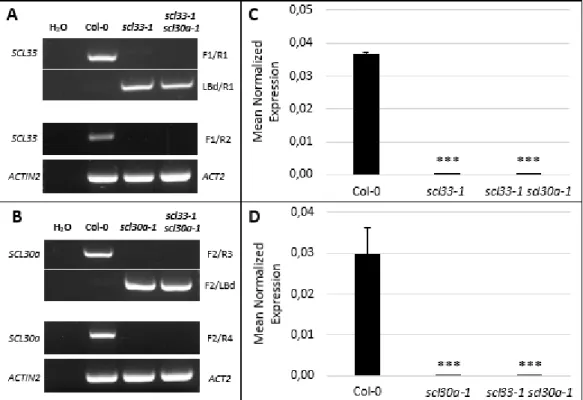
Figure 1.1 Isolation and molecular characterization of the scl33-1, scl30a-1 and scl33-1 scl30a-1 mutants
(A-B) PCR-based genotyping of the T-DNA insertion mutant lines scl33-1, scl30a-1 and scl33-1 scl30a-1. The disruption of the SCL33 (A) and SCL30a (B) genes was assessed using gene-specific primers flanking the insertion (F1/R1 for SCL33; F2/R3 for SCL30a). Presence of the T-DNA insertion was assessed using a primer annealing to the left border of the T-DNA (LBd) in combination with the appropriate gene-specific primer. Disruption of the SCL33 (A) and SCL30a (B) transcripts was confirmed by RT-PCR using gene-specific primers annealing to the coding region flanking the T-DNA (F1/R2 for SCL33; F2/R4 for SCL30a). The Actin-2 gene was used as a loading control. (C-D) Quantitative real-time RT-qPCR analysis, using primers flanking the T-DNA insertion in the SCL33 (C) and the SCL30a (D) genes, in the wild type (Col-0) and scl33-1, scl30a-1 and scl33-1 scl30-1 mutant backgrounds. EF1α was used as a reference gene. The results are shown for two independent biological and technical replicates and the values represent means ± SE (n = 4). Asterisks indicate significant differences (*** p < 0.001; Student’s t-test) from the corresponding wild type.
1.2. Functional characterization of the Arabidopsis scl33-1, scl30a-1 and scl33-1 scl30a-1 mutants
1.2.1. Expression patterns of SCL33 and SCL30a during plant development
To obtain clues on the biological role(s) of the SCL33 and SCL30a proteins and aid in the phenotypic analyses of the corresponding mutants, we examined the expression profiles of SCL33 and SCL30a in different tissues/organs and development stages of Arabidopsis using RT-qPCR. As shown in Figures
12 1.2.A and 1.2.B, these genes were ubiquitously expressed in the different vegetative and floral tissues analyzed, suggesting a general role for these proteins in plant development. During early developmental stages, we noticed a higher expression level of SCL33 in seeds (dry and imbibed seeds) in comparison to the other stages analyzed (Fig. 1.2.C), while the SCL30a gene showed a more stable expression level (Fig. 1.2.D). Given the previously described role for SCL30a during germination and early seedling development, and the high expression level of SCL33 in seeds, we decided to focus our study of these two genes during germination and post-germination events.
Figure 1.2 Expression profile of the SCL33 and SCL30a genes in different tissues and during early development
Quantitative real-time RT-qPCR analysis of total transcript levels of SCL33 (A and C) and SCL30a (B and D) in different tissues/organs and developmental stages of wild type (Col-0) plants, using EF1α as a reference gene. Results are from two independent biological and technical replicates and values represent means ± SE (n = 4). Different letters indicate statistically significant differences (p < 0.05; Student’s t-test).
1.2.2. Phenotypic characterization of the scl33-1, scl30a-1 and scl33-1 scl30a-1 mutants
Unpublished work from the PMB lab has shown that the SCL30a protein is a negative regulator of the ABA pathway, relying on this hormone to regulate seed-specific traits, such as size and dormancy, as well as germination under high salt and osmotic conditions (Carvalho & Richardson et al., manuscript in preparation). Indeed, the scl30a-1 loss-of-function mutant exhibits enhanced seed dormancy and hypersensitivity in the response to ABA-related stresses. We therefore asked whether its close paralog, SCL33, could share the same functions as SCL30a during ABA-related stress responses.
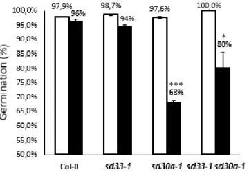
13 To this end, we first assessed the seed dormancy of the scl33-1, scl30a-1 and scl33-1 scl30a-1 mutants (Fig. 1.3). Seeds that had been stratified (to break dormancy) were used as positive controls for each genotype to ensure that the observed dormancy phenotypes were not due to any seed viability related problem. Consistent with previous results from our lab (Carvalho & Richardon et al., manuscript in preparation), after 7 days of germination under control conditions, scl30a-1 seeds exhibited a strong impairment in germination rates when compared to non-stratified wild type seeds. By contrast, germination of scl33-1 mutant seeds was unaffected, suggesting that dormancy is not affected by the lack of a functional SCL33 gene. Surprisingly, we found that germination of the scl33-1 scl30a-1 double mutant was significantly higher than that of the scl30a-1 single mutant. Thus, loss of SCL33 function can partially rescue the hyperdormant phenotype of scl30a-1 mutant by negatively regulating seed dormancy. Together, these results point to opposite roles between SCL33 and SCL30a during seed dormancy, where SCL33 would act as a positive regulator of this process.
Figure 1.3 Germination rates of scl33-1, scl30a-1 and scl33-1 scl30a-1 mutant seeds
Germination of freshly-harvested wild type (Col-0), scl33-1, scl30a-1 and scl33-1 scl30a-1 seeds scored after 7 days of incubation under control conditions with (white bars) or without (black bars) seed stratification (means ± SE, n = 3). Asterisks indicate significant differences (* p < 0.05; *** p < 0.001; Student’s t-test) from the corresponding non-stratified wild type.
We then evaluated seed germination and early seedling development of the scl33-1, scl30a-1 and scl33-1 scl30a-1 mutants under different ABA-related stresses. For this, germination of wild type and mutant seeds was assessed under ABA, salt (NaCl), and drought (mimicked by high concentrations of mannitol) stresses. Consistent with previous observations in the lab, we detected a hypersensitive phenotype for the scl30a-1 mutant, with germination being strongly altered in response to all stresses tested (Fig. 1.4). By contrast, no significant differences were observed between wild type seeds and the scl33-1 mutant. Interestingly, the strong scl30a-1 stress hypersensitivity observed during germination was suppressed in the scl33-1 scl30a-1 double mutant. Again, this result suggests that the SCL33 and SCL30a genes play opposite roles during germination of the seed, with SCL33 being a negative regulator of seed germination under stress conditions. Furthermore, as the scl33-1 single mutant exhibits the same germination pattern under stress conditions as wild type seeds, we conclude that the function of SCL33 becomes relevant only when SCL30a is altered or absent.
14 Figure 1.4 Germination rates of scl33-1, scl30a-1 and
scl33-1 scl30a-1 mutant seeds under ABA-related
stresses
Germination rates of Col-0 (black), scl33-1 (blue), scl30a-1 (yellow) and scl33-1 scl30a-1 (green) stratified seeds, grown under standard light conditions in the presence of different concentrations of ABA (A), mannitol (B) or NaCl (C), were scored 7 days after stratification (means ± SE, n = 3). Asterisks indicate statistically significant differences from the wild type (*** p < 0.001; Student’s t-test).
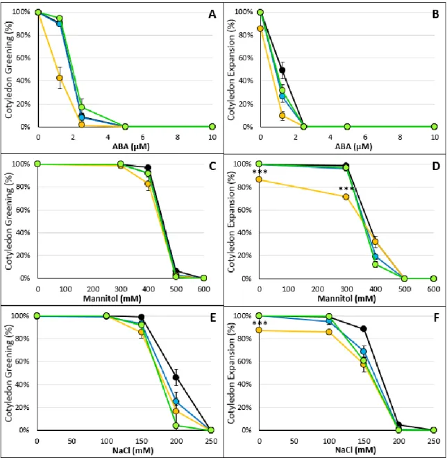
As ABA-related stresses are also known to affect later stages of seedling development, such as cotyledon development, we also compared this developmental processes in wild type and scl33-1, scl30a-1 and scl33-1 scl30a-1 seedlings. As shown in Figure 1.5, only the scl30a-1 mutant showed significant differences in cotyledon greening or expansion from the wild type under the different treatments. Indeed, this mutant was slightly more sensitive (p < 0.05) than the other mutant lines during the majority of the treatments.
Interestingly, we noticed a striking difference in cotyledon expansion of the scl30a-1 mutant under control conditions and in response to all the stress treatments for 9% of the scl30a-1 mutant population (Fig.1.5 and 1.6). Although this population showed normal cotyledon greening, cotyledon shape and development was altered, giving rise to an unorganized mass of cells similar to plant calli. These seedlings were able to survive at least one month after germination under control conditions.
Germination, cotyledon greening and cotyledon expansion assays were conducted on seeds from five consecutive generations and similar results were obtained.
15 Figure 1.5 Cotyledon greening and expansion rates of scl33-1, scl30a-1 and scl33-1 scl30a-1 mutant seeds
Cotyledon greening (A, C and E) and cotyledon expansion (B, D and F) rates of Col-0 (black), scl33-1 (blue), scl30a-1 (yellow) and scl33-scl30a-1 scl30a-scl30a-1 (green) stratified seeds, grown under standard light conditions in the presence of different concentrations of ABA (A-B), mannitol (C-D) and NaCl (E-F) (means ± SE, n = 3). All three parameters were scored 7 days after seed stratification. Asterisks indicate statistically significant differences from the wild type (*** p < 0.001; Student’s t-test).
Figure 1.6 Part of the scl30a-1 population is incapable of proper development under control conditions
Representative images of abnormal scl30a-1 seedlings 4 (A and D) and 30 (B and E) days after stratification and transfer to light. A 4-day old normally developed seedling (C) is shown as representative of the rest of the population
16 Given the ABA-related phenotypes of scl30a-1 mutant seeds, and because ABA is also known to play a key role in vegetative tissues in the regulation of stomatal apertures [6], we next asked whether the Arabidopsis SCL33 and SCL30a SR proteins would play an ABA-dependent role in stomatal movements and hence in the control of leaf water loss under drought stress conditions. To address this, we measured the transpiration rates of detached rosette leaves from wild type plants and the three mutant lines. As shown in Supplementary Figure 3, we could not detect any significant (p > 0.05) changes in water loss rates for the mutant lines. This observation suggests that neither the SCL30a nor SCL33 genes are involved in the regulation of stomatal movements in response to stress.
Thus far, we have been able to confirm the previously described phenotype of the scl30a-1 mutant during germination under stress conditions. On the other hand, we have found that disruption of a close paralog of SCL30a, SCL33, does not seem to affect seed dormancy, germination or early development of scl33-1 mutant plants. In fact, our results point to a role for SCL33 only in the absence of SCL30a function. This suggested that there are compensatory mechanisms between the two genes, whereby one gene directly or indirectly controls the expression/activity of the other. To test this hypothesis, we determined the expression levels of SCL33 (Fig. 1.7.A) and SCL30a (Fig. 1.7.B) genes in wild type and mutant backgrounds by RT-qPCR. As shown in Figure 1.7.A, we detected a two-fold induction of SCL33 transcript levels in the scl30a-1 mutant background. By contrast, expression of the SCL30a gene was not altered in the scl33-1 mutant (Fig. 1.7.B). These results suggest that SCL30a directly or indirectly regulates SCL33 expression.
Figure 1.7 SCL33 and SCL30a expression in the scl33-1, scl30a-1, and scl33-1 scl30-1 mutants
Quantitative real-time RT-qPCR analysis of total transcript levels of SCL33 (A) and SCL30a (B) in wild type (Col-0), scl30a-1, scl33-1, and scl30a-1 scl33-1 mutant rosette leaves, using EF1α as a reference gene. Results are from two independent biological and technical replicates and values represent means ± SE (n = 4). Asterisks indicate statistically significant differences from the wild type (** p < 0.01 and *** p < 0.001; Student’s t-test).
17 2. Functional characterization of the Arabidopsis SR34 gene during early development
2.1. Expression and splicing pattern of SR34 during plant development
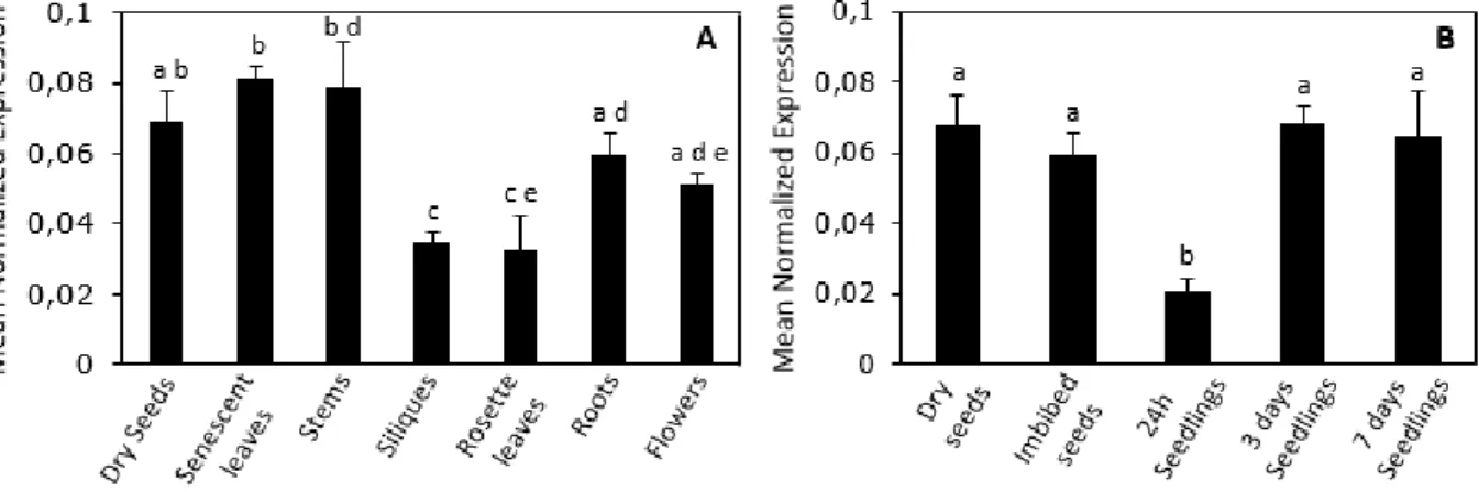
As a first approach to characterize the functional role of the Arabidopsis SR34 gene, we analyzed its developmental and tissue-specific expression profile by RT-qPCR. As seen in Figure 2.1, the SR34 gene is ubiquitously expressed in Arabidopsis thaliana vegetative tissues and during early developmental, suggesting a global role for this gene throughout plant development.
Figure 2.1 Expression profile of the SR34 gene
Quantitative real-time RT-qPCR analysis of total SR34 transcript levels in different tissues (A) and early development stages (B) of wild type (Col-0) plants, using EF1α as a reference gene. Results are from two independent biological and technical replicates and values represent means ± SE (n = 4). Different letters indicate statistically significant differences (p < 0.05; Student’s t-test).
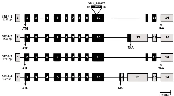
Both animal and plant pre-mRNAs encoding SR proteins are themselves particularly prone to alternative splicing. This prompted us to examine the splicing pattern of the SR34 gene. Our RT-PCR analyses in both rosette leaves and young seedlings resulted in two distinct bands of different relative intensities. Sequencing these two gel bands revealed that one contains two different already annotated (www.Arabidopsis.org) splice variants (SR34.1 and SR34.3), which differ only in four base pairs in the 5’UTR due to an alternative 3’ splice site event. The higher molecular weight band corresponded to a novel splice variant, which we named SR34.4 (Fig. 2.2). Under our experimental conditions, we were unable to detect the annotated SR34.2 splice variant (www.Arabidopsis.org). The RT-PCR results obtained here also conflict with previous studies from Reddy and co-workers who reported the presence of six different SR34 splice variants [61, 67]. Indeed, it is clear that cDNAs from other developmental stages/environmental conditions would need to be tested to enable a comprehensive view of the SR34 splice variants.
Comparison of the three alternative SR34-specific sequences with the genomic fragment revealed that the gene contains at least fourteen exons. The most expressed SR34.1 transcript arises from
18 constitutive splicing of the pre-mRNA and, as SR34.3, is predicted to encode the full-length SR34 protein. By contrast, SR34.4 results from a partial intron retention and harbors a PTC.
Taken together, the above results indicate that the SR34 gene displays ubiquitous expression in embryonic vegetative tissues, being highly expressed during seed imbibition/germination, and produces at least three alternative transcripts. While the SR34.1 and SR34.3 mRNAs encode the full-length protein, the SR34.4 transcript is either a putative NMD target or may be translated into a non-functional or truncated (lacking a part of the SR domain) protein with an altered function.
Figure 2.2 Schematic diagram of the Arabidopsis SR34 splice variants
Schematic representation of four SR34 splice variants (the length of each transcript is indicated). Exons are represented as rectangles and introns as thin lines. Black rectangles represent the coding exons, UTRs are shown in grey. The T-DNA insertion in the SALK_106067 line is also shown.
2.2. Isolation and phenotypic characterization of the sr34-1 mutant
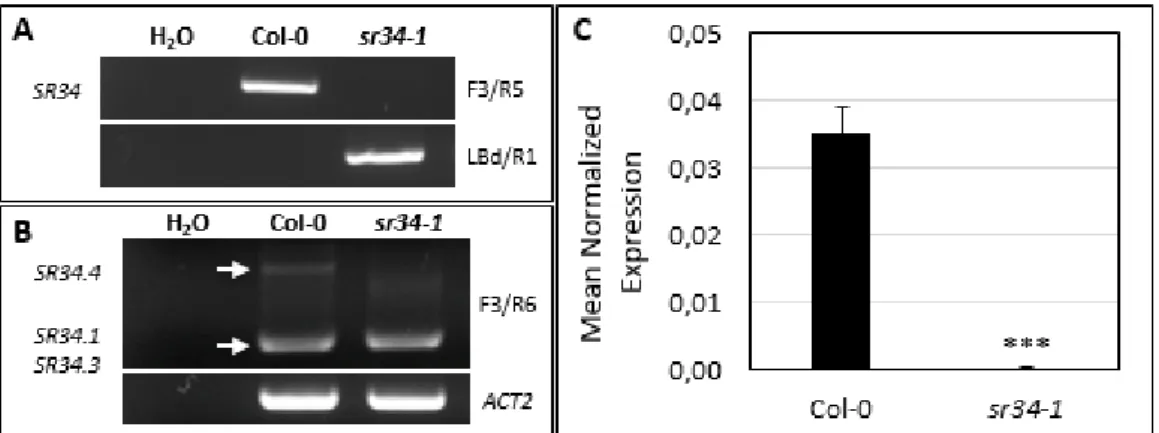
To initiate the functional characterization of the SR34 gene, we obtained four T-DNA insertion mutant lines, SALK_ 106067, SALK_102166, SALKseq_105967 and later SALK_010894, expected to carry a T-DNA insertion in the gene’s tenth exon (SALK_ 106067 and SALKseq_105967), fourth exon (SALK_102166) or in the 5’ UTR (SALK_010894). The different lines were genotyped using the same PCR-based method as described previously. Of the four independent mutant lines, we were only able to isolate homozygous mutant plants for SALK_106067 (Fig. 2.3.A). This was hence the insertion line used for further functional characterization.
To discern whether the expression of SR34 was disrupted at the mRNA level, we performed semi-quantitative RT-PCR experiments using gene-specific primers flanking the T-DNA insertion. We detected residual mRNA expression in sr34-1 when using these primers (Fig. 2.3.B). To more precisely