Carina Isabel Correia Crucho
Mestre em BioorgânicaSynthesis of Polymeric Nanoparticles for
biomedical delivery applications
Dissertação para obtenção do Grau de Doutor em
Química
Orientador: Professora Doutora Maria Teresa Barros, Professor
Associado com Agregação, FCT-UNL
Júri: Presidente: Professora Doutora Maria João Lobo de Reis Madeira Crispim Romão
Arguentes: Professor Doutor Christopher David Maycock Professora Doutora Maria de Lurdes dos Santos Cristiano
Vogais: Professora Doutora Ana Paula da Assunção Esteves Professor Doutor Marco Diogo Richter Gomes da Silva Professor Doutor António Gil de Oliveira Santos Professor Doutor Alcino Jorge Lopes Leitão Doutora Maria Rita Mendes Bordalo Ventura Centeno Lima Doutora Krasimira Todorova Markova-Petrova
iii
Carina Isabel Correia Crucho
Mestre em BioorgânicaSynthesis of Polymeric Nanoparticles for
biomedical delivery applications
Dissertação para obtenção do Grau de Doutor em
Química
v
“
Begin at the beginning and go on until you come to the end; then stop."
vii
Copyright
ix
Acknowledgments
This thesis was a very lonely work! Although, many people gave me generous help and support for which I am extremely thankfully.
I wish to express my gratitude to my supervisor, Professor Maria Teresa Barros, who has taught me the value of carbohydrate research. I am also grateful to her for granting me the freedom to choose my research direction. This experience will serve me well in future activities for which I will always be indebted.
I also thank Professor Christopher Maycock for sharing his experience and knowledge. His diligence, erudition and sympathy were a vital part for the completion of my research. Along the same lines, all committee members were very helpful.
I am very thankful to Photochemistry and Supramolecular Chemistry research group from Department of Chemistry, FCT-UNL, for an invaluable source of cooperation and for giving me the opportunity to use their DLS instrument for size characterization.
I would like to thank Professor Jorge Caldeira for his valuable scientific support in the introduction and use of AFM.
A special word of gratitude goes to my friend and colleague Dr. Paula Correia-da-Silva for valuable advice, kindness and friendship.
xi
Abstract
Polymeric nanoparticles (PNPs) have attracted considerable interest over the last few years due to their unique properties and behaviors provided by their small size. Such materials could be used in a wide range of applications such as diagnostics and drug delivery. Advantages of PNPs include controlled release, protection of drug molecules and its specific targeting, with concomitant increasing of the therapeutic index.
In this work, novel sucrose and cholic acid based PNPs were prepared from different polymers, namely polyethylene glycol (PEG), poly(D,L-lactic-co-glycolic acid) (PLGA) and PLGA-co-PEG copolymer. In these PNP carriers, cholic acid will act as a drug incorporation site and the carbohydrate as targeting moiety.
The uptake of nanoparticles into cells usually involves endocytotic processes, which depend primarily on their size and surface characteristics. These properties can be tuned by the nanoparticle preparation method. Therefore, the nanoprecipitation and the emulsion-solvent evaporation method were applied to prepare the PNPs. The influence of various parameters, such as concentration of the starting solution, evaporation method and solvent properties on the nanoparticle size, size distribution and morphology were studied.
The PNPs were characterized by using atomic force microscopy (AFM), scanning electron microscopy (SEM) and dynamic light scattering (DLS) to assess their size distribution and morphology. The PNPs obtained by nanoprecipitation ranged in size between 90 nm and 130 nm with a very low polydispersity index (PDI < 0.3). On the other hand, the PNPs produced by the emulsion-solvent evaporation method revealed particle sizes around 300 nm with a high PDI value. More detailed information was found in AFM and SEM images, which demonstrated that all these PNPs were regularly spherical. ζ-potential measurements were satisfactory and evidenced the importance of sucrose moiety
on the polymeric system, which was responsible for the obtained negative surface charge, providing colloidal stability.
The results of this study show that sucrose and cholic acid based polymeric conjugates can be successfully used to prepare PNPs with tunable physicochemical characteristics. In addition, it provides novel information about the materials used and the methods applied. It is hoped that this work will be useful for the development of novel carbohydrate based nanoparticles for biomedical applications, specifically for targeted drug delivery.
xiii
Resumo
Nanopartículas poliméricas (NPPs) têm atraído um considerável interesse nos últimos anos devido às suas propriedades únicas e comportamentos proporcionados pelo seu pequeno tamanho. Estes materiais podem ser utilizados numa vasta gama de aplicações, tais como em diagnóstico e na veiculação de fármacos. As vantagens das NPPs incluem a libertação controlada, protecção do fármaco e o seu direcionamento a um alvo específico, com concomitante aumento do índice terapêutico.
Neste trabalho, novas NPPs baseadas em unidades de sacarose e de ácido cólico foram preparadas a partir de diferentes polímeros, nomeadamente polietileno glicol (PEG), poli(ácido
láctico-co-ácido glicólico ) (PLGA) e do copolímero PLGA-co-PEG. Nestas NPPs a unidade de ácido cólico irá actuar como o local de incorporação do fármaco e o açúcar como a unidade de direccionamento.
A incorporação das nanopartículas pelas células envolve geralmente processos de endocitose, que dependem primariamente do seu tamanho e superficie. Estas propriedades podem ser ajustadas de acordo com o método de preparação das nanopartículas. Deste modo, o método de nanoprecipitação e a técnica de emulsificação-evaporação do solvente foram utilizados para preparar as NPPs. A influência de vários parâmetros, tais como a concentração da solução inicial, o método de evaporação e as propriedades do solvente no tamanho médio das nanoparticulas, na distribuição de tamanhos e na morfologia foram estudados.
As NPPs foram caracterizadas por microscopia de força atómica (AFM), microscopia electrónica de varrimento (SEM) e dispersão dinâmica de luz (DLS) para avaliar a distribuição de tamanhos e a morfologia. O tamanho das NPPs obtidas pelo método de nanoprecipitação variou entre 90 nm a 130 nm, com um baixo índice de polidispersão (PDI <0,3). Por outro lado, as NPPs obtidas pelo método de emulsificação-evaporação do solvente revelou partículas com um tamanho de cerca de 300 nm e com elevado valor de PDI. Informação mais detalhada foi encontrada nas imagens obtidas por AFM e SEM, que demonstraram que todas as NPPs eram regularmente esféricas. As medidas de potencial-ζ foram satisfatórias e evidenciaram a importância da unidade de sacarose no sistema polimérico, que foi responsável pela carga superficial negativa obtida, proporcionando maior estabilidade coloidal.
Os resultados deste estudo mostram que os conjugados poliméricos baseados em unidades de sacarose e ácido cólico, podem ser utilizados com sucesso na preparação de NPPs com características físico-químicas ajustáveis. Além disso, fornecem novas informações sobre os materiais utilizados e os métodos aplicados. Espera-se que este trabalho seja útil para o desenvolvimento de novas NPPs à base de glúcidos para aplicações biomédicas, especificamente para sistemas de veiculação direcionada de fármacos.
xv
List of Contents
CHAPTER I: SYNTHESIS OF POLYMERIC NANOPARTICLES: AN OVERVIEW OF THE
PREPARATION AND CHARACTERIZATION METHODS
1
I.1 Introduction 3
I.2 Preparation of Polymeric Nanoparticles 5
I.2.1 Two-step procedures based on Emulsification 6
I.2.1.1 Emulsification-solvent evaporation 7
I.2.1.2 Emulsification-solvent diffusion 10
I.2.1.3 Emulsification–reverse salting-out 12
I.2.2 One-step procedures 14
I.2.2.1 Nanoprecipitation or solvent displacement method 14
I.2.2.2 Dialysis 17
I.2.2.3 Supercritical fluid technology 19
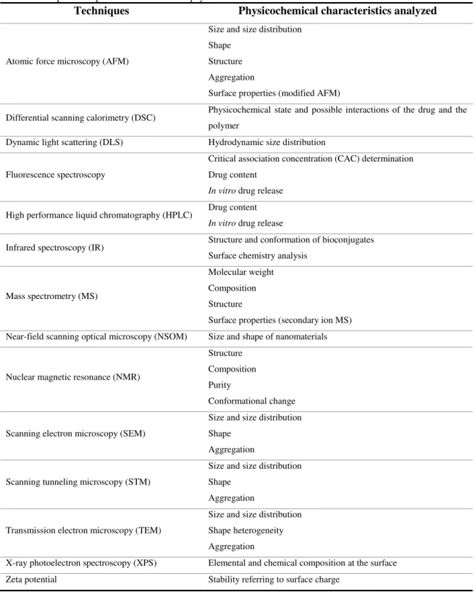
I.3 Characterization methods of Polymeric Nanoparticles 20
I.3.1 Size, shape, surface properties and stability 22
I.3.2 Drug-polymer interactions 25
I.4 Concept of this thesis 27
CHAPTER II: SYNTHESIS OF PEG-BASED POLYMERIC NANOPARTICLES
29
II.1 Introduction 31
II.2 Results and Discussion 32
II.2.1 Synthesis and characterization of PEG-based polymer conjugates 32
II.2.1.1 Chemoselective derivatization of 6’-hydroxyl group of sucrose 33
II.2.1.2 Chemoselective derivatization at the 3’ position of cholic acid 37
II.2.1.3 Synthesis of Suc-PEG-Chol polymer conjugates 39
II.2.1.4 Physicochemical characterization of Suc-PEG-Chol polymer conjugates 41
II.2.1.5 Synthesis of Cholic-PEG polymer conjugates 43
II.2.2 Preparation and physicochemical characterization of PNPs 44
II.2.2.1 PNPs prepared from Suc-PEG-Chol polymer conjugates 44
II.2.2.2 PNPs prepared from Cholic-PEG polymer conjugates 51
xvi
CHAPTER III: SYNTHESIS OF PLGA-BASED POLYMERIC NANOPARTICLES
57
III.1 Introduction 59
III.2 Results and Discussion 60
III.2.1 Synthesis and characterization of PLGA-based polymer conjugates 60
III.2.2 Preparation and physicochemical characterization of PNPs 63
III.2.2.1 PNPs prepared by the emulsion-solvent evaporation method 63
III.2.2.2 PNPs prepared by nanoprecipitation 65
III.3 Conclusion 71
CHAPTER IV: SYNTHESIS OF PLGA-PEG-BASED POLYMERIC NANOPARTICLES
73
IV.1 Introduction 75
IV.2 Results and Discussion 76
IV.2.1 One-pot synthesis of PLGA-co-PEG-based polymer conjugates 76
IV.2.2 Preparation and physicochemical characterization of PNPs 78
IV.3 Conclusion 80
CHAPTER V: CONCLUSIONS AND FUTURE PERSPECTIVES
83
CHAPTER VI: EXPERIMENTAL PART
89
VI.1 Materials and Instruments 91
VI.2 Synthesis and characterization of polymer conjugates 92
VI.2.1 Chemoselective derivatization of the 6’ position of the sucrose 92
VI.2.1.1 6’-O-tert-butyldiphenylsilyl-sucrose (2) 92 VI. 2.1.2 1’,2,3,3’,4,4’,6-Hepta-O-benzyl-6’-O-tert-butyldiphenylsilyl sucrose (4) 93
VI. 2.1.3 1’,2,3,3’,4,4’,6-Hepta-O-benzyl-sucrose (5) 94
VI. 2.1.4 1’,2,3,3’,4,4’,6-Hepta-O-benzyl-6´-O-succinyl-sucrose (6) 95
VI. 2.2 Chemoselective derivatization at the 3’ position of cholic acid 95
VI. 2.2.1 Methyl 3α,7α,12α-trihydroxy-5β-cholan-24-ate (8) 95
VI. 2.2.2 Methyl 3α-O-benzyl, 7α, 12α-dihydroxy-5β-cholan-24-oate (9) 96
VI. 2.2.3 3α-O-benzyl, 7α, 12α-dihydroxy-5β-cholic acid (10) 97
VI.2.3 General procedure 1 for DCC-mediated coupling reactions 97
xvii
VI.2.4.1 Benzylated Suc-PEG2000-OH (11) 98
VI.2.4.2 Benzylated Suc-PEG4000-OH (12) 98
VI.2.4.3 Benzylated Suc-PEG6000-OH (13) 99
VI.2.4.4 Benzylated Suc-PEG2000-Chol (14) 100
VI.2.4.5 Benzylated Suc-PEG4000-Chol (15) 101
VI.2.4.6 Benzylated Suc-PEG6000-Chol (16) 102
VI.2.4 General procedure 2 for hydrogenation reactions 103
VI.2.5 Deprotection of PEG-based conjugates 103
VI.2.5.1 Suc-PEG2000-Chol (17) 103
VI.2.5.2 Suc-PEG4000-Chol (18) 104
VI.2.5.3 Suc-PEG6000-Chol (19) 105
VI.2.6 General procedure 3 for the synthesis of Cholic-PEG conjugates 107
VI.2.6.1 Cholic-PEG2000 (20) 107
VI.2.6.2 Cholic-PEG4000 (21) 108
VI.2.6.3 Cholic-PEG6000 (22) 109
VI.2.7 Synthesis of PLGA-based conjugates 111
VI.2.7.1 Benzylated Suc-PLGA-OH (23) 111
VI.2.7.2 Benzylated Suc-PLGA-Chol (24) 111
VI.2.7.3 Suc-PLGA-Chol (25) 112
VI.2.8 Synthesis of PLGA-PEG based conjugates 112
VI.2.8.1 One-pot synthesis of benzylated Suc/Chol-PLGA-PEG-Chol/Suc (26) 112
VI.2.8.2 Benzyl deproctection of Suc/Chol-PLGA-PEG-Chol/Suc 26 (27) 113
VI.2.9 Measurement of fluorescence spectroscopy (pyrene) 113
VI.3 Preparation of polymeric nanoparticles 114
VI.3.1 Emulsion-solvent evaporation method 114
VI.3.2 Nanoprecipitation method 114
VI.4 Characterization of polymeric nanoparticles 115
VI.4.1 Particle size and zeta potential 115
VI.4.2 Surface morphology – scanning electron microscopy (SEM) and atomic force microscopy (AFM) 115
xix
List of Figures
FIGURE 1 TYPES OF NANOCARRIERS FOR DRUG DELIVERY SYSTEMS. POLYMERIC NANOPARTICLES: A,
NANOCAPSULES AND B, NANOSPHERES. C, LIPOSOMES. D, POLYMERIC MICELLES. E, DENDRIMERS. F,
CARBON NANOTUBES. ... 4
FIGURE 2 SCHEMATIC REPRESENTATION OF SEVERAL TECHNIQUES FOR THE PREPARATION OF PNPS. ... 5
FIGURE 3 SCHEMATIC DIAGRAM OF EMULSIONS FABRICATED FROM OIL, WATER AND SURFACTANT... 6
FIGURE 4 SCHEMATIC REPRESENTATION OF THE EMULSIFICATION-SOLVENT EVAPORATION METHOD FOR THE
PRODUCTION OF NANOSPHERES. ... 7
FIGURE 5 NANOCAPSULES WITH DIFFERENT MORPHOLOGIES PREPARED BY ASHJARI ET AL. CORE-SHELL OR HALF-MOON MORPHOLOGY WAS OBSERVED WHEN SLOW EVAPORATION WAS USED COMPARED TO
FAST EVAPORATION. [ADAPTED FROM REF. 31] ... 8
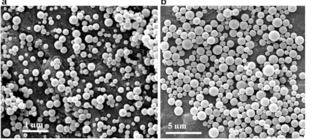
FIGURE 6 SEM IMAGES OF PARTICLES OBTAINED FROM A POLYESTER (PCL) AND A POLYACRYLATE POLYMER
(POLY(MMA-AA)). THE SOLVENT USED FOR POLYMER SOLUBILIZATION INFLUENCED THE PARTICLE SIZE
AND MORPHOLOGY. [ADAPTED FROM REF. 34] ... 9
FIGURE 7 SCHEMATIC REPRESENTATION OF THE EMULSIFICATION-SOLVENT DIFFUSION METHOD FOR THE
PREPARATION OF NANOCAPSULES. ... 10
FIGURE 8 SEM IMAGES OF THE PLA NANOPARTICLES PREPARED BY TRIMAILLE ET AL. A) 2% PLA; B) 10% PLA. [ADAPTED FROM REF. 39] ... 11
FIGURE 9 SCHEMATIC REPRESENTATION OF THE EMULSIFICATION-REVERSE SALTING-OUT TECHNIQUE. ... 13
FIGURE 10 SCHEMATIC ILLUSTRATION OF THE NANOPRECIPITATION METHOD FOR THE PREPARATION OF
NANOSPHERES. FOR THE PREPARATION OF NANOCAPSULES AN OIL IS INTRODUCED IN THE ORGANIC
PHASE. ... 14
FIGURE 11 INFLUENCE OF POLYMER CONCENTRATION IN THE ORGANIC PHASE ON THE MEAN DIAMETER OF
PNPS. ABBREVIATION: BNZ, BENZYLAMINE; FITC, FLUORESCEIN ISOTHIOCYANATE. [ADAPTED FROM REF.
63] ... 16
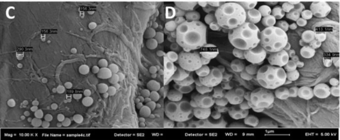
FIGURE 12 FESEM MICROGRAPHS OF POLYSTYRENE PNPS PREPARED BY NANOPRECIPITATION. POLYMER
SOLVENT: TETRAHYDROFURAN. NON-SOLVENT: C, ACETONE; D, WATER AND PLURONIC-F68. [ADAPTED
FROM REF. 65] ... 17
FIGURE 13 SCHEMATIC REPRESENTATION OF THE DIALYSIS METHOD FOR THE PREPARATION OF
NANOSPHERES. ... 17
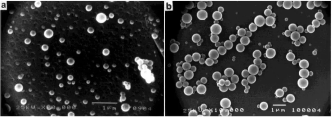
FIGURE 14 SEM IMAGES OF PLGA 50:50 NANOPARTICLES PREPARED BY JEONG ET AL. A) DMAC OR B) ACETONE
AS THE POLYMER SOLVENT. [ADAPTED FROM REF. 72] ... 18
FIGURE 15 SEM IMAGES OF DIFFERENT MORPHOLOGIES OBTAINED FOR HYALURONIC ACID BASED
NANOSTRUCTURES. EXPERIMENTAL CONDITIONS: T = 4 ºC; CONC. [MG/ML] = 0.5; SOLVENT PAIR: H, H2O/ETOH; I, H2O/MEOH; L, DMSO/MEOH; M, H2O/ACETONE. MORPHOLOGY: H, FLOWERLIKE; I AND L,
SPHERES AND M, DANDELION-LIKE. [ADAPTED FROM REF. 70] ... 19
xx
FIGURE 17 SCANNING ELECTRON MICROGRAPHS OF A CANTILEVER TIP (A) AND SCHEMATIC OF AFM
OPERATION (B). ... 23
FIGURE 18 FESEM IMAGE (LEFT) AND AFM IMAGES (MIDDLE (2D IMAGE) AND RIGHT (ZOOM IN 3D IMAGE)) OF PLA-TWEEN 80-10 PNPS. ... 23
FIGURE 19 SURFACE MORPHOLOGY OF PEG-G-PLA PNPS BY AFM. SCALE X-AXIS: 0.200 µM/DIV. ... 24
FIGURE 20 DSC RESULTS OF A) KETOPROFEN LOADED PLGA NANOPARTICLES, B) DRUG FREE PLGA NANOPARTICLES, C) PHYSICAL MIXTURE OF PLGA AND KETOPROFEN AND D) PURE PLGA POLYMER [ADAPTED FROM REF. 103]. ... 26
FIGURE 21 TYPICAL END-GROUPS IN POLYETHYLENE GLYCOL MODIFICATIONS. ... 31
FIGURE 22 SYNTHETIC STRATEGY FOR PREPARING THE SUC-PEG-CHOL POLYMER CONJUGATES. ... 32
FIGURE 23 DIFFERENT REACTIVITY OF PRIMARY/SECONDARY HYDROXYLS OF SUCROSE. ... 33
FIGURE 24 HMQC SPECTRUM EXPANSION OF 6’-O-TERT-BUTYLDIPHENYLSILYL-SUCROSE 2 IN DMSO-D6, FOCUSED ON SUCROSE PROTONS. ... 35
FIGURE 25 HMQC SPECTRUM EXPANSION OF 1’,2,3,3’,4,4’,6-HEPTA-O-BENZYL-6’-O-TERT-BUTYLDIPHENYLSILYL SUCROSE 5 IN DMSO-D6, FOCUSED ON SUCROSE PROTONS... 36
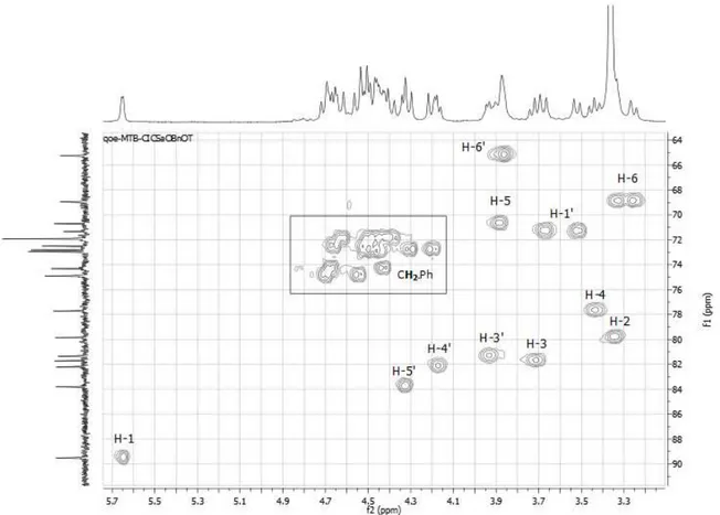
FIGURE 26 HMQC SPECTRUM EXPANSION OF 3Α-O-BENZYL, 7Α, 12Α-DIHYDROXY-5Β-CHOLIC ACID 10 IN CDCL3, FOCUSED ON THE STEROID SKELETON. ... 39
FIGURE 27 A) FLUORESCENCE EMISSION SPECTRA OF PYRENE/SUC-PEG2000-CHOL AGAINST CONCENTRATION OF SUC-PEG2000-CHOL IN DISTILLED WATER. B) PLOTS OF THE OVERALL INTENSITY OF THE PYRENE EMISSIONS SPECTRA VS. POLYMER CONCENTRATIONS. ... 42
FIGURE 28 1H-NMR SPECTRUM OF SUC-PEG4000-CHOL 18 IN D 2O. ... 43
FIGURE 29 PARTICLE SIZE DISTRIBUTIONS OF SUC-PEG-CHOL PNPS PREPARED BY THE EMULSION-SOLVENT EVAPORATION METHOD. ... 45
FIGURE 30 AFM HEIGHT IMAGE OF SUC-PEG4000-CHOL PNPS PREPARED BY O/W EMULSION-SOLVENT EVAPORATION METHOD WITH THE EMULSION FORMULATED BY VORTEX MIXING. THE SAMPLE WAS PREPARED FROM A PNP SOLUTION OF 0.1 MG/ML. ... 46
FIGURE 31 PARTICLE SIZE DISTRIBUTIONS OF SUC-PEG-CHOL PNPS PREPARED BY NANOPRECIPITATION. ... 47
FIGURE 32 A) CONCENTRATION DEPENDENT PARTICLE SIZES OF SUC-PEG-CHOL PNPS AT ROOM TEMPERATURE PREPARED BY NANOPRECIPITATION. B) STUDY ON THE SUC-PEG4000-CHOL PNPS STABILITY, STORED AT 5ºC OVER ONE MONTH. ... 48
FIGURE 33 AFM HEIGHT IMAGE OF SUC-PEG-CHOL PNPS FROM A PNP SOLUTION OF 0.1MG/ML DRIED AT 5 ºC. A) SUC-PEG2000-CHOL PNPS. B) SUC-PEG4000-CHOL PNPS. C) SUC-PEG6000-CHOL PNPS. ... 49
FIGURE 34 AFM HEIGHT IMAGE OF LYOPHILIZED SUC-PEG-CHOL PNPS FROM A PNP SOLUTION OF 0.1MG/ML DRIED AT 5 ºC. A) SUC-PEG4000-CHOL PNPS. B) SUC-PEG6000-CHOL PNPS. ... 50
FIGURE 35 AFM PHASE IMAGE OF LYOPHILIZED SUC-PEG2000-CHOL NANOPARTICLES FROM A PNP SOLUTION OF 0.1MG/ML DRIED BY FREEZE-DRYING. ... 50
xxi
FIGURE 37 AFM PHASE IMAGE OF LYOPHILIZED SUC-PEG4000-CHOL NANOPARTICLES FROM A PNP SOLUTION
OF 0.1MG/ML DRIED UNDER PHOSPHORUS PENTOXIDE. VISUALIZATION OF NANOMETRIC CRYSTALLITES
OF PEG. ... 51
FIGURE 38 SEM IMAGE OF A) SUC-PEG4000-CHOL PNPS AND B) SUC-PEG6000-CHOL PNPS. ... 52
FIGURE 39 PARTICLE SIZE DISTRIBUTIONS OF CHOLIC-PEG CONJUGATES PNPS PREPARED BY THE EMULSION-SOLVENT EVAPORATION METHOD... 53
FIGURE 40 AFM PHASE IMAGE OF LYOPHILIZED CHOLIC-PEG2000 NANOPARTICLES FROM A PNP SOLUTION OF 0.1MG/ML PREPARED BY THE EMULSION-SOLVENT EVAPORATION METHOD. ... 53
FIGURE 41 CHEMICAL STRUCTURE OF POLY(D,L-LACTIC-CO-GLYCOLIC ACID) AND ITS MONOMERS. ... 59
FIGURE 42 SYNTHETIC STRATEGY FOR PREPARING THE SUC-PLGA-CHOL POLYMER CONJUGATE. ... 61
FIGURE 43 1H-NMR SPECTRUM OF BENZYLATED SUC-PLGA-OH 23. ... 62
FIGURE 44 1H-NMR SPECTRUM OF BENZYLATED SUC-PLGA-CHOL 24... 63
FIGURE 45 PARTICLE SIZE DISTRIBUTIONS OF PLGA AND SUC-PLGA-CHOL PNPS PREPARED BY THE EMULSION-SOLVENT EVAPORATION. METHOD... 64
FIGURE 46 SEM MICROGRAPHS OF LYOPHILIZED PLGA PNPS PREPARED BY THE EMULSION SOLVENT EVAPORATION METHOD. ... 65
FIGURE 47 SEM MICROGRAPHS OF LYOPHILIZED SUC-PLGA-CHOL PNPS PREPARED BY THE EMULSION SOLVENT EVAPORATION METHOD. ... 65
FIGURE 48 EFFECT OF VARYING THE ORGANIC SOLVENT IN THE PARTICLE SIZE (GREY BAR) AND PDI (STRIPED BAR) OF SUC-PLGA-CHOL PNPS OBTAINED BY NANOPRECIPITATION. ... 66
FIGURE 49 INFLUENCE OF THE EVAPORATION RATE ON THE PARTICLE SIZE DISTRIBUTION OF PLGA AND SUC-PLGA-CHOL PNPS. SOLVENT EVAPORATION METHOD: A) AND C) EVAPORATION UNDER REDUCE-PRESSURE; B) AND D) ATM. PRESSURE EVAPORATION. ... 67
FIGURE 50 CORRELATION OF PNP MEAN PARTICLE SIZE WITH SUC-PLGA-CHOL POLYMER CONCENTRATION OBTAINED BY NANOPRECIPITATION. ALL THE SAMPLES WERE BROUGHT TO THE SAME CONCENTRATION BEFORE ANALYZED BY DLS. ... 68
FIGURE 51 SEM MICROGRAPHS OF LYOPHILIZED PLGA PNPS PREPARED BY NANOPRECIPITATION. ... 68
FIGURE 52 SEM MICROGRAPHS OF LYOPHILIZED SUC-PLGA-CHOL PNPS PREPARED BY NANOPRECIPITATION. . 69
FIGURE 53 SEM MICROGRAPHS OF LYOPHILIZED SUC-PLGA-CHOL PNPS PREPARED BY NANOPRECIPITATION LOADED WITH 0.25% RHODAMINE B. ... 69
FIGURE 54 SEM MICROGRAPHS OF LYOPHILIZED PLGA PNPS PREPARED BY NANOPRECIPITATION. THE SAMPLE WAS FREEZE-DRIED IN THE PRESENCE OF 10% (W/W) GLUCOSE AS A CRYOPROTECTOR. ... 70
FIGURE 55 SEM MICROGRAPHS OF LYOPHILIZED SUC-PLGA-CHOL PNPS PREPARED BY NANOPRECIPITATION AND DRIED UNDER VACUUM OVER PHOSPHORUS PENTOXIDE AS AN ALTERNATIVE TO FREEZE-DRYING. 70 FIGURE 56 SEM MICROGRAPHS OF LYOPHILIZED SUC-PLGA-CHOL PNPS PREPARED BY NANOPRECIPITATION WITHOUT PURIFICATION BY CENTRIFUGATION. ... 71
FIGURE 57 SYNTHETIC STRATEGY FOR PREPARING THE SUC-PLGA-CO-PEG-CHOL POLYMER CONJUGATES. ... 76
xxii
FIGURE 59 EFFECT OF VARYING THE ORGANIC SOLVENT IN THE PARTICLE SIZE (GREY BAR) AND PDI (STRIPED
BAR) OF SUC-PLGA-CO-PEG-CHOL PNPS OBTAINED BY NANOPRECIPITATION. ... 78 FIGURE 60 PARTICLE SIZE DISTRIBUTIONS OF PLGA-CO-PEG AND SUC-PLGA-CO-PEG-CHOL PNPS PREPARED BY
NANOPRECIPITATION. ... 79
FIGURE 61 CORRELATION OF PNP MEAN PARTICLE SIZE WITH SUC-PLGA-CO-PEG-CHOL POLYMER
CONCENTRATION, OBTAINED BY NANOPRECIPITATION. ALL THE SAMPLES WERE BROUGHT TO THE SAME
CONCENTRATION BEFORE MEASURED BY DLS. ... 79
FIGURE 62 SEM MICROGRAPHS OF LYOPHILIZED SUC-PLGA-CO-PEG-CHOL PNPS PREPARED BY
NANOPRECIPITATION. ... 80
xxiii
List of Schemes
SCHEME 1 PROTECTION/DEPROTECTION SEQUENCE FROM SUCROSE. 33
SCHEME 2 MECHANISM OF THE SILYLATION OF SUCROSE WITH TBDPSCL. 34
SCHEME 3 PENTACOORDINATE INTERMEDIATE INVOLVED IN THE DEPROTECTION OF TBDPS PROMOTED BY
TBAF. 35
SCHEME 4 CHEMOSELECTIVE DE-O-BENZYLATION OF OCTABENZYLATED SUCROSE PROPOSED BY YIN ET AL. 36 SCHEME 5 SYNTHESIS OF THE SUCROSE SUCCINATE DERIVATIVE 1’,2,3,3’,4,4’,6-HEPTA-O-BENZYL-6´-O
-SUCCINYL-SUCROSE 6. 37
SCHEME 6 PROPOSED MECHANISM FOR THE ESTERIFICATION REACTION OF 1’,2,3,3’,4,4’,6-HEPTA-O-
BENZYL-6’-O-TERT-BUTYLDIPHENYLSILYL SUCROSE 5 WITH SUCCINIC ANHYDRIDE. 37 SCHEME 7 SYNTHETIC STRATEGY FOR PREPARING THE SELECTIVELY 3Α-OH PROTECTED CHOLIC ACID
DERIVATIVE 10. 38
SCHEME 8 SYNTHESIS OF THE BENZYLATED SUC-PEG-OH CONJUGATES BY A STEGLICH ESTERIFICATION. 39
SCHEME 9 THE REACTION MECHANISM OF STEGLICH ESTERIFICATION. 40
SCHEME 10 SYNTHESIS OF THE BENZYLATED SUC-PEG-CHOLIC CONJUGATES BY A STEGLICH ESTERIFICATION. 41
SCHEME 11 SYNTHESIS OF THE SUC-PEG-CHOL CONJUGATES BY PD/C CATALYZED HYDROGENOLYSIS. 41
SCHEME 12 SYNTHESIS OF THE CHOLIC-PEG POLYMER CONJUGATES BY A STEGLICH ESTERIFICATION. 43
SCHEME 13 SYNTHESIS OF THE BENZYLATED SUC-PLGA-OH CONJUGATES BY ESTERIFICATION REACTION
THROUGH A PLGA-NHS INTERMEDIATE. 61
xxv
LIST OF TABLES
TABLE 1 EFFECT OF STABILIZER TYPE ON PARTICLE SIZE, POLYDISPERSITY INDEX (PDI) AND ENTRAPMENT
EFFICIENCY, FOR PLGA NANOPARTICLES PREPARED BY JAIN ET AL. 12
TABLE 2 PRINCIPAL TECHNIQUES FOR EVALUATION OF THE PHYSICOCHEMICAL CHARACTERISTICS OF PNPS. 21
TABLE 3 INFLUENCE OF THE PEG CHAIN LENGTH ON THE MELTING TEMPERATURE (TM) AND ON THE CRITICAL
ASSOCIATION CONCENTRATION (CAC) OF THE SUC-PEG-CHOL CONJUGATES. 42
TABLE 4 INFLUENCE OF THE PEG CHAIN LENGTH ON THE MELTING TEMPERATURE (TM) OF THE CHOLIC-PEG
POLYMER CONJUGATES. 44
TABLE 5 MEAN PARTICLE SIZE AND Ζ-POTENTIAL OF THE SUC-PEG-CHOL PNPS PREPARED BY O/W
EMULSION-SOLVENT EVAPORATION METHOD WITH THE EMULSION FORMULATED BY VORTEX MIXING. 44
TABLE 6 MEAN PARTICLE SIZE OF THE SUC-PEG-CHOL PNPS PREPARED BY O/W EMULSION-SOLVENT
EVAPORATION METHOD WITH THE EMULSION FORMULATED BY SONICATION. 46
TABLE 7 SIZE DISTRIBUTION AND Ζ-POTENTIAL OF THE SUC-PEG-CHOL PNPS PREPARED BY
NANOPRECIPITATION. 47
TABLE 8 SIZE DISTRIBUTION OF LYOPHILIZED SUC-PEG-CHOL PNPS PREPARED BY NANOPRECIPITATION AFTER
VORTEXING FOR 5 MIN. 48
TABLE 9 MEAN PARTICLE SIZE OF THE CHOLIC-PEG PNPS PREPARED BY O/W EMULSION-SOLVENT
EVAPORATION METHOD WITH THE EMULSION FORMULATED BY SONICATION. 52
TABLE 10 PHYSICOCHEMICAL CHARACTERISTICS OF THE SUC-PEG-CHOL PNPS OBTAINED BY
NANOPRECIPITATION AND EMULSION-SOLVENT EVAPORATION TECHNIQUE. 54
TABLE 11 GLASS TRANSITION TEMPERATURES (TG) OF PLGA AND PLGA-BASED CONJUGATES. 63
TABLE 12 MEAN PARTICLE SIZE AND POLYDISPERSITY INDEX (PDI) OF PLGA AND SUC-PLGA-CHOL PNPS
PREPARED BY O/W EMULSION-SOLVENT EVAPORATION METHOD WITH THE EMULSION FORMULATED BY
VORTEX MIXING. 64
TABLE 13 EFFECT OF EVAPORATION METHOD IN THE MEAN PARTICLE SIZE AND PDI OF PLGA AND
SUC-PLGA-CHOL PNPS OBTAINED BY NANOPRECIPITATION. 67
TABLE 14 PHYSICOCHEMICAL CHARACTERISTICS OF THE PLGA AND SUC-PLGA-CHOL PNPS OBTAINED BY
NANOPRECIPITATION AND EMULSION-SOLVENT EVAPORATION TECHNIQUE. 71
TABLE 15 SIZE DISTRIBUTION AND Ζ-POTENTIAL OF THE PLGA-CO-PEG AND SUC-PLGA-CO-PEG-CHOL PNPS
xxvii
List of Symbols and Abbreviations
ADME Absorption, distribution, metabolism and excretion
AFM Atomic force microscopy
atm Atmospheric pressure
Bn Benzyl
Bnz Benzylamine
CAC Critical aggregation concentration Cisplatin cis-Diamminedichloroplatinum (II) CMC Critical micelle concentration COSY Correlation spectroscopy
CTAB Cetyltrimethylammonium bromide
Dh Hydrodynamic diameter
DCM Dichloromethane
DCC N,N-dicyclohexylcarbodiimide
DCU Dicyclohexylurea
DDS Drug delivery systems
DLS Dynamic light scattering
DMAB Didodecyldimethylammonium bromide
DMAc Dimethylacetamide
DMAP 4-(Dimethylamino)pyridine
DMF Dimethylformamide
DMSO Dimethylsulfoxide
DSC Differential scanning calorimetry
Dt Diffusion coefficient
EC Ethyl cellulose
EO Ethylene oxide
EPR Enhanced permeability and retention effect FDA Food and Drug Administration
FESEM Field Emission Scanning Electron Microscope FITC Fluorescein isothiocyanate
GAS Gas antisolvent process
HMBC Heteronuclear multiple-bond correlation spectroscopy HMQC Heteronuclear multiple quantum correlation spectroscopy HPLC High performance liquid chromatography
xxviii HOMO Highest occupied molecular orbital
IR Infrared spectroscopy
LED Light-emitting diode
LUMO Lowest unoccupied molecular orbital
MALDI-TOF Matrix-assisted laser desorption ionization time-of-flight
m.p. Melting point
MPS Mononuclear phagocyte system
MS Mass spectrometry
MWCO Molecular weight cut-off NMPy N-methyl-2-pyrrolidinone NMR Nuclear magnetic resonance
NSOM Near-field scanning optical microscopy
o/w Oil-in-water
P20 Polysorbate 20
PBA Polybutyladipate
PBLG Poly(γ-benzyl-L-glutamate) PCL Poly(ԑ-caprolactone)
PCS Photon correlation spectroscopy PDI Polydispersity index
PDLLA Poly(D,L-lactic acid)
PEG Polyethylene glycol
PGA Poly(glycolic acid) PGGA Poly(γ-glutamic acid)
PGSS Particles from gas-saturated solution
PLA Poly(lactic acid)
PLC Poly(D,L-lactide-co-caprolactone) PLGA Poly(D,L-lactic-co-glycolic acid) PLLA Poly(L-lactic acid)
PMMA Poly(methyl methacrylate) PNPs Polymeric Nanoparticles
Poly(MMA-AA) Poly(methylmethacrylate-acrylic acid) PPA Poly(phenyl acetylene)
PS Polystyrene
PVA Poly (vinyl alcohol)
RESS Rapid expansion of supercritical solutions
xxix Rf Ratio to solvent front
SAS Supercritical antisolvent process
SC Sodium cholate
scCO2 Supercritical carbon dioxide SCF Supercritical fluids
SDS Sodium dodecyl sulfate SEM Scanning electron microscopy SN2 Bimolecular nucleophilic substitution SPM Scanning probe microscopy
STM Scanning tunneling microscopy
T Temperature
TBAI Tetra-n-butylammonium iodide TBAF Tetra-n-butylammonium fluoride TBDPSCl Tert-butyldiphenylsilyl chloride
Tc Critical temperature
TEM Transmission electron microscopy
Tg Glass transition temperature
Tm Melting temperature
Pc Critical pressure
THF Tetrahydrofuran
TLC Thin layer chromatography
TOF Time of flight
w/o Water-in-oil
w/o/w (water-in-oil)-in-water
XPS X-ray photoelectron spectroscopy
λ Wavelength
1
Chapter I
Synthesis of Polymeric Nanoparticles: An overview of the
preparation and characterization methods
*Since the emergence of Nanotechnology in the past decades, the development and design of
nanomaterials has become an important field of research. An emerging component in this field is nanomedicine, wherein nanoscale materials are being developed for use as imaging agents or for drug
delivery applications. Much work is currently focused in the preparation of well-defined nanomaterials in terms of size and shape. These factors play a significantly role in the nanomaterial behavior in vivo. In this context, the first part of this chapter gives a general but non-exhaustive overview of the preparation methods of nanoparticles, while in the second part the characterization methods are
discussed. In particular atomic force microscopy, dynamic light scattering and scanning electron microscopy will be discussed in detail.
3
I.1 Introduction
Nanotechnology has generated a significant impact in nearly every aspect of science. It deals with the characterization, synthesis and application of materials with new properties due to their small size: scales at which unique phenomena enable novel applications.1
Nature continues to be the ultimate in nanotechnology, where polymeric nanometer-scale architectures play a central role in biological systems.2 Inspired by the way nature forms functional supramolecular assemblies, researchers are trying to make nanostructures and to incorporate these into macrostructures as nature does.2
Recent advances and progress in nanoscience have demonstrated the great potential that nanomaterials have for applications in healthcare.3 In the realm of drug delivery, targeted nano-encapsulated medicinal drugs (nanomedicines)4 have captured the attention of many scientists who have
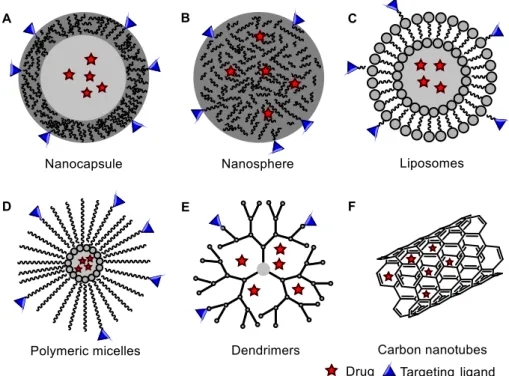
tried to realize the concept of the “magic bullet”, proposed by Paul Ehrlich almost a century ago.5 Complications arise because drugs have to overcome natural biological hurdles and most infections are localized. Nevertheless, by delivering pharmacologically active agents more effectively and more selectively, nanomedicines aim to improve the pharmacokinetic and pharmacodynamic properties of therapeutic molecules, enhancing efficacy while reducing toxicity.6 Many different nanostructures such as quantum dots, nanoparticles, cyclodextrin, dendrimers, liposomes and lipid-based nanocarriers, nanofibers, nanowires and carbon nanotubes have been developed and demonstrated increasing potential in diagnostics and therapeutics.7 However, high tissue accumulation of non-biodegradable nanocarriers as rendered some of them as not-so-popular therapeutic and diagnostic systems due to toxicity problems.8
Polymeric nanoparticles (PNPs) also play an important role in the “Room at the Bottom” and
this family may be considered amongst the most well studied nanomedicines to date.9These are solid colloidal particles generally varying in size from 10 to 1000 nm, although there is some degree of ambiguity regarding the upper size limit. In medical applications well-defined sizes are of great importance, as they play a crucial role in mediating biological effects and in vivo fate of the drug delivery system.10 Therefore to assure the highest potential for in vivo applications, PNPs of intermediate size (20 - 100 nm) have the highest potential, given their ability to circulate in the bloodstream for long periods of time as well as their biodistribution patterns.11
4
drug is confined and surrounded by a polymer membrane. A nanosphere, however, has a matrix-like structure consisting of the drug and the polymer uniformly dispersed.
Surface functionality is another critical parameter in the development of ideal PNPs for drug delivery systems (DDS). PNPs can be conjugated to a bio-specific ligand or coupled to macromolecules that could target the PNPs to the desired site of action.8,13 This allows efficient delivery of small-molecular-weight drugs, as well as macromolecules to target sites.14 Targeting the desired site of action not only increases the therapeutic efficiency of a drug, but also increases its cellular uptake through receptor-mediated endocytosis. The surface of PNPs can also be suitably modified to avoid uptake by
the body’s mononuclear phagocyte system (MPS) after opsonization, thereby increasing their circulation half-life in the body.15
The main advantages of nanomedicines are enhanced stability, controlled release, improved bioavailability, significant decreased toxicity, as well as improved therapeutic effects due to a greater drug fraction able to reach the target site.8 In addition, expensive potentially active substances could be applied efficiently in small amounts. In general, molecules susceptible to enzymatic (nuclease and protease) degradation, particularly protein, peptide and nucleic acid drugs, are better preserved when they are entrapped in nanocarriers.16
In drug delivery, the biodegradability and biocompatibility of the polymers are among the most important properties. A variety of materials have been used for the preparation of PNPs, such as proteins or other natural macromolecules, biodegradable polymers and non-biodegradable, but pharmaceutically acceptable polymers.17 To self-assemble these materials into PNPs, a variety of preparation techniques has been successfully developed.18
Figure 1 Types of nanocarriers for drug delivery systems. Polymeric nanoparticles: A, Nanocapsules and B, Nanospheres. C,
5
I.2 Preparation of Polymeric Nanoparticles
Polymeric nanoparticles have been synthesized by various methods depending on the needs of application and the physicochemical characteristics of the drug. The choice of the most suitable method plays a vital role in order to obtain PNPs with the desired properties for a particular application.19
Several preparation methods have been developed and can be divided into two groups, namely, those based on polymerization of monomers and those taking advantage of preformed polymers (Fig. 2).18 This methods can be further classified into two categories: two-step procedures involving the preparation of an emulsification system followed by formation of nanoparticles in the second step of the process and one-step procedures where emulsification is not required previous to the formation of nanoparticles.
In polymerization methods, the monomers are polymerized to form the encapsulating polymer. This process can be carried out in two ways, either as emulsion polymerization techniques or interfacial polymerization.9 Some drawbacks have been reported which have limited the use of polymerization methods for the synthesis of PNPs.9 Not only are most PNPs formed from slowly biodegradable or nonbiodegradable monomers, but also non-biocompatible byproducts may be generated with these methods. Toxic residues such as monomers and initiators may persist which require extensive purification work to result in a pharmaceutically acceptable product. Another challenge is the requirement for free-radical polymerization or UV light to trigger polymerization, which prevents the addition of proteins or peptides during polymerization. Considering the limitations of polymerization techniques, attention is focused on describing the methods involving preformed polymers, as many of the problems involved in the former method can be avoided.
Preformed polymers
Emulsification - solvent evaporation
Emulsification - solvent diffusion
Salting-out
Nanoprecipitation
Dialysis
Supercritical fluid technology
Polymerization of monomers
Emulsion
Mini Emulsion
Micro Emulsion
Interfacial Polymerization
Controlled/Living radical
6
I.2.1 Two-step procedures based on Emulsification
Colloidal delivery systems based on emulsions are widely used in food and pharmaceutical industries to encapsulate, protect, and deliver bioactive components.
The term emulsion is defined basically as a mixing of one liquid phase into another totally or partially immiscible, through the use of amphiphilic surface-active molecules (surfactants), which reduce the interfacial tension between the two liquids in order to achieve stability.
Emulsions can also be classified based on composition (oil, water, surfactants) or morphology.20 Generally, emulsions may be of the oil-in-water (o/w) or water-in-oil (w/o) types depending on whether the oil is dispersed as droplets in water, or vice versa. In addition, more complex systems such as (water-in-oil)-in-water (w/o/w) can also be obtained. Depending on the droplet size, the emulsion formed can be classified into three main categories: nanoemulsions, miniemulsions and macroemulsions (Fig. 3).20 In contrast to more common microscale emulsions, nanoemulsions have some interesting physical properties. For instance, nanoemulsions exhibit optical transparency, while microscale emulsions typically exhibit strong multiple scattering of visible light, and, as a result, have a white appearance.20
A common mistake reported in the scientific literature is to describe nanoemulsions as
“microemulsions”, thereby confusing an emulsion system with a thermodynamic phase.21 Microemulsions are not emulsions in the classical sense, but rather nanoscale self-assembled equilibrium phases (lyotropic phases). In contrast, nanoemulsions do not form spontaneously, as an external shear must be applied to rupture larger droplets into smaller ones. Although these two kinds of colloidal dispersion can be comprised of three simple components (oil, water and surfactants) and have many structural similarities, the distinguishing difference is not one of composition, but rather one of thermodynamics: microemulsions are thermodynamically stable, whereas nanoemulsions are not.21
In two-step emulsification/solvent removal methods the polymer organic solution is emulsified in an aqueous phase. Low- and high-energy emulsification techniques can be used to produce nanodroplets and consequently nanoparticles.18 In emulsion method the droplet formation step is fundamental because it determines the size and size distributions of the resulting PNPs. Polymer precipitation on preformed nanodroplets is achieved by removing the organic solvent by different
7
methods such as solvent evaporation, fast diffusion after dilution or salting out. A similarity between these techniques is the drug encapsulation process in which the drug is generally added in the polymer solution.9b
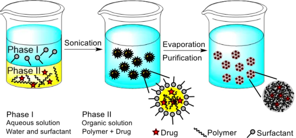
I.2.1.1 Emulsification-solvent evaporation
Solvent evaporation was the first method developed to prepare PNPs from a preformed polymer.22 In a pioneer work, Gurny et al. applied it successfully in the preparation of drug carriers from biocompatible polymers.23 In this method, the polymer is first dissolved in a volatile solvent (see Fig. 4). Dichloromethane and chloroform have been widely used in the past. However, due to their toxicity they have been replaced by ethyl acetate which displays a better toxicological profile and therefore more suitable for biomedical applications.24 The resulting organic solution is emulsified in the aqueous phase and the mixture is typically processed using a surfactant and high-speed homogenization or ultrasonication, yielding a dispersion of nanodroplets. Afterwards, a suspension of nanoparticles is formed by evaporation of the polymer solvent, which is allowed to diffuse through the continuous phase of the emulsion.24 The solvent is evaporated either by continuous magnetic stirring at room temperature or under reduced pressure, which is a slow process. After the solvent has evaporated, the solidified nanoparticles can be washed and collected by centrifugation, followed by freeze-drying for long term storage.25
The emulsification-solvent evaporation method has been widely applied to prepare PNPs with the desired characteristics by adjusting different experimental parameters. For example, poly(lactic acid) (PLA) PNPs with an average size around 200 nm were prepared by Zambaux et al. using dichloromethane and polyvinyl alcohol (PVA) as the solvent and stabilizing agent, respectively.26 The authors adopted the double-emulsion method (w/o/w) to prepare the PNPs and studied the influence of experimental parameters such as the preparation temperature, solvent evaporation method, internal aqueous phase volume, surfactant concentration and polymer molecular weight on the physicochemical
8
properties of the obtained PNPs. In another report, Bilati et al. studied the effect of the sonication process on the characteristics of poly(D,L-lactic-co-glycolic acid) (PLGA) nanocapsules prepared by the w/o/w solvent evaporation method.27 They concluded from their study that the duration of the second mixing step (leading to the w/o/w emulsion), had a greater influence on the final mean particle size than the first step (w/o emulsion). In order to study the effect of the polymer solvent on the PNP properties, Mainardes
et al. prepared PLGA nanoparticles applying either of two organic solvents (dichloromethane and ethyl acetate) as the dispersed phase.28 They demonstrated that the size of the PNPs prepared with dichloromethane was larger than those prepared with ethyl acetate. Manchanda and coworkers formulated PLGA nanoparticles using methanol and dichloromethane (1:2 v/v) as the solvent system and PVA as the stabilizing agent.29 It was evidenced that an increase in polymer amount leads to larger nanoparticles. This was attributed to an increase in the viscous resistance of the emulsion mixture, thereby absorbing the agitation energy which in turn leads to reduction in shear stress resulting in droplets with larger size. When the concentration of PVA was taken into account, the particle size decreased from 159 nm (1 % PVA) to 113 nm (5 % PVA) due to an improvement in the emulsification process. PLGA and PLA nanoparticles were prepared by Budhian et al. by employing dichloromethane as the solvent and PVA as the stabilizing agent.30 It was shown that decreasing the organic solvent volume resulted generally in a decrease in particle size. The method used to remove the organic solvent can also affect the final properties of the PNPs prepared by the solvent evaporation method. This effect was studied by Ashjari et al. who prepared magnetic/cis-Diamminedichloroplatinum (II) (cisplatin) - loaded PLGA nanocapsules through a w/o/w double emulsion-solvent evaporation technique.31 They observed a change in the morphology and a decrease in the particle size of nanocapsules when slow evaporation at room temperature was used in comparison with evaporation at reduced pressure (Fig. 5). An extensive study was reported by Khoee et al. on the physicochemical properties of cisplatin loaded polybutyladipate (PBA) nanoparticles prepared from w/o/w emulsion.32 The obtained PNPs showed a size dependence on polymer concentration, decreasing when the polymer concentration decreases. The size of PNPs was also influenced by other process parameters such as volume of oil phase, power of sonication and drug concentration in the internal water phase. Ethyl cellulose (EC) nanospheres were prepared by Wachsmann et al. to study the influence of surface properties on the accumulation
Figure 5 Nanocapsules with different morphologies prepared by Ashjari et al. Core-shell or half-moon morphology was
9
selectivity of nanoparticles in murine experimental colitis.33 For this purpose, PNPs of similar sizes (212 - 258 nm) were prepared using different surfactants (polysorbate 20 (P20), SDS, sodium cholate (SC), cetyltrimethylammonium bromide (CTAB) and PVA). It was shown that the accumulation of PNPs in the inflamed areas as well as in the healthy tissue was dependent on surfactant type. The targeting pattern for P20 and CTAB particles showed a distinctly increased accumulation in the inflamed tissue compared to SDS particles with slightly higher values for P20. However, as CTAB particles also exhibited a significantly higher accumulation in healthy tissue compared to the other two preparations, the highest selectivity was obtained with P20 particles. More recently, Barba and coworkers developed a preparation technique based on multiple emulsion system to produce polymeric nano and microparticles.34 Different polymers, such as polyesters (PCL, PLA and poly(D,L-lactide-co -caprolactone) 70:30 (PLC)) and poly(methylmethacrylate-acrylic acid) (Poly(MMA-AA)) 73/27, loaded with different model molecules, were explored. Depending on type of polymer and, consequently, on solvent used for solubilization (dichloromethane), polyester-based nanoparticles with round shape and smooth surface were obtained (Fig. 6). Conversely, polyacrylates yielded microparticles with high porosity and lower yield of encapsulation, due to the presence of hydrophilic co-solvents (ethanol and isopropyl alcohol) that caused an easy coalescence between the oil and the water phases (Fig. 6).
Over the past decade, methods for emulsion preparation with nanoscale droplets have been considerably developed due to the technological improvement of emulsification devices, which has prompted the development of the solvent evaporation technique. Although, this method is simple and versatile, it can only be applied mainly to liposoluble drugs, it is time consuming and there is also the possibility of nanoparticle coalescence during evaporation. In addition, for scale-up production alternative methods using low-energy requirements in homogenization are preferred.
10
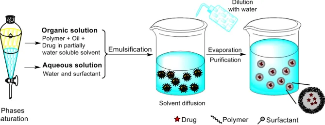
I.2.1.2 Emulsification-solvent diffusion
The emulsification-solvent diffusion method was first introduced by Leroux et al..35 It consists in the formation of a conventional o/w emulsion between a partially water-miscible solvent containing the polymer and the drug, and an aqueous solution, containing a surfactant (Fig. 7). For the success of this method, the polymer solvent and water are mutually saturated at room temperature to ensure the initial thermodynamic equilibrium of both liquids. The subsequent dilution with an extensive amount of water, induces solvent diffusion from the dispersed droplets into the external phase, resulting in the formation of colloidal particles. Such diffusion process is milder than the direct evaporation of the organic solvent from the nanodroplets. In contrast with methods based on solvent evaporation, in this tecnique the droplet size decreases suddenly in a millisecond time scale during solvent diffusion.36 Generally, nanospheres are produced by this method but nanocapsules can be obtained just by adding a small amount of oil, for example miglyol, in the organic phase. Finally, depending on its boiling point, the solvent can be eliminated by evaporation or filtration.
Several formulation parameters can affect the size of the obtained PNPs by solvent diffusion. For example, it has been shown that most properties of nanocapsules are determined at the emulsification step.36 Guinebretière et al. prepared PCL nanocapsules using ethyl acetate and PVA as the solvent and stabilizing agent, respectively. They observed that the final size of the nanocapsules was influenced by the concentration of oil in the organic phase, volume of the solvent in the emulsion and nature and concentration of the surfactant.37 Also, the thickness of the nanocapsule was linked to the polymer concentration in the organic phase. In another study, PLA nanospheres were prepared by Quintanar-Guerrero et al. by employing propylene carbonate as the solvent and PVA or Pluronic F68 as surfactants.38 It was evidenced that high concentrations of polymer leads to larger particle sizes with an increase in the polydispersity index. On the other hand, an increase in stirring rate and in the surfactant concentration were found to reduce moderately the size of the PNPs. Similar results were obtained by
11
Trimaille et al. who prepared PLA nanoparticles using ethyl acetate and Pluronic F68 as the solvent and stabilizing agent, respectively.39 It was shown that increasing the PLA concentration resulted in an increase of the mean particle size from approximately 260 to 530 nm (Fig. 8). The same experimental parameters were applied by Surassmo et al. for the preparation PCL nanocapsules.40 The authors observed that an increase of the surfactant amount resulted in a decrease of the mean particle size. Although, it seems that above some level further significant size reduction is no longer possible as the excess surfactant remains in the continuous phase, and does not play any significantly role in the emulsification. In a different approach, Colombo and coworkers designed with chemical engineering equipment, a pilot plant to study the process of emulsification-diffusion.41 It was demonstrated that the agitation time, stirrer type and rotational speed were the most important parameters in the emulsification
step. In contrast, during the dilution step, the agitation has no influence on the final size distribution. Only sufficient mixing is needed in order to homogenize the mixture. In another report, Sahana and coworkers used a modified emulsification-diffusion method by using a non-saturated organic solvent and water. In their work they studied the influence of the type of solvent and surfactant on particle size distribution and entrapment efficiency of PLGA nanoparticles.42 When didodecyldimethylammonium bromide (DMAB) and PVA were compared, the former gave the smallest particles but with the lowest encapsulation efficiency, regardless of the type of organic solvent. For both stabilizers, dichloromethane in combination with ethyl acetate yielded the highest entrapment efficiency. Similar results were obtained by Jain et al. who compared the influence of different stabilizers (Pluronic F68, DMAB and PVA) in the preparation of PLGA PNPs from ethyl acetate organic solutions.43 DMAB, when used as surfactant, led to smaller particle sizes as compared to PVA, but on the other hand, PVA produced particles with higher entrapment efficiency (Table 1). The authors also studied the effect of droplet size reduction by using both homogenization and sonication. It was demonstrated that sonication resulted in smaller particles (165 nm) as compared to homogenization (around 225 nm). Hallouard et al. studied the oil nature on the physicochemical characteristics of nanocapsules.44 For PCL-mPEG diblock copolymer, it was shown that the nature of the oil had no influence on the encapsulation rate. Though, it influenced the particle size and polydispersity, with macroglycerides, appearing to be the lipid
12
structure best suited to obtain the smallest monodisperse nanocapsules. More recently, Musazzi and coworkers prepared PNPs by a modified solvent-diffusion method without surfactant from a combination of PLGA and PCL-PEG copolymer.45 The latter was used due to its amphiphilic nature, which stabilizes the nanodroplets surface in the emulsification step.
Table 1 Effect of stabilizer type on particle size, polydispersity index (PDI) and entrapment efficiency, for PLGA nanoparticles
prepared by Jain et al.43
Surfactants Size (nm) Polydispersity Index (PDI)
Entrapment
efficiency (%)
2% PVA 165.6 0.085 86.20 1% DMAB 120.0 0.015 15.30 Pluronic F68 130.0 0.136 38.56
The experimental parameters related to the solvent diffusion step do not seem to affect particle size.46 Free solvent diffusion is guaranteed as long as the organic solvent solubility condition is satisfied. This could explain some contradictory results reported by Song et al. in which the highest particle size was obtained at the lowest volume of water for dilution.47 In their study, the lowest volumes of water used did not lead to complete solubility of the organic solvent. In addition, difficult solvent diffusion can be expected due to the barrier effect of the stabilizing agent on the emulsion droplet. This could also explain the results reported by Kwon et al. where the size of submicron particles prepared using PVA as a stabilizing agent is influenced by the temperature of the dilution water.48 It was shown that the particle size decreased as the temperature of dilution water increases. In this case, reducing the viscosity of the external phase promoted solvent diffusion, and consequently a decrease in particle size.
The emulsification-diffusion technique presents clear advantages such as high yields, easy in scaling-up, no need for high-pressure homogenizers or ultrasonication, batch-to-batch reproducibility and generally good encapsulation efficiencies.49 However, there are also disadvantages. For instance, high volumes of water to be eliminated from the suspension and possible leakage of water soluble drugs into the external phase throughout emulsification step.9b
I.2.1.3 Emulsification
–
reverse salting-out
Ouzo-13
effect51, without employing any high-shear forces. The miscibility of acetone and water is reduced by saturating the aqueous phase, which allows the formation of an o/w emulsion from the otherwise miscible phases. A reverse salting-out effect is obtained by dilution of the formed o/w emulsion with an excess of water to promote the diffusion of acetone into the aqueous phase, which leads to the precipitation of the polymer dissolved in the emulsified nanodroplets. The remaining polymer solvent and salting-out agent are eliminated by cross-flow filtration.52 The condition of complete miscibility between the organic solvent and water is not essential, but simplifies the execution process.50 If it is not the case, there is a need for a greater water/solvent volume ratio during the formation of the nanoparticles.
PNPs have been prepared successfully by employing the emulsification-reverse salting-out method with several polymers, solvents and salting-out agents. The only condition which should be met is the need for a two-phase system in the presence of the salting-out agent. In a typical process carried out by Allémann et al. the influence of several process parameters on particle size was studied.53 Eudragit® S PNPs were first prepared using acetone and magnesium chloride as the solvent and salting-out agent, respectively. In addition, PVA was also added to the aqueous phase as a viscosity-increasing agent and emulsion stabilizer. It was shown that by changing the molecular weight and concentration of PVA in the external phase, the size of the PNPs could be controlled within a wide range (186 – 1130 nm). Additionally, an increase in stirring rate also allowed a slight decrease in particle size. PNPs with a particle size of 200 to 500 nm were obtained by varying the polymer concentration or the internal/external phase ratio. When Eudragit® E was studied, magnesium acetate was selected as salting-out agent due to the solubility of the polymer in acidic medium. For PLA PNPs when an acidic aqueous phase (magnesium chloride) was changed to a basic one (magnesium acetate), a slight increase on the particle size was observed (228 to 247 nm). Zweers and coworkers also concluded in their studies that the particle size can be best controlled by adjusting the polymer concentration in the external phase.54 In another report, Song et al. prepared PLGA nanoparticles by employing sodium chloride as the
14
out agent instead of magnesium chloride or magnesium acetate.55 Although acetone is the most commonly used organic solvent, other solvents have been reported. For example, Konan et al. prepared PLGA and PLA nanoparticles with a mean particle size below 200 nm using tetrahydrofuran (THF) as the polymer solvent.52
The main advantages of the salting-out method are the avoidance of chlorinated solvents, which are hazardous to the environment as well as to the physiological systems. The greatest disadvantages are the exclusive application in encapsulating lipophilic drugs and the need for intensive purification steps due to the use of salts. The latter can be responsible for the few reports that have been published in the recent years quoting the salting-out method.
I.2.2 One-step procedures
I.2.2.1 Nanoprecipitation or solvent displacement method
The nanoprecipitation method, also called solvent displacement, was firstly developed by Fessi
et al.56 The basic principle of this technique is based on the interfacial deposition of a polymer after displacement of the organic solvent from a lipophilic solution to the aqueous phase (Fig. 10).9 The polymer is dissolved in a water-miscible solvent of intermediate polarity and this solution is added into a stirred aqueous solution in one shot, stepwise, dropwise or by controlled addition rate.46 Due to the
15
fast spontaneous diffusion of the polymer solution into the aqueous phase, the nanoparticles form instantaneously in an attempt to avoid the water molecules. This process appears to be governed by the Marangoni effect57, wherein a decrease in the interfacial tension between the two phases, increases the surface area due to the rapid diffusion and leads to formation of small droplets of organic solvent. As the solvent diffuses out from the nanodroplets, the polymer precipitates in the form of nanocapsules or nanospheres. In general, the organic phase is added to the aqueous phase but the protocol could also be reversed without compromising the nanoparticle formation. The most common used organic solvent is acetone, because it is miscible with water and easy to remove by evaporation. Though, ethanol and binary solvent blends, such as acetone with a small amount of water, ethanol or methanol can also be used.58 It is also possible to use either two organic phases or two aqueous phases as long as solubility, insolubility and miscibility conditions are satisfied.46 Usually, surfactants could be included in the process to guarantee the stability of the colloidal suspension, but their presence is not required to ensure formation of nanoparticles. The obtained nanoparticles are typically characterized by a well-defined size (around 200 nm in diameter) and a narrow size distribution, which is better than those produced by the emulsification solvent evaporation procedure.
The key variables that are conditioning the final nanoparticle properties are those related with the experimental design. By carefully adjusting the nature and concentration of the components, organic phase/aqueous phase ratio, organic phase injection rate, fluid dynamics and mixing speed, it is possible to control the PNP physicochemical properties. For example, increasing the polymer concentration or the polymer molecular weight generally results in an increase on particle size. These findings are explained by a higher organic phase viscosity, which hinders solvent diffusion and results in larger nanodroplets.59 In a typical process carried out by Chancón et al. the polymer concentration, organic phase injection rate and needle gauge, were identified as the principal size determinants on the preparation of PLGA nanoparticles.60 The smallest particles (46 nm) were obtained by using the lowest polymer concentration, the highest injection rate and the lowest needle gauge. Similar results were obtained by Simsek and coworkers in the preparation of PLGA-b-PEG nanoparticles.61 The average hydrodynamic diameter of these particles could be controlled between 30 to 172 nm by the choice of polymer concentration and PEG content. Chorny et al. prepared nanospheres of PLA by employing acetone and dichloromethane (39:1 v/v) as the solvent system and Pluronic F-68 as the stabilizing agent.59 It was demonstrated that replacing acetone in the organic phase by equal volumes of ethanol resulted in particle size reduction from 115 to 70 nm. The authors also observed that the organic solvent evaporation rate and an increase of aqueous phase volume had no influence on the nanosphere size. In another report, Dong et al. prepared PEG-PLA nanoparticles by using acetonitrile as the solvent and Pluronic F-68 as the surfactant.62 They found that the surfactant concentration slightly influenced the nanoparticle size. Likewise, a slight smaller size were obtained by increasing the organic phase volume. PNPs were prepared by Özcan and coworkers by a modified nanoprecipitation method using several
16
tetrahydrofuran at 30 ˚C and this solution was added to an aqueous phase by dripping without the presence of any surfactant. As shown in Figure 11, the polymer concentration in the organic phase strongly influenced the mean diameter of the nonpegylated nanoparticles. In contrast, very small PNPs were obtained for the PBLG-PEG copolymer and almost no influence of the concentration was observed
due to the amphiphilic nature of this copolymer. The mixing rate is also important in the preparation of PNPs by nanoprecipitation. Asadi et al. prepared nanoparticles from PLA-PEG-PLA tri-block copolymer.64 It was shown that increasing the mixing rate led to a decrease in particle size. Furthermore, smaller particles were also obtained by decreasing organic/aqueous phase ratio due to a better dispersion of the solvent and faster diffusion rate. This is a conflicting result to that obtained by Dong et al.62 Nevertheless, the polymer nature and the experimental parameters are different and could have influenced the results. More recently, Bukhari et al. studied the effect of solvent/non-solvent dispersion medium on the preparation of polystyrene (PS) nanoparticles.65 Chloroform and tetrahydrofuran were explored as solvents for polystyrene and several dispersion phases (methanol, chloroform, acetone and water) were investigated. The results revealed that the combination of tetrahydrofuran with acetone and water, as well as chloroform with methanol and acetone leads to the formation of nanoparticles. They concluded from their study that the dielectric constant difference plus the affinity of the solvent for the non-solvent were responsible for nanoprecipitation. In addition, the morphology was remarkably dependent on the nature of the dispersion phase (Fig. 12).
Overall, the challenge in nanoprecipitation is to find a suitable drug/polymer/solvent/non-solvent system, which allows successful nanoparticle production and drug encapsulation. Though, this method is widely used due to its simplicity, rapidity and reproducibility. One of the difficulties is the mixing process during nanoprecipitation. A microfluidic platform66 could be a promising tool for the controlled synthesis of PNPs, where the hydrodynamic flow ensures a fast and tunable mixing of solvent/non-solvent in the microfluidic channels. Other recent development is the advent of automation to nanoprecipitation with high throughput experimentation (pipetting robot, inkjet printing).67 Another drawback is the poor encapsulation efficacy of hydrophilic drugs, because the drug can diffuse to the
Figure 11 Influence of polymer concentration in the organic phase on the mean diameter of PNPs. Abbreviation: Bnz,
17
aqueous phase during polymer precipitation. By modifying the solubility of the drug through changes in the pH or varying the solvent composition are among other means to improve encapsulation efficiency.68
I.2.2.2 Dialysis
The dialysis method has been applied successfully in the preparation of small PNPs with narrow size distribution.69 It is governed by a mechanism resembling that previously described for the nanoprecipitation technique, but with a slightly different experimental setup. In this method, dialysis tubes or semipermeable membranes with a suitable molecular weight cut-off (MWCO) are used as a physical barrier for the polymer.9a Generally, the polymer is dissolved in an organic solvent, placed inside the dialysis membrane and dialyzed against a non-solvent (Fig. 13). Basic prerequisites are the miscibility of the solvents and the existence of dilute polymer solutions. The displacement of the solvent inside the membrane causes the mixture to be progressively less able to dissolve the polymer. In addition, an increase in interfacial tension results in polymer aggregation and leads to the formation of a colloidal suspension of nanoparticles. Although dialysis is a simple and common method, the large
Figure 12 FESEM micrographs of polystyrene PNPs prepared by nanoprecipitation. Polymer solvent: tetrahydrofuran.
Non-solvent: C, Acetone; D, water and Pluronic-F68. [Adapted from ref. 65]
18
volume of counter dialyzing medium could arouse a premature release of the nanoparticle payload due to the long duration of the process.
The morphology and particle size distribution of the obtained PNPs can be modulated by several experimental parameters, such as solvent/non-solvent pair, dialysis MWCO, the temperature at which the procedure is carried out, polymer concentration and speed of solvent mixing.70 The influence of the solvent was examined in a work of Akagi et al. in which either of four organic solvents (dimethylsulfoxide (DMSO), dimethylformamide (DMF), dimethylacetamide (DMAc), N -methyl-2-pyrrolidinone (NMPy)) were applied as the polymer solvent to prepare PNPs based on poly(γ-glutamic acid) (PGGA).71 They concluded that the particles prepared with DMSO were smaller, with a narrower size distribution than those prepared with NMPy. A similar approach was used by Jeong et al. to prepare PLGA nanoparticles from DMAc, DMF, DMSO and acetone as the polymer solvent.72 The size of the PNPs prepared from DMAc, DMF and DMSO were in the range of 200 – 300 nm and not significantly different. On the other hand, acetone yielded larger particles with a mean size of 642 nm (Fig. 14). This change in particle size could be explained by the difference in solvent viscosity, miscibility with water and in the solubility behavior of the polymer. In another report, Chronopoulou et al. studied the influence
of several experimental parameters on the size and morphology of nanoparticles prepared from natural and synthetic polymers.70 For poly(methyl methacrylate) (PMMA) nanoparticles, a linear correlation between polymer concentration and the size of the nanospheres was determined. The same behavior was observed for poly(phenyl acetylene) (PPA) nanospheres. In addition, at low concentrations, PPA nanoparticles with smaller diameters were obtained at low temperature than those obtained at room temperature. The influence of the MWCO was also studied for this synthetic polymer. It was shown, that reducing the membrane MWCO induces a decrease of the mean particle size. The biopolymers under study also followed the same trend. This is due to a decrease in the mixing rate of the solvents, thus favoring thermodynamic factors over kinetic ones. Different morphologies were observed for hyaluronic acid based nanostructures by changing the chemical properties of the solvent/nonsolvent pair (Fig. 15).
Figure 14 SEM images of PLGA 50:50 nanoparticles prepared by Jeong et al. a) DMAc or b) acetone as the polymer solvent.