Diana Alves Afonso
T
HE IMPACT OF CHOROID PLEXUS DERIVED FACTORS IN THE
SUBVENTRICULAR ZONE NEURAL PROGENITOR CELLS NICHE
Dissertação de Mestrado
Mestrado em Ciências da Saúde
Trabalho efetuado sob a orientação do:
Prof. Doutor João Carlos Sousa
e da:
Prof. Doutora Fernanda Marques
DECLARAÇÃO Nome: Diana Alves Afonso
Endereço eletrónico: [email protected] Telefone: 936845708
Número do Cartão de Cidadão: 14396751
Título da dissertação:
The impact of choroid plexus derived factors in the subventricular zone neural progenitor cells niche
Orientadores:
Prof. Doutor João Carlos Sousa
Prof. Doutora Fernanda Marques
Ano de conclusão: 2016
Designação do Mestrado: Ciências da Saúde
É AUTORIZADA A REPRODUÇÃO INTEGRAL DESTA DISSERTAÇÃO APENAS PARA EFEITOS DE INVESTIGAÇÃO, MEDIANTE DECLARAÇÃO ESCRITA DO INTERESSADO, QUE A TAL SE COMPROMETE.
Universidade do Minho, 31 de Outubro de 2016 Assinatura:
Funding
“O que dá o verdadeiro sentido ao encontro é a busca e que é preciso andar muito para alcançar o que está perto” José Saramago
Agradecimentos
O trabalho realizado ao longo desta tese foi o esforço conjunto de muitos pessoas a quem gostaria de deixar umas pequenas palavras de agradecimento.
Gostaria de começar por agradecer ao João. Obrigada por teres sido meu orientador, pelos conselhos e pelas criticas que me fizeram crescer não só de forma profissional, mas também pessoal. Obrigada também por me ensinares que a melhor maneira de lidar com as adversidades é fazê-lo com um sorriso na cara.
À Fernanda, obrigada pela tua orientação e disponibilidade constantes. Obrigada pelas sugestões, discussões e apoio dado ao longo deste ano que foram essenciais para me guiar neste trajeto. Levo comigo tudo o que me ensinaste e todos os conhecimentos que me transmitiste.
À Ana, porque “Rocky e a amiga” já diz bastante, mas não o suficiente, obrigada por teres sempre tempo para ajudar ou para descontrair. Fico feliz de acabar esta tese sabendo que levo daqui alguém com quem vou poder contar.
À Catarina, mini chefe, obrigada pela ajuda indispensável que foste ao longo desta tese, pelas discussões e pelo que me ensinaste. Obrigada também, pelos momentos de descontração e boa disposição proporcionadas em conjunto com os nossos companheiros de mesa, Marco e Dinis.
Ao António Salgado, ao Fábio e à Bárbara pela ajuda que foram com a cultura de células. À Ana Pereira e ao Luís Jacinto pelo que me ensinaram.
Ao Master Gang, por partilharmos juntos as graças e desgraças deste ano. Um especial obrigado à Joana e à Sara pelo apoio e por perceberem que “sometimes you just need to dance it out”.
Aos meus Nerds bioquímicos, Ângela, Augusto, Carla, Cristina, Daniela, Liliana e Patrícia por provarem que mesmo à distância e com pouco compatibilidade de agenda conseguiram apoiar-me sempre. É muito bom ter amigos como vocês!
À Ariana e à Rita um pedido de desculpas por todas as vezes que troquei a vossa companhia pela de outros mamíferos menos evoluídos filogeneticamente.
Aos meus pais, ao Filipe e à minha família porque sem eles nada disto era possível. Obrigada que mesmo não compreendendo tenham aceitado todos os meus momentos de ausência. Vocês sabem que só o fiz para dar o meu melhor porque foi isso que vocês me ensinaram a fazer.
The impact of choroid plexus derived factors in the subventricular zone neural
progenitor cells niche
Abstract
Neurogenesis is the complex process of generating new neurons from their progenitor cells. It occurs both in developmental stages and in adulthood (in the latter case mainly in two brain areas, the subventricular zone (SVZ) and the subgranular zone). Neural stem cells (NSCs) and their progeny are regulated by a proper balance of intrinsic and extrinsic factors. One source of extrinsic factors is the choroid plexuses (CPs), located inside the cerebral ventricles. CPs play active roles in barrier function, secretion and as major regulator of the cerebrospinal fluid (CSF) composition. The SVZ neurogenic niche and the lateral ventricles are in close proximity and NSCs project their apical process into the lateral ventricles being under direct influence of CSF signaling. Thereby, it is plausible to assume that CP, through the modulation of CSF composition, can impact neurogenesis at embryonic and adult SVZ. Thus, the aim of this master thesis was to provide new insights on how molecules secreted by the CP affect the SVZ neurogenic niche. For this purpose, we selected two candidate proteins to study their hypothetic effect in SVZ cells: amphiregulin (AREG) and insulin growth factor binding protein 2 (IGFBP2), both known to be secreted by the CP towards the CSF. For this we used two in vitro techniques, a clonal assay and a differentiation assay, allowing us to study the impact of these proteins in proliferation and differentiation of human neuro progenitor cells. We also evaluated the effects of each protein in the SVZ of adult rats in terms of proliferation, cell cycle index and formation of new immature neurons. Moreover, as an additional aim, we intended to analyze the CP in key milestones of development to determine the temporal changes in the CP transcriptome and then correlate it with developmental changes known to occur in the SVZ cell population. For the transcriptome analysis, we extracted RNA from CP of animals in postnatal day 1, 4, 7, 10 and 60 and sequenced it by RNA-Seq.
Our results indicate that AREG increases cell proliferation associated with a superior number of cells reentering the cell cycle and increases the number of newborn neurons. Regarding the experiments performed with IGFBP2 they indicate that this protein can increase proliferation rate and the number of cells reentering the cell cycle. This is accompanied with an augment in the number of newly born neuroblasts.
In summary, the data presented here provides novel insights for two factors, secreted by the CP, that are playing a role in the modulation of SVZ neurogenic niche.
Impacto de fatores secretados pelo plexo coroide no nicho de células
neuroprogenitoras da região subventricular
Resumo
A neurogénese é um processo complexo que leva à formação de novos neurónios a partir de células progenitoras. Este processo acontece tanto em estadios de desenvolvimento embrionário como no estado adulto (neste caso ocorre maioritariamente em duas áreas do cérebro, a região subventricular e a região subgranular). As células neuronais estaminais e a sua descendência são reguladas pelo balanço adequado de fatores intrínsecos e extrínsecos. Uma das fontes de fatores extrínsecos é o plexo coroide, localizado no interior dos ventrículos cerebrais. O plexo coroide tem um papel ativo em funções de barreira e secreção, bem como na modulação da composição do liquido cefalo-raquidiano (LCR). O nicho neurogénico da região subventricular e os ventrículos laterais são estruturas próximas e as células neuronais estaminais projetam o seu processo apical para o ventrículo lateral, estando desta forma sob a influência direta da sinalização do LCR. Assim, é plausível assumir que o plexo coróide, através da modulação da composição do LCR, consegue modular a neurogénese na região subventricular tanto no estado embrionário como no adulto.
Desta forma, nesta tese de mestrado foi objetivo explorar de que forma moléculas secretadas pelo plexo coroide afetam o nicho neurogénico da região subventricular. Para tal, selecionámos duas proteínas para estudar o seu potencial impacto nas células da região subventricular: a anfiregulina (AREG) e a proteína de ligação 2 ao fator de crescimento insulina (IGFBP2), ambas secretadas pelo plexo coróide para o LCR. Para este estudo aplicamos duas técnicas in vitro, um ensaio clonal e um ensaio de diferenciação, que nos permitiram avaliar o efeito das proteínas na proliferação e diferenciação de células neuronais progenitoras humanas. Avaliamos também, in vivo, o efeito de cada proteína na região subventricular de ratos adultos em termos de proliferação, índice do ciclo celular e formação de novos neurónios imaturos. Como outro objetivo do trabalho propusemo-nos ainda a analisar as alterações temporais do transcriptoma do plexo coróide em alturas específicas do desenvolvimento, para posteriormente as correlacionar com alterações que ocorrem na população de células da SVZ. Para a análise do transcriptoma, extraímos RNA do CP de animais no dia pós-natal 1, 4, 7, 10 e 60 e sequenciámo-lo por RNA-Seq.
Resultados similares foram observados quando testámos os efeitos da IGFBP2, uma vez que observámos um aumento da proliferação acompanhado de um número superior de células a reentrarem no ciclo celular e no número de novos neuroblastos.
Em suma, os dados apresentados aqui demonstram o papel desempenhado por dois fatores secretados pelo plexo coróide na modulação do nicho neurogénico da região subventricular.
Funding ... iii
Abstract... ix
Resumo... xi
Table of contents ... xiii
List of abbreviations ... xvii
List of figures ... xix
List of tables ... xxi
1. Introduction ... 1
1.1. Neurogenesis ... 3
1.2. Embryonic neural stem cells ... 3
1.2.1. The VZ microenvironment ... 4
1.2.2. Cellular features of RGCs ... 5
1.2.3. Extrinsic regulation of embryonic VZ-SVZ ... 6
1.3. Adult neurogenesis ... 8
1.3.1. Adult SVZ ... 9
1.3.2. Cellular composition and structural organization of the aSVZ ... 9
Type E cells ... 10
Type B cells ... 11
Type C cells ... 11
Type A cells ... 12
1.3.3. Extrinsic regulation of the adult SVZ ... 12
1.4. Functional relevance of neurogenesis ...14
1.5. Choroid plexus ...14
1.5.1. Choroid plexus-CSF impact in embryonic neurogenesis ... 16
1.5.2. Choroid plexus-CSF impact in adult SVZ ... 17
2. Aims ... 19
3. Materials and methods ... 23
3.1. Ethics statement ...25
3.2. Animals ...25
3.3. Study the impact of CP-derived proteins in human neural progenitor cells. ...25
3.3.1. Clonal assay ... 26
3.3.2. Differentiation assay ... 26
3.3.3. Immunocytochemistry for BrdU and βIII-Tubulin ... 27
3.4. Study the impact of CP-derived proteins in the SVZ cell population ...27
3.4.1. Surgeries ... 28
3.4.2. BrdU and proteins administration ... 28
3.4.3. BrdU, Ki67 and Doublecortin immunohistochemistry ... 29
3.4.4. Confocal imaging and quantitative analysis... 30
3.5. Transcriptome analysis of CP in different stages of development ...31
3.5.1. CP collection ... 31
3.5.2. RNA extraction and quality analysis ... 31
3.5.3. Library preparation ... 32
3.5.4. RNA sequencing ... 33
3.5.5. Data analysis and interpretation ... 33
3.6. Statistical analysis ...33
4. Results ... 35
4.1. Impact of AREG in neurogenesis ...37
4.1.1.1. Clonal assay ... 37
4.1.1.2. Differentiation assay ... 38
4.1.2. Does AREG have an effect in the adult rat SVZ? ... 40
4.1.2.1. Analysis of cell proliferation rates along the SVZ ... 40
4.1.2.2. Analysis of cell cycle index along the SVZ ... 44
4.1.2.3. Analysis of new neuroblasts in the SVZ ... 45
4.2. Impact of IGFBP2 in neurogenesis ...47
4.2.1. IGFBP2 can impact growth of hNPCs? ... 47
4.2.1.1. Clonal assay ... 47
4.2.1.2. Differentiation assay ... 48
4.2.2. IGFBP2 impact in SVZ neurogenesis ... 50
4.2.2.1. Effects of IGFBP2 in cell proliferation in the SVZ... 50
4.2.2.2. Effects of IGFBP2 in cell cycle index ... 54
4.2.2.3. Effects of IGFBP2 in the number of newly formed neuroblasts at the SVZ ... 55
4.3. Analysis of CPs transcriptome in key developmental stages ...57
4.3.1. RNA quality control ... 57
4.3.2. cDNA library quality control ... 58
4.3.3. Quality control of RNA seq data: MultiQC report ... 59
4.3.3.1. Per base sequence quality ... 59
4.3.3.2. Per sequence quality score... 60
4.3.3.3. Per base N content ... 61
5. Discussion ... 63
5.1. Amphiregulin impact in the neural stem cells modulation ...66
5.1.1. AREG enhances proliferation ... 66
5.1.3. AREG increase the number of immature neurons ... 68
5.2. IGFBP2 impact in neural stem cells modulation ...69
5.2.1. IGFBP2 enhances cells proliferation ... 70
5.2.2. IGFBP2 alters cell cycle index at the SVZ ... 71
5.2.3. IGFBP2 increases the number of newborn neurons ... 71
5.3. Choroid plexus transcriptome analysis in key milestones of development ...72
6. Conclusion and future perspectives ... 73
7. References ... 79
List of abbreviations
A
aCSF- Artificial cerebrospinal fluid AREG- Amphiregulin
Ascl1- achaete-scute complex homolog 1 aSVZ- Adult subventricular zone
B
BDNF- Brain derived neurotrophic factor bHLH- Basic helix loop helix
BLBP- Brain lipid binding protein BMPs- Bone morphogenic proteins BrdU- 5-bromo-2’-deoxyuridine C
cDNA- Complementary deoxyribonucleic acid CNS- Central nervous system
CPs- Choroid plexuses CSF- Cerebrospinal fluid D DAPI- 4’,6-diamidino-2-phenylindole DCX- Doublecortin Dlx2- Distal-less homeobox E
EGF- Epidermal growth factor
EGFR- Epidermal growth factor receptor ERK- Extracellular signal-regulated kinases Ex- Embryonic day x
F
FACS- Fluorescence-activated cell sorting FBS- Foetal bovine serum
FGF- Fibroblast growth factor FITC- Fluorescein isothiocyanate G
GDNF- Glial cell derived neurotrophic factor GFAP- Glial fibrillary acidic protein
GLAST- Glutamate aspartate transport H
HCl- Hydrochlorid acid I
ICV- Intracerebroventricular injection IGFs- Insulin growth factors
IGFBP2- Insulin growth factor binding protein 2 IGFR- Insulin growth factor receptor
IL- Interleukin
IPCs- Intermediate progenitor cells J
L
LVs- Lateral ventricles M
MAP- Microtubule associated protein MAPK- Mitogen activated protein kinase N
NECs- Neuroepithelial cells Ngn- Neurogenin
NICD- Notch intracellular domain NPC- Neural progenitor cells NSCs- Neural stem cells NT- Neurotrophin O
OBs- Olfactory bulbs P
PBS- Phosphate buffered saline
PBS-T- Phosphate buffered saline-Triton PCR- Polymerase chain reaction
PEDF- Pigment epithelium derived factor PFA- Paraformaldehyde
Pi3K- Phosphoinositide 3-kinase Prom1- Prominin 1
PSA-NCAM- polysialylated neural cell adhesion molecule
Px- Postnatal day
Q
qNSCs- Quiescent neural stem cells R
RGCs- Radial glial cells
RMS- Rostral migratory stream RNA- Ribonucleic acid
RQI- RNA quality índex RT- Room temperature
RT-PCR- Real time- polymerase chain reaction S
SEM- Standard error of the mean SDF- Stromal derived factor Shh- Sonic hedgehog SVZ- Subventricular zone T
TAPs- Transit amplifying cells TGF- Transforming growth factor TNF- Tumor necrosis factor V
List of figures
Figure 1: Representation of the cellular components of the embryonic neurogenic niche. ... 4
Figure 2: Embryonic origin of neural stem cells of adult subventricular zone.. ... 9
Figure 3: The structural and cellular organization of the adult subventricular zone. ... 10
Figure 4: Structural organization of the choroid plexus. ... 15
Figure 5: Schematic representation of clonal assay. ... 26
Figure 6: Schematic representation of differentiation assay. ... 26
Figure 7: Schematic representation of protein administration procedure and postmortem analysis. ... 29
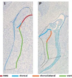
Figure 8: Division of the subventricular zone along the dorsal-ventral axes. ... 31
Figure 9: Schematic representation of total RNA extraction protocol using SPLIT RNA Extraction Kit. . 32
Figure 10: Clonal assay in hNPCs exposed to AREG. ... 37
Figure 11: Differentiation assay in hNPCs exposed to AREG. ... 39
Figure 12: BrdU+ cells in the rostral migratory stream (RMS), dorsolateral and ventral areas of the subventricular zone of animals injected with 25, 100 or 200ng/mL of AREG. ... 41
Figure 13: Ki67+ cells of animals injected with 200ng/mL of AREG in the rostral migratory stream (RMS), dorsolateral and ventral areas of the subventricular zone. ... 43
Figure 14: Cell cycle index analysis of animals injected with 200ng/mL of AREG in the rostral migratory stream (RMS), dorsolateral and ventral areas of the subventricular zone. ... 44
Figure 15: DCX+BrdU+ cells in the rostral migratory stream (RMS), dorsolateral and ventral areas of the subventricular zone of animals injected with 25, 100 or 200ng/mL of AREG. ... 46
Figure 16: Clonal assay in hNPCs exposed to IGFBP2.. ... 48
Figure 18: BrdU+ cells in the rostral migratory stream (RMS), dorsolateral and ventral areas of the
subventricular zone of animals injected with 25, 100 or 200 ng/mL of IGFBP2. ... 51
Figure 19: Ki67+ cells of animals injected with 200ng/mL of IGFBP2 in the rostral migratory stream
(RMS), dorsolateral and ventral areas of the subventricular zone. ... 53
Figure 20: Cell cycle index of animals injected with 200ng/mL of IGFBP2 in rostral migratory stream (RMS), dorsolateral and ventral areas of the subventricular zone. ... 54
Figure 21: DCX+BrdU+ cells in the rostral migratory stream (RMS), dorsolateral and ventral areas of the
subventricular zone of animals injected with 25, 100 or 200ng/mL of IGFBP2. ... 56 Figure 22: Summary of the quality control of the multiplexed cDNA library. ... 60
Figure 23: Per base sequence quality. Provides an overview of the quality values of all sequenced bases in each sample file. ... 59
Figure 24: Per sequence quality score. Plot of the average quality of a read according to the distribution of this average quality. ... 61
Figure 25: Per base N content. This graph represents the percentage of base calls at each position for which an N was identified. ... 62
Figure 26: Representation of hypothetical temporal alterations of the CPs transcriptome and its effects in the SVZ cellular dynamics. ... 77
List of tables
Table 1: Signaling pathways involved in the modulation of the aSVZ.. ... 13
Table 2: Factors involved in the modulation of the aSVZ. ... 13
Table 3: Summary of the CP transcriptome analysis performed by array or high throughput sequencing. ... 16
Table 4: List of primary and secondary antibodies used for the immunocytochemistry ... 27
Table 5: List of primary and secondary antibodies used for the immunohistochemistry. ... 30
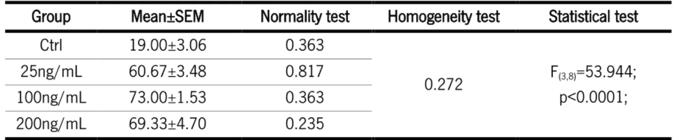
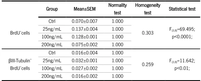
Table 6: Statistical analysis of the clonal assay with AREG. ... 38
Table 7: Statistical analysis of the differentiation assay of cells exposed to AREG. ... 40
Table 8: Statistical analysis of BrdU+ cells in the subventricular zone of animals injected with AREG. .. 42
Table 9: Statistical analysis of Ki67+ cells in the subventricular zone areas of animals injected with AREG. ... 44
Table 10:Statistical analysis of the cell cycle index in the subventricular zone of animals injected with AREG.. ... 45
Table 11: Satatistical analysis of DCX+BrdU+ cells in the subventricular zone of animals injected with AREG. ... 47
Table 12: Statistical analysis of the clonal assay with IGFBP2. ... 48
Table 13: Statistical analysis of the differentiation assay with IGFBP2. ... 50
Table 14: Statistical analysis of BrdU+ cells in the subventricular zone of animals injected with IGFBP2. ... 52
Table 15: Statistical analysis of Ki67+ cells in the subventricular zone of animals injected with IGFBP2. ... 54
Table 16: Statistical analysis of cell cycle index in the subventricular zone of animals injected with IGFBP2. ... 55
Table 17: Statistical analysis of DCX+BrdU+ cells in the subventricular zone of animals injected with IGFBP2. ... 57
Table 18: Concentration and quality of the extracted RNA assessed with the RNA StdSenses Analysis Kit. ... 58
Table S 1: Post hoc test for the statistical analysis of clonal assay with AREG ... 93
Table S 2: Post hoc test for the statistical analysis of differentiation assay with AREG. ... 94
Table S 3: Post hoc tests for the Statistical analysis of BrdU+ cells in the subventricular zone of animals
injected with AREG. ... 95
Table S 4: Post hoc tests for the Statistical analysis of DCX+BrdU+ cells in the subventricular zone of
animals injected with AREG. ... 96
Table S 5: Post hoc test for the statistical analysis of clonal assay with IGFBP2. ... 97
Table S 6: Post hoc test for the statistical analysis of differentiation assay with IGFBP2. ... 98
Table S 7: Post hoc tests for the Statistical analysis of BrdU+ cells in the subventricular zone of animals
injected with IGFBP2. ... 99
Table S 8: Post hoc tests for the Statistical analysis of DCX+BrdU+ cells in the subventricular zone of
1.1. Neurogenesis
Neurogenesis encompasses the set of events that leads to the formation of new neurons from their stem or progenitor cells. This process includes cell division, production of migratory precursors, differentiation, and lastly, integration of functional neurons into existing circuits. It occurs massively during embryonic development to construct the nervous system and it persists throughout adulthood in restricted areas. For over a century it was thought that the capacity to generate new neurons was restricted to embryonic development, and that no new neurons were added to the adult mammalian brain1. Even though, some
suggestions regarding the existence of dividing cells in the postnatal brain were raised2. Nevertheless, the
first evidence of adult neurogenesis was only published in 1958 reporting the existence of mitosis in the lateral wall of brain ventricles of young mice3. After that many evidences of adult neurogenesis on the
adult mammalian brain were communicated4,5. Currently, the major efforts on this field relay on sorting
the molecular mechanisms involved in the formation and integration of new neurons, and in unraveling the heterogeneity of the neurogenic niches. Due to the scope of this thesis we will next make a summary of the major aspects of both embryonic and adult neurogenesis at the lateral walls of the brain ventricles. In the next sections we will give an overview on the neurogenesis in the embryonic and in the adult brain
1.2. Embryonic neural stem cells
The embryonic central nervous system (CNS) contains two distinct proliferative areas where the stem/progenitor cells are retained6. The first to be generated is the ventricular zone (VZ); it is constituted
of neuroepithelial cells (NECs) with stem cell properties that appear approximately at embryonic day 8 (E8) and from which all the mature cells of the embryonic and adult CNS will be generated6. After a period
of symmetric division of the NECs, a second proliferative area appears, the subventricular zone (SVZ), generated by the asymmetric division of the progenitor cells from the VZ that migrate basally7. The cells
that compose the embryonic SVZ are known as intermediate progenitors (IPCs) or basal progenitors and produce neurons or glia by symmetric division7 (Figure 1).
The VZ microenvironment
When neural induction happens, the walls of the neural tube are composed of a monolayer of NECs. These cells are multipotent, radially elongated and present epithelial features: apical-basal polarity and lateral connections through tight and adherent junctions. During cell cycle, the nuclei of the NECs migrates up and down through the apical-basal axis, being located at the apical side in mitosis and basally during S phase. This phenomenon is called interkinetic nuclear migration and gives a pseudostratified appearance to the neuroepithelium layer8.
The NECs are highly proliferative at the anterior end of the central canal, where they divide fast and symmetrically to expand the precursor structure of the future brain. Due to this foundational process, the lumen of the developing brain starts to be surrounded by several layers, and the progenitor cells are located in the innermost apical layer, the VZ8.
At the onset of neurogenesis, between E10-E12 in the mice, NECs transform into radial glial cells (RGCs)9.
This transition from NECs to RGCs is characterized by the downregulation of Golgi derived apical
Figure 1: Representation of the cellular components of the embryonic neurogenic niche. Appearance and ontogenic maturation of the embryonic niche components derived from neuroepithelial cells. SVZ, subventricular zone; VZ, ventricular zone; NECS, neuroepithelial cells; RGCs, radial glia cells; IPCs, intermedia progenitor cells; CSF, cerebrospinal fluid.
trafficking and loss of tight junctions. Moreover, the cells initiate the expression of astroglial markers (glutamate aspartate transporter [GLAST], glial fibrillary acidic protein [GFAP], and brain lipid binding protein [BLBP])10. The mechanisms involved in this transition are not fully elucidated, however some
candidate molecules involved in the process were identified. For instance, it was reported that Hes1 and Hes5 deficient mice present normal NECs at E8 but impaired RGCs differentiation at E9.511. Another
study, demonstrated a transient expression of fibroblast growth factor (Fgf)10 by NECs coincident with this transition period, where targeted deletion of Fgf10 causes a delay in the transition12.
1.2.2. Cellular features of RGCs
As mentioned above, RGCs are characterized by the expression of astroglial markers. However, these cells also share epithelial features with the NECs, such as the expression of the intermediate filament protein nestin, interkinetic nuclear migration and the presence of adherent junctions.
Adherent junctions consist of cadherins and catenins that connect the intracellular network of actin and are present at basal to apical and at the subapical membrane of RGCs, mediating cell-cell adhesion10.
Newborn neurogenic cells downregulate cadherins, a process mediated by proneural genes, allowing cells to withdraw their apical endfoot and migrate away from the ventricle. Abscission of the apical endfoot is characterized not only by the dowregulation of cadherins but also by the loss of ciliary proteins and cell cycle exit, which in turn leads to cell differentiation10.
The apical plasma membrane of NECs and RGCs faces the ventricular surface of the developing brain10.
As described with further detail in section 1.5, brain ventricles are filled with cerebrospinal fluid (CSF) that contains signaling molecules produced by the choroid plexus (CP)13. These molecules include, FGFs,
insulin growth factors (IGFs), retinoic acid, bone morphogenic proteins (BMPs), sonic hedgehog (Shh) and Wnt, molecules that have already been described as playing a role in brain development, and its effects are likely to be mediated by receptors at the apical membrane14-16. Interestingly, both NECs and
RGCs extend their primary cilium into the brain ventricles, receiving signals from the molecules present at the CSF14,15,17. The primary cilium also plays an essential role in the maintenance of NECs and RGCs
polarity18.
On the other hand, the basal process of RGCs, besides serving as a scaffold for migrating newborn neurons, plays an important role in the maintenance of RGCs proliferation, since active transportation and translation of cyclin D2, a regulator of G1-S phase, induces asymmetric division of RGCs10,19.
One other particular feature of both NECs and RGCs is their complex mitotic behavior, the interkinetic nuclear migration20. It is hypothesized that this process regulates neurogenesis exposing RGCs nucleus
to signals present along the apical-basal gradient, specifically to Notch, that can regulate proliferation21,22
or block cell differentiation23,24.
In vitro and in vivo data suggest that RGCs divide asymmetrically to produce another RGCs and a neuron or IPCs25,26. This IPCs accumulate above the VZ producing a second germinal zone, the SVZ8. The
expansion of the SVZ, constituted of IPCs and RGCs, is thought to cause the majority of cerebral cortex expansion. In accordance, a Cre/loxP fate mapping study using the promoter of BLBP, a marker of RGCs, revealed that all neuronal populations of the CNS derived from BLBP expressing progenitor cells27.
Moreover, the transition of RGCs to neurogenic state is spatiotemporal dynamic, since RGCs from the ganglionic eminence achieve earlier their neurogenic stage when compared with their counterparts in neocortex27. This dynamic is tightly regulated by the combination of intrinsic and extrinsic factors that
provide unique signaling to the different regions of the VZ-SVZ. Next we will summarize the impact of different factors on the embryonic VZ-SVZ.
1.2.3. Extrinsic regulation of embryonic VZ-SVZ
The neurogenic niche is the term used to describe the highly complex environment that supports NECs and RGCs and their progeny. This is a dynamic microenvironment composed of several cells and factors that together provide signals that determine the fate of these cells. This microenvironment guides the neurogenic niche through its structural and functional changes observed from embryonic development into adulthood8,10.
During embryonic development the progenitor cells receive diffusible signals from different sources, including ventricular fluid, blood vessels, meninges and components that mediate cell to cell and cell-extracellular matrix interactions (ECM). These molecules form gradients within the brain and can signal far from their source. As a result, progenitor cells located in different positions of the CNS experience a unique combination of signals that might induce a region specific behavior. Growth factors are particular examples of these molecules, and represent a group of major key factors involved in the regulation of stem cells proliferation and differentiation20. We will next summarize existing evidence of the role of the
BMPs, members of the transforming growth factor-β (TGF-β) family, are implicated in neuronal development given that it was shown that BMP is present at the VZ and triggers neuronal differentiation. Specifically, BMP4 treatment increased the number of microtubule associated protein 2 (MAP-2) and TUJ-1 positive cells within the VZ28. Another report, shows that BMP7 regulates cortical neurogenesis trough
two independent sources, i.e. the meninges and the CP29. In particular, it was hypothesized that the
secretion of BMP7 by the CP will impact proliferation and neurogenin 2 expression (Ngn2, a proneural gene)29.
Fgf2 is expressed by microglia in the embryonic brain and plays a crucial role in the regulation of cortical development8. Fgf2 and FGF receptors (FGFR) are transiently expressed along the pseudostratified
epithelium during early neurogenesis30. 5-bromo-2’-deoxyuridine (BrdU) studies in embryos that received
a FGF2 injection in brain ventricles suggests that this growth factor increases the number of proliferating cells without affecting its cell cycle31. Moreover, mice that lost function of all FGFR at E12.5 lead to a
faster production of neurons causing a severe loss of RGCs and early end of neurogenesis30. Associated
with FGFR loss of function it was observed a decreased in the production of NICD (active Notch), Notch1 mRNA and the Notch downstream gene Hes1, together with a upregulation of Ngn2, Dll1 and Mash130.
These results indicate cooperation between FGFR and Notch signaling to regulate RGCs self-renewal. The Notch pathway is essential in the modulation of both embryonic and adult neurogenesis. Activation of this signaling pathway has the ability to induce self-renewal by inhibiting neuronal differentiation trough lateral inhibition, and by promoting symmetric division to promote expansion of the stem cells pool32,33.
Insulin and insulin like peptides are involved in the regulation of growth and maintenance of stem cells in several organisms, including Drosophlia melanogaster, Caenorhaditis elegans and Danio Rerio34. Similar
to the findings on invertebrates and zebrafish, IGF signaling is involved in the modulation of mammalian stem cells. IGF1 regulates cell proliferation at the VZ during cortical neurogenesis by the modulation of the cell cycle length, decreasing specifically the length of G1 phase35. Another member of the insulin family
involved in the modulation of the embryonic VZ-SVZ is IGF2. This growth factor is expressed by the CP and secreted into the CSF in an age dependent manner, presenting different expression levels in rat CSF at the following ages: E13, E15, E17, E19, E21, P0, adult. It binds to the IGF1 receptor (IGF1R) present at the apical domain of neural stem cells (NSCs) promoting progenitor cells15.
The Wnt signaling pathway plays a critical role in the regulation of proliferation and differentiation in the developing brain. During early neurogenesis, Wnt ligands promote symmetric division of RGCs causing a
delay in IPCs formation. Conversely, at later neurogenic stages, Wnt promotes IPCs formation and differentiation through the upregulation of its downstream target N-myc36,37.
Other members of the complex microenvironment that modulates the fate of RGCs and their progeny are Shh ligands. A crosstalk between Shh and Notch signaling has been shown to promote symmetric division of RGCs to extend their pool38. Moreover, Shh signaling has been shown to promote RGCs proliferation
and expansion of GABAergic neurons39.
Despite the fact that we focused our attention on summarizing the modulation of the embryonic SVZ by extrinsic factors, there are several intrinsic factors that also control their behavior. These intrinsic factors comprehend transcription factors and cell cycle components that modulate processes as epigenetic modifications and post translational modifications10. Given the scope of this thesis, we will not refer here
to the specific embryonic intrinsic factors regulating neurogenesis, or their role in this process.
1.3. Adult neurogenesis
Active neurogenesis in the adult mammalian brain occurs at a much slower rate when compared to embryonic neurogenesis and it is constrained to specific brain areas: the subgranular zone of the dentate gyrus, which produces neurons to the granular zone of the hippocampus, and the adult SVZ (aSVZ)40. The
aSVZ, considered the major neurogenic niche in the adult mammalian brain, underlies the lateral walls of the lateral ventricles and, under physiological conditions, it produces neurons that integrate existing circuits of the olfactory bulbs (OBs). It is suggested that the NSCs of the aSVZ are derived from RGCs. A Cre-lox technique that specifically and permanently labeled RGCs revealed that at postnatal day 0 (P0), the aSVZ is mainly composed of RGCs and few immature neurons, however the number of RGCs at this region decreases with time and it correlated with an increase in GFAP+ cells (P6). Moreover, a technique
that specifically labels RGCs progeny revealed that infected RGCs produce GFAP+ aSVZ astrocytes,
indicating that NSCs of the aSVZ derive from RGCs41. What mediates this embryonic to adult change in
the cell phenotype of NSCs at the aSVZ is unknown. In the context of the present thesis, in the next sections, we will only explore the aSVZ niche.
1.3.1. Adult SVZ
A large population of multipotent cells with self-renewing capacities is present in the aSVZ lining the CSF-filed ventricles of the adult mammalian brain. This region is typically seen as a thin layer of proliferative cells separated of the lateral ventricles by a layer of ependymal cells. However, NSCs are not exclusively restricted to the lateral ventricles, being also found in the rostral migratory stream (RMS)42, subcallosal
zone43 and in the ventral third ventricle44.
The embryonic origin of NSCs from the aSVZ, determined by a Cre-lox model, revealed that these cells are derived from telencephalic neuroepithelum, specifically from the medial ganglionic eminence, lateral ganglionic eminence, and cerebral cortex45. Moreover, depending on their embryonic origin, NSCs will
have different positions in the aSVZ, specifically NSCs derived from the cortex and lateral ganglionic eminence settle in the ventral aSVZ, while stem cells derived from the medial ganglionic eminence are located in the dorsolateral aSVZ45 (Figure 2).
1.3.2. Cellular composition and structural organization of the aSVZ
The aSVZ is composed mainly of three cell types (type A, B and C) that are differently distributed into three layers, below an ependymal cell layer (type E cells)40 (Figure 3). Quiescent type B cells (that may
be further sub-divided into type B1 and type B2 cells), or progenitor cells, when activated, divide asymmetrically, giving rise to another type B cell and a type C or transit-amplifying cell (TAPs). These TAPs cells undergo rapid divisions producing neuroblasts (type A cells) or glia (astrocytes or
Figure 2: Embryonic origin of neural stem cells of adult subventricular zone. Contributions of the different regions of the embryonic ventricular zone to the adult subventricular zone. Emx1-Cre was used to label cerebral cortex, Gsh2-Cre to label lateral and medial ganglionic eminence, Nkx2.1-Cre used to label medial ganglionic eminence and Dbx1-Cre to label corticostriatal sulcus. Adapted from Young et al. 200745.
oligodendrocytes). Neuroblasts are immature neurons that migrate along the RMS through chains of astrocytes and vasculature towards the OBs. We will next describe with further detail these cell types.
Type E cells
A monolayer composed of ependymal cells separates the aSVZ cells from the lateral ventricles. Two types of ependymal cells are present at this monolayer, E1 and E2 cells. E1 cells are multiciliate, and are organized in a pinwheel like structure around the apical process of type B1 cells. E2 cells are biciliated cells characterized by extraordinary complex bodies and constitute less than 5% of cells on the ventricular surface46. These two cells, located within all regions of the ventricle, show no evidence of proliferation46
and are commonly marked with the S100β marker47.
Figure 3: The structural and cellular organization of the adult subventricular zone. The adult subventricular zone, located below an ependymal layer is composed of mainly three cell types (Type B, C and A). Type B or progenitor cells, that project their apical process into the brain ventricles, give rise to type C cells or transit amplifying cells, that express molecular markers such as Mash1 (a basic helix loop helix transcription factor) or Dlx2. These C cells divide into neuroblasts (type A) that express proteins associated with neuronal migration and adhesion (DCX and PSA-NCAM). The adult subventricular zone also contains type B2 cells and blood vessels from the blood brain barrier. Adapted from Falcão et al. 201216.
Type B cells
In 1997 Doetsch and colleagues first identified GFAP expressing-astrocytic cells as the stem cells of aSVZ48. Entitled type B cells, these can be divided into two sub-types: B1 cells, which are located in close
contact to the ependymal layer and considered to be the NSCs of the adult brain; and B2 cells, characterized by a highly branched morphology which are located deeper in the brain parenchyma48. As
mentioned above (section 1.3), type B1 cells are thought to be the direct successors of RGCs41, presenting
a radial morphology49 and the expression of the astrocytic markers GFAP, GLAST and BLBP50. Moreover,
B1 cells express Id1 transcription factor, which is responsible for the self-renewing capacity of this cell type51. The main features that recognize B1 cells as the NSCs are based on the following evidences: i)
they are structurally similar to their ancestors, that also extend an apical process into the brain ventricle; ii) they express prominin1 (Prom1 or CD133) and nestin, which are markers of NSCs; and iii) they are multipotent cells giving rise to neurons and glial cells. aSVZ NSCs extend their apical processes trough the center of the pinwheel structures formed by the ependymal cells and directly contact with the CSF present in the lateral ventricles being exposed to signals present in this fluid46,52,53. On the other hand,
their basal processes extend into the perivascular niche, which also represents an important source of signals to the aSVZ8. These cells exist as quiescent NSCs or in an activated state NSCs. A recent study
revealed that quiescent NSCs are largely dormant, rarely form colonies in vitro and do not express nestin. However, when activated, B1 cells upregulate nestin and epidermal growth factor receptor (EGFR) expression, becoming highly proliferative54. Moreover, this study revealed that quiescent NSCs can be
prospectively isolated as GFAP+CD133+ and activated NSCs can be isolated as GFAP+CD133+EGFR+54.
Type C cells
Type C cells, or TAPs, present throughout the aSVZ and characterized by their abundant free ribosomes, deeply invaginated nucleus and absence of intermediate filaments, were first identified by electron microscopy48. These cells express distal-less homeobox 2 (Dlx2)55, implicated in the development of
GABAergic neurons and oligodendrocytes during embryonic development. They also express achaete-scute complex homolog 1 (Ascl1 or Mash1), a basic helix loop helix (bHLH) transcription factor described as essential for neuronal differentiation during embryogenesis40. These cells are the most frequently
labeled after injection of a proliferation marker, such as BrdU, constituting the largest pool of dividing cells in adult SVZ. Type C cells express EGFR56. Injection of exogenous EGF into the brain ventricle
significantly increased cell proliferation at the SVZ associated with an increase in the number of newborn astrocytes at the OB and reduction of neurogenesis57, indicating multipotency of C cells that can be
prompted by EGF. In vitro studies suggest that TAPs give rise to multipotent neurospheres when stimulated with EGF, supporting the idea that these cells can be reprogrammed in response to EGF55.
Type A cells
Derived from the TAP cells, neuroblasts or type A cells are the immature neurons that express doublecortin (DCX) and polysialylated neural cell adhesion molecule (PSA-NCAM), two proteins associated with neuronal migration56. Neuroblasts are not homogeneous and express a variety of different markers.
The pattern of markers expressed by these cells is associated with their site of origin at the SVZ and will determine the type of interneurons that will integrate different layers of the OB58. During migration trough
the RMS, type A cells continue to divide and initiate the maturation process59. When approaching OBs the
proliferation rate of these cells significantly decreases and the cell cycle length is enhanced59.
1.3.3. Extrinsic regulation of the adult SVZ
The stability of the aSVZ cell population is maintained due to the tight balance of intrinsic and extrinsic factors that control NSCs proliferation and self-renewal or differentiation into neuroblasts or glial cells. Next, we will briefly summarize the regulation of the aSVZ by extrinsic factors. Although not being the scope of this thesis, intrinsic factors also play a relevant role in the modulation of this neurogenic niche40.
Several extrinsic factors that regulate SVZ population comprehend growth and trophic factors, hormones, cytokines, morphogens and neurotransmitters. FGF2 and EGF are the principal mitogens used in the in vitro culture of SVZ NSCs. Receptors for this growth factors are expressed in the aSVZ and mice with the ablation of the gene for Fgf2 or for TGF-α (an EGF receptor ligand), present impaired neurogenesis in the aSVZ40. Ciliary neurotrophic factor, is the example of another factor involved in the modulation of the
aSVZ, given that its ventricular infusion increases proliferation. Other factors have been involved in the regulation of aSVZ. Brain derived neurotrophic factor (BDNF), is implicated in aSVZ proliferation and neurogenesis at the OBs. Several other growth factors and signaling pathways are described as participating in the modulation of neurogenesis and are summarized in table 1 and 240,60. These signaling
molecules have their origin in diverse sources, mostly from neurons, NSCs, ependymal cells, blood vessels, microglia and the CP-CSF system60. Furthermore, neurotransmitters released by neurons are key
members of the SVZ niche, with different neurotransmitters playing different roles60. For instance,
serotonin induces NSCs proliferation while dopamine can either increase or decrease proliferation. A key component of the microenvironment that surrounds the neurogenic niche is the CP-CSF system16.
Further information on this system, and its influence in the SVZ will be provided bellow (Section 1.5).
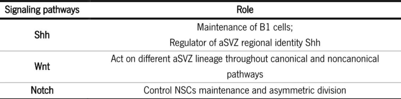
Table 1: Signaling pathways involved in the modulation of the aSVZ.
Signaling pathways Role
Shh Maintenance of B1 cells;
Regulator of aSVZ regional identity Shh
Wnt Act on different aSVZ lineage throughout canonical and noncanonical pathways
Notch Control NSCs maintenance and asymmetric division
Table 2: Factors involved in the modulation of the aSVZ.
Source Factor Effect
Ependymal cells
Noggin (BMP antagonist) Inhibits or promotes neurogenesis Pigment epithelium
derived factor (PEDF) Promotes self-renewal of NSCs Fractones (ECM extensions) FGF2 Promotes proliferation of NSCs BMP4, BMP7 Inhibit neurogenesis Microglia NT3, GDNF, FGF2, BDNF
and cytokines Enhance neurogenesis IL-1α, IL-1β, IL-1 and
TNF-α,β
Decrease differentiation and proliferation of NSCs
Vascular endothelial cells
PEDF NSCs self-renewal
NT3 NSCs maintenance
SDF1 Neuronal migration
Choroid plexus-CSF
SLIT1/2 Neuronal migration
Amphiregulin Mitogen
IGF2 Mitogen, NSCs self-renewal CSF flow Neuronal migration
Neurons
Serotonin NSCs proliferation
ChAT+ proliferation
Nitric oxide Effect on proliferation on a dose dependent manner
1.4. Functional relevance of neurogenesis
Under physiological conditions the major function of aSVZ NSCs is the production of new neurons to the OBs, promoting plasticity of the existing circuitry by making new synaptic contacts with mature neurons56.
Behavioral studies propose that adult neurogenesis is important for memory consolidation dependent on the OBs61. New OB interneurons play important roles in short-term memory and flexible olfactory
associative learning.
Under pathological or challenging conditions, adult SVZ neurogenesis seems to play diverse roles dependent on the condition. For instance, in rodent ischemia models enhanced neurogenesis occurs in both adult neurogenic niches. Specifically, in the SVZ after ischemic stroke, cells migrate to the injury site expressing markers of resident neurons. In the case of neurodegenerative diseases, for example Alzheimer’s disease models based on amyloid precursor protein, present reduced neurogenesis62. Also,
Parkinson models based on the overexpression of wild-type human synuclein present reduced survival of new neurons on both neurogenic niches, while expression of mutant α-synuclein inhibited cell proliferation. CNS response to inflammation is often related with neurodegenerative diseases and brain injury62. For instance, endotoxin induced inflammation leads to a down-regulation of adult neurogenesis,
restored by anti-inflammatory treatments62. Studies that examined the role of immune system in the
regulation of adult neurogenesis suggest that different mechanisms of the immune response might have a different impact on adult neurogenesis62. Taking these evidences into account NSCs provide a source
of potential endogenous therapies to several CNS disorders.
1.5. Choroid plexus
The choroid plexuses are folded epithelial structures residing at the brain ventricles. They are a highly vascularized secretory structure that develops from different locations along the dorsal axis of the neural tube63. There are four CPs in the mammalian brain, being the CP of the fourth ventricle the first to be
formed, followed by the CPs of the lateral ventricles and later the one lining the third ventricle63. The
structure of the CPs consists of a monolayer of epithelial cells surrounding a stromal core. Neighboring epithelial cells are bounded together by tight junctions, forming the blood-CSF barrier that prevents the free passage of molecules from the systemic circulation into the CSF8 (Figure 4). The stroma of this
structure is highly vascularized, consisting of pericytes, connective tissue and blood vessels63. Contrarily
barrier, these are fenestrated and leaky63. CPs have dual embryonic origin, being the stromal cells
originated from the mesoderm and the epithelial cells having origin from the ectoderm8.
Functionally, the CPs display several roles: i) regulation and protection of brain environment, acting as the blood-CSF barrier; ii) secretion and modulation of CSF composition trough protein secretion by their epithelial cells; iii) removal of brain metabolites and waste via CSF renewal; and, iv) during embryonic development it creates an expansive pressure trough the secretion of CSF16.
The production and secretion of molecules by the CPs varies in a time dependent manner and it is regionally different, i.e. the pattern of expression is different among the four CPs, which contributes to the dynamic signaling gradients across the brain64,65.
Table 3 summarizes the several CP transcriptome analyses performed both in rat and mouse that revealed the expression of several factors by the CP that influence neurogenesis at the SVZ both during embryonic development and adulthood.
Figure 4: Structural organization of the choroid plexus. The choroid plexuses are a monolayer of epithelial cells that surround a stromal core. The epithelial cells are bounded by tight junctions, preventing the free passage of molecules from the systemic circulation into the CSF. The stroma of the choroid plexuses is highly vascularized and contains fenestrated capillaries, macrophages dendritic cells and fibroblasts. Adapted from Falcão et al. 201216.
Table 3: Summary of the CP transcriptome analysis performed by array or high throughput sequencing.
1.5.1. Choroid plexus-CSF impact in embryonic neurogenesis
As mentioned before, embryonic progenitor cells, that line the ventricular surface, project their apical process into lateral brain ventricles, being in direct contact with the CSF. This apical process contains receptors that receive signals from the CSF. Therefore, being the CP the major responsible for the regulation of CSF production and for the modification of its composition, either by alterations in the transference of molecules from blood into CSF or by variations in the secretion of molecules produced by the CP, it is expected that the dynamic CSF composition will somehow have an impact on neurogenesis. In fact, an in vitro study revealed that CSF alone was capable of maintaining the viability and proliferation of cortical cells66. Additionally, this study demonstrated that CSF isolated from different ages affected
differently the growth of cortical cultures66. Similar results were observed when rat neurospheres were
exposed to CSF isolated from different embryonic ages; specifically, cells cultured with CSF collected from E17 proliferated more when compared with neurospheres exposed to CSF from E1915. Moreover, a
proteome analysis of embryonic CSF revealed the presence of 423 proteins in E12.5 CSF, 318 in E14.5 and 382 in E17.5 of which only 137 proteins where common among the three ages, highlighting the dynamic range in CSF composition67. IGF2 constitutes an example of these molecules that vary in a time
depend manner in the CSF, present in different ages of embryonic CSF, but at different levels. Moreover, some of the proliferative effects of the CSF in the SVZ can be attributed to this CP-secreted protein, that
Species Age Sample Type accession GEO
Rattus
norvegicus E15
LV- CP epithelial
cells
Expression profiling by high
throughput sequencing GSE44072 Rattus
norvegicus E19, P2, adult LV-CP Expression profiling by array GSE44056 Mus musculus E15, 10 weeks
LV-CP epithelial
cells Expression profiling by array GSE33009 Mus musculus E18.5 Fourth and
LV-CP
Expression profiling by high
throughput sequencing GSE66312 Mus musculus 8-9 weeks CP from all ventricles Expression profiling by array GSE23714 Mus musculus 8 weeks and 72
establishes its effects trough IGF1R present at the apical process of RGC15,68. Nevertheless, CSF has a
milieu of signaling molecules with the ability to influence neurogenesis, for example, Shh, FGF, retinoic acid and leukemia inhibitory factor constitute some of the molecules with confirmed presence in embryonic CSF and described as capable of modulating neurogenesis. Hence, the proper balance of CSF composition is essential for the correct interaction of multiple signaling activities that reach the embryonic SVZ. In vivo studies of CSF composition manipulation support the idea that CSF regulation has an impact on neurogenesis. For example, intraventricular injection in rat and mice embryos of recombinant IGF1 resulted in increased cell proliferation69. Another experiment, revealed that removal of Fgf2 from chick
embryonic CSF lead to a decrease in cell proliferation and increase in neuronal differentiation70.
Taking this into account, the CPs, via modulation of CSF composition, are potential regulators of embryonic NSCs. However, until now little is known on how these molecules are regulated and secreted by the CPs. The improvement of region and time specific deletion techniques will provide new strategies to target CP-derived molecules and study its effect on neurogenesis. For example, deletion of Shh from the 4th ventricle-CP resulted in a decrease of proliferation and an impair in the production of GABAergic
neurons39. Another study revealed that inhibition of Otx2 from 4th ventricle-CP resulted in increased
proliferation caused by alterations in Wnt signaling14. This studies evidence that the CPs produce
modulators of essential signaling involved in the regulation of embryonic NSCs and their progeny.
1.5.2. Choroid plexus-CSF impact in adult SVZ
The proximity of the aSVZ with the ventricular surface and the fact that NSCs project their apical process into the lateral ventricles makes the CP-CSF system a key component of the aSVZ neurogenic niche. Recent adult CP transcriptome analysis revealed the presence of molecules, such as IGF1, IGF2, EGF, that as mentioned before, are involved in signaling pathways that modulate cell cycle, cell survival and cell differentiation71. Adult CSF is capable of supporting the growth of adult rat neurospheres15. Alterations
of CSF composition in a time dependent manner are also observed during aging, where heterochronic infusion of “old” CSF into young mice resulted in a decrease of NSCs at the aSVZ and conversely the injection of “young” CSF into old mice increases the number of NSCs at the aSVZ65. Additionally, this
study revealed that not all types of aSVZ cells are sensitive to aged CSF, with NSCs being affected but not TAPs. Interestingly, transcriptome analysis of young and adult mice lateral ventricle CPs revealed that BMP5 and IGF1 are enriched in young CP (2 months), suggesting that this factors might be involved in
the age dependent effects that CP-derived factors promote in the SVZ. Moreover, taking into account its location between blood and CSF, the CP is capable of altering its secretion pattern in response to local changes of the niche as well as peripheral alterations.
The maintenance of the aSVZ cytoarcithecure is fundamental for the proper production of NSCs and their progeny. CP-secreted interleukin-1β (IL-1β) has been proposed as being involved in the modulation of aSVZ cytoarchitecture and proliferation of quiescent NSCs. By binding to IL-1 receptors on the surface of B cells, IL-1 β regulates the expression of VCAM153. In vivo disruption of this adhesion molecule resulted
in disturbed cytoarchitecture and proliferation of NSCs resulting in increased neurogenesis at the OBs. Being IL-1 β secreted by the CP, this study revealed the key role of the CP to act as an environmental sensor, responding to chemokines involved in tissue repair. One other example of CP-derive molecule is neurotrophin 3, which regulates quiescence and long term maintenance of aSVZ-NSCs, inducing the phosphorilation of nitric oxide synthase leading to the production of nitric oxide. Moreover, SLIT proteins, secreted by the CPs are chemorepulsive factors that repel C cells in the direction of the OBs72.
Based on a previous transcriptome analysis of the adult CP in this work we selected two proteins secreted by the CP to explore their impact, which is currently unknown, in SVZ neurogenic niche: Amphiregulin (AREG) and Insulin Growth Factor Binding Protein 2 (IGFBP2)71. AREG is an EGF family protein that binds
to the EGFR activating signaling cascades of cell survival, proliferation and motility. Previous work reported that AREG is expressed in the CP and hippocampus of adult mice73. IGFBP2 belongs to a family of proteins
responsible for the bioavailability and transport of IGF proteins. Interestingly, IGFBP2 is the predominant IGF binding protein present in the CSF34.
Altogether, the information summarized above provides some evidences of the role of CP-derived molecules in the modulation of the SVZ cellular dynamics. Nevertheless, several aspects are still unexplored. Namely, what proteins secreted by the CPs play a role in the regulation of SVZ? What is the impact in the SVZ caused by alterations in the CPs transcriptome due to aging or immune response? Can CPs transcriptome provide a possible source of endogenous therapy for neurodegenerative diseases?
Given the proximity between the SVZ neurogenic niche and the CP, the central hypothesis of the work developed under this thesis is that CP-derived molecules play a role in modulating SVZ dynamics. So, to gain further insights in SVZ cell population dynamics, the goal of this project is to understand how molecules secreted by the CP into the CSF influence the SVZ cell population.
Specifically, we aimed at:
i) determining the effects of candidate CP-derived proteins using in vitro cultures of neural progenitor cells. For this, we used neurospheres cultures exposed to different concentrations of selected molecules secreted by the CP and evaluated cell proliferation and differentiation.
ii) evaluating how candidate CP-derived proteins impact in the SVZ cell population dynamics in vivo. For this we performed ventricular injections of the selected proteins to determine its putative effects in SVZ cells, namely SVZ cells proliferation and differentiation.
iii) characterizing the ontogenic expression pattern of the CP, from early postnatal stages to early adulthood. For this we determined the transcriptome of the CP by using high throughput sequencing RNA-seq. This will potentially enable the identification of key molecules that contribute to structural and functional temporal changes occurring in the SVZ.
3.1. Ethics statement
This project was part of a study approved by the Ethics Comission of the University of Minho (SECVS-001/2013) and by the Portuguese national authority for animal experimentation, Direcção Geral de Veterinária (DGV9457). Animals were kept and handled in accordance with the guidelines for the care and handling of laboratory animals in the Directive 2010/63/EU of the European Parliament and of the Council.
3.2. Animals
For this project we used Wistar Han rats. Due to previously described specificities related to hormonal fluctuation, namely at gene expression level, females were not used. Animals were maintained in 12h light/dark cycles at 22 to 24ºC and 55% humidity and fed with regular rodent’s chow and tap water ad libitum.
3.3. Study the impact of CP-derived proteins in human neural progenitor cells.
To study the impact of CP-derived proteins in neural progenitor cells (NPCs) in vitro we used human NPCs (hNPCs), a kind gift from Prof. Leo A. Behie (University of Calgary, Canada). The cells were isolated, in the lab of Prof. Behie, from the telencephalon region of a 10 week post-conception fetus according with the protocols and strict ethical guidelines previously established and approved by the Conjoint Health Research Ethics Board (CHREB, University of Calgary, Canada; ID:E-18786)74. In all the experiments,
cells were maintained at 37°C in a humidified atmosphere containing 5% CO2.
hNPCs were thawed and the content placed in T-75 culture flasks containing 15mL of serum-free medium PPRF-h274. After three days, cells were mechanically dissociated (25-30 times) into a single cell
suspension and cultured in fresh PPRF-h2 medium. After 10 days of growth, cells were collected and enzymatically dissociated with 0.05% Trypsin-EDTA (Invitrogen, Carlsbad, California, USA) during 10min at 37°C followed by mechanic dissociation. At this point, cells were plated under specific conditions to perform clonal and differentiation assays that will be explained bellow in detail.
3.3.1. Clonal assay
Dissociated cells were plated under non-adherent conditions in serum-free medium (PPRF-h2), at a density of 10cells/µL75,76. At the second day of culture, cells were exposed to one of the following
concentrations of either AREG (Biolegend, San Diego, California, USA) or IGFBP2 (Biolegend): 25ng/mL, 100ng/mL or 200ng/mL. After 10 days in culture, we evaluated the effects of the exposure to the different concentrations of each protein by counting the number of formed neurospheres (Figure 5).
3.3.2. Differentiation assay
Dissociated hNPCs were plated on pre-coated coverslips [poly-D-lysine (100μg/mL, Sigma-Aldrich, St Louis, Missouri, USA) and laminin (10μg/mL, Sigma-Aldrich)] in 24-well plates at a density of 50.000cells/well77. At the second day of culture cells were exposed to the same three concentrations of
AREG or IGFBP2 as for the clonal assay. Simultaneously, cells were also exposed to BrdU, a thymidine analogue that incorporates newly generated DNA during the S-phase of the cell cycle, to also evaluate cell proliferation (10μM, Sigma-Aldrich)78. After 5 days in culture, cells were fixed during 30 minutes with 4%
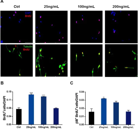
paraformaldehyde (PFA) for posterior immunocytochemistry for BrdU and βIII-Tubulin (Figure 6).
Figure 5: Schematic representation of clonal assay.
3.3.3. Immunocytochemistry for BrdU and βIII-Tubulin
Fixed cells were permeabilized in phosphate buffered saline (PBS) with 0.1% Triton X-100 (PBS-T; Sigma-Aldrich) for 5min at room temperature (RT). The acidification, required for the denaturation of DNA strands allowing the binding of the antibody against BrdU, was achieved immersing cells in 1M hydrochlorid acid (HCl) during 30min. Blockage of non-specific binding was performed using 10% fetal bovine serum (FBS, Invitrogen, Carlsbad, USA) for 1h at RT. Cells were then incubated with the primary antibodies (Table 4) diluted in 10% FBS for 1h at RT and then with the appropriate secondary antibodies (Table 4) diluted in 10% FBS for 1h at RT. The cells were counterstained with 4’,6-diamidino-2-phenylindole (DAPI, 1:1000, Sigma-Aldrich), a compound that labels the nucleus, for 10min at RT. Coverslips were mounted on slides using immu-mount (Thermo Scientific, Waltham, Massachusetts, USA)77. For quantification analysis,
samples were observed using a fluorescence microscope (BX61, Olympus, Tokyo, Japan), and for this purpose, three coverslips per condition and 20-25 representative fields were chosen and analyzed. The results are presented as the number of positive cells for the respective markers (Table 4), divided the total number of cells in the field (DAPI-positive cells)77.
Table 4: List of primary and secondary antibodies used for the immunocytochemistry.
Primary Antibody Dilution Company
BrdU – Rat 1:20 Abcam
βIII-Tubulin – Mouse 1:500 Millipore
Secondary Antibody Dilution Company
Alexa Fluor 594 – Goat α-Rat 1:1000 Invitrogen Alexa Fluor 488 – Goat α-Mouse 1:1000 Invitrogen
3.4. Study the impact of CP-derived proteins in the SVZ cell population
All experiments were conducted in 8-weeks-old Wistar Han rats. To reduce stress-induced changes in the hypothalamus-pituitary axis associated with the surgery procedure, animals submitted to intraperitoneal and cannula injections were handled daily during 1 week prior to surgery.
3.4.1. Surgeries
Animals were anesthetized with medetomidine (0.5mg/kg) and ketamine (75mg/kg). Once animals were anesthetized the skin over the implantation site was shaved and animals were immobilized in a stereotaxic apparatus. A small incision was made from between the eyes to the base of the neck to expose the scalp. A sterilized stainless-steel guide cannula (26gauge; Plastics One, Virginia, USA) was implanted in the right lateral ventricle (0.24 posterior, 1.5 lateral and 4 depth), through a burr-hole. The cannula was fixed to the scalp with four screws and dental acrylic cement and the skin sutured around it. To prevent contamination, it was inserted a dummy cannula into the guide cannula. At the end of the surgical procedures, the anesthesia was reverted with atipamezole hydrochloride (1mg/kg) and animals were monitored until they were fully recovered. After surgery animals were treated with anti-inflammatory (Carprofen, 5mg/kg) and antibiotic (Enrofloxacin, 5mg/kg) and were allowed to recover for one week79
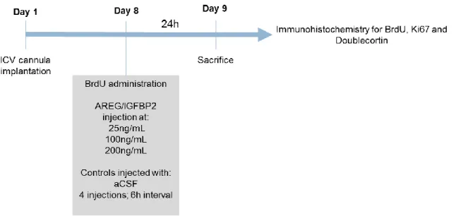
(Figure 7).
3.4.2. BrdU and proteins administration
For the analysis of AREG (Biolegend) and IGFBP2 (Biolegend) impact in the SVZ cell population, four groups of animals were compared (n= 5/group). To evaluate the effects of protein administration in cells proliferation, all animals received a single injection of BrdU (50mg/kg), 30min before the first administration of the protein. Three groups of animals received 4 injections (6h interval), during a 24h period, of protein at 25ng/mL, 100ng/mL or 200ng/mL of concentration. A sham control group was injected with the vehicle solution, artificial CSF (Figure 7).
For the protein injection animals were anesthetized with 4% sevoflurane in 100% oxygen. The proteins were administered through a 33gauge injection cannula (Plastics One), using a 5.0µL Hamilton syringe connected to the injection cannula through a polyethylene catheter (PE-10; Plastics One). The injection volume was 4µL injected at a velocity of 2µL/min. To prevent reflux of the protein back to the injection cannula, the injection cannula was left in place for one additional minute. Animals were sacrificed 24h after the first injection of the protein.