Raquel Alexandra Gonçalves Peixoto
Development of bacterial cellulose membranes
coated with hydroxyapatite for bone regeneration
14 R aq uel Ale xandr a Gonçalv es P eix ot o De
velopment of bacterial cellulose membranes coated wit
h h
ydro
xy
Dissertation for the M.Sc. degree in Biomedical Engineering:
Clinical engineering
Supervisors:
Miguel Gama, PhD
Anabela Alves, PhD
Raquel Alexandra Gonçalves Peixoto
Development of bacterial cellulose membranes
coated with hydroxyapatite for bone regeneration
Nome Raquel Alexandra Gonçalves Peixoto Endereço eletrónico: [email protected] Número do Bilhete de Identidade: 13915044
Título dissertação: Development of bacterial cellulose membranes coated with hydroxyapatite for bone regeneration. Orientador(es): Francisco Miguel Portela da Gama e Anabela Alves
Ano de conclusão: 13915044
Designação do Mestrado: Mestrado Integrado em Engenharia Biomédica, Ramo de Engenharia Clínica
1. É AUTORIZADA A REPRODUÇÃO INTEGRAL DESTA DISSERTAÇÃO APENAS PARA EFEITOS DE INVESTIGAÇÃO, MEDIANTE DECLARAÇÃO ESCRITA DO INTERESSADO, QUE A TAL SE COMPROMETE;
Universidade do Minho, ___/___/______
“Science never solves a problem without creating ten more.” George Bernard Shaw “Somewhere, something incredible is waiting to be known.” Carl Sagan
This is probably the most difficult part to write down on my thesis. Due to tiredness, due to inertia or due to difficulty in expose my feelings, but essentially due to the inherent difficulty at summing up all people and how they have contributed to my grow as person, as student and as scientist.
First of all, I have to recognize complete gratitude to my supervisor, Prof. Miguel Gama and my co-supervisor, Anabela Pinto: my scientific models and the ones that I want to follow being or not a scientist.
Despite have accepted me immediately as a member of his team, Prof. Miguel have always believed in my capabilities, showing patience for all my doubts, as well as interest for my work being an active part on the decisions made over it. The serenity, gaiety and leadership concept with emphasis on relationships between people will be marks that I would never forgot.
To Anabela Pinto, I must confess that first I was really afraid of you. All those questions and discussions that I took home and made search instead of resting, all that pressure the made me think that I was going crazy and when you asked me for an index at the end of 2 days of work? However, as soon as you were not present anymore, I immediately understand the way that you tailored me and that I am absolutely sure that it will be useful independently the way that I will follow. I am thankful for the teaching and the patience, for always have fight by my side and for the camaraderie.
To Dr. Rui Coelho, for proposing this work and to trust their development to a simple graduate student, despite being supported by amazing researchers.
Fun, relaxation and distraction moments, but also learning and companionship were complimentary of LTEB and of people who constitute it.
Vera Carvalho is the first person I would like to thank. You were a fundamental piece of my work and your help was untiring in the fight that are cell’s assays! Thank you for your follow up, for the questions raised and for you practical sense, for your willingness, for alter your routines because of me, for all conversations.
A special acknowledge to Jorge Padrão. Your help was crucial through all my journey because you accompanied me in madness, you accompanied me over and over to the autoclave and you wake up all statistical asleep inside my brain.
the laughter.
An especial acknowledgment to Electroactive Smart Materials Research Group from University of Minho, particularly to Vitor Sencadas, for complimentary on mechanical assays realization and help during them.
William Penn said “A true friend freely, advises justly, assists readily, adventures boldly, takes all patiently, defends courageously, and continues a friend unchangeably.”
For these and more thousand countless reasons a special thank to my second family Rita, Liliana, Ivo, Campos and João. Despite the distance, friendship that brought us together 5 years ago continues to help overcome all obstacles.
The same is also valid to old school buddies, Rui, Isac, Pedro, Micael, Jorge, Cláudia, Eduarda, André, Tânia, Sónia, and Anas. No one of us is easy to handle up, but we still together and united, which only show love! "Bem hajam"!
To the entire class of Clinical Engineering of 2012-2014, for the companionship during these two years and with a special appreciation to Carina and Juliana, who received us in the best possible way in this new academy and with who I have learned a lot.
To my beloved boyfriend and truest friend Hugo! For everything! For the love, friendship, support, comprehension! You were always there for me, understanding my absences and giving me strength for the future.
My final words are addressed inevitably to my family, to whole I surrender my complete and most cherished gratitude. To those who gave all of them for me since my first day, since my first breath.
To my mum, dad and my brother! You are my driving forces, you understood my absences, you gave me your unconditional support and patience to all my questions and existential crises and specially, you have never gave up on me. There are not enough words to expound what I fell for you and how grateful I am.
To my aunt and cousins for the support during this last step.
There was a role of people that I would like to thank for their contribution in some way for me to achieve my goals and to my happiness.
Abstract
Bone, the major constituent of skeletal system, is susceptible to several pathological scenarios that could compromise its correct function. Advanced solutions to overcome these handicaps have always been a desire of the medical community, being guided bone regeneration (GBR) one of the approaches under development. Non-resorbable GBR membranes are mainly composed of synthetic materials. However, bacterial cellulose (BC) arises as an attractive natural origin, non-resorbable solution. In fact, this natural polysaccharide presents interesting properties for the medical field, which lead to increased research efforts into tailoring it with specific features to fit the requirements of the envisaged applications.
The development of BC membranes coated with hydroxyapatite (HA) to be used both as a barrier in GBR and as a promoter of bone regeneration was the main goal of the present work. Two different types of BC, a commercial one (cBC) and in-house produced (gxBC), in three drying states, named never dried, air dried and freeze-dried, where used. BC composites (BC-HA) were prepared following a biomimetic approach involving materials pre-rehydration and chemical modification with sodium hydroxide 3M (NaOH 3M), followed by immersion in specific simulated body fluid (SBF) solutions, NaCl-free SBF and complete SBF. The influence of BC origin, drying procedures and parameters associated with the mineralization protocol were evaluated and deeper characterization was performed on a set constituted by freeze dried cBC and gxBC membranes produced with the defined and optimized protocol, namely modification with NaOH 3M for 1 min, NaCl-free SBF for 1 day and SBF for 7 days.
Scanning electron microscopy (SEM) results denote that air-drying and freeze-drying promote modifications in BC microstructure, and higher porosity of gxBC freeze-dried samples, both before and after rehydration, is evident. This differentiating feature influences the mineralization of the samples, allowing internal formation of a ceramic filler, only observed on gxBC. On the other hand, in the particular case of cBC membranes, this ceramic layer is only observed on the surface of the membranes. Nevertheless, successful formation of a ceramic coating grants the effectiveness of the mineralization protocol. Elemental analysis (EDS) and x-ray diffraction (XRD) reveal the nature of this phase, identified as calcium deficient hydroxyapatite (CDHA). Material’s microstructure and the alkali treatment also influence water uptake and degradation profiles, being the highest values associated with gxBC, especially to modified
assays revealed promising results, with long-term cytocompatibility for all sets of BC membranes. At the end of 21 days, 3T3 and MC 3T3-E1 cells present higher metabolic active and higher viability levels when seeded in gxBC in the absence and presence of coating, respectively.
Therefore, material-related and methodological variables influence both the biomimetic mineralization and final behaviour of BC membranes. The herein presented results demonstrate an effective biomimetic mineralization, with formation of CDHA phase, resulting in a final material with interesting characteristics that can be considered for GBR applications.
Resumo
O osso, um dos principais constituintes do sistema esquelético, é uma estrutura suscetível a várias patologias que podem comprometer a sua função. Soluções avançadas capazes de solucionar estes problemas sempre foram um objetivo da comunidade médica, sendo a regeneração guiada de osso (RGO) uma das abordagens em desenvolvimento. As membranas não reabsorvíveis usadas em RGO são maioritariamente compostas por materiais sintéticos. Contudo, a celulose bacteriana (CB) também tem sido estudada, surgindo como uma solução atrativa, natural e não reabsorvível. De facto, este polissacarídeo de origem natural é dotado de características interessantes para a medicina, o que tem estimulado a sua investigação e interesse em adaptá-las para que melhor se enquadre na aplicação desejada.
O desenvolvimento de membranas de CB revestidas com hidroxiapatite (HA) para serem usadas em RGO, tanto como membrana barreira como material promotor de regeneração óssea, é o principal objetivo deste trabalho.
Neste trabalho foram usados dois tipos distintos de CB, comercial (cCB) e produzida em laboratório (gxCB), em três estados de secagem, nomeadamente nunca secas, secas ao ar e liofilizadas. Compósitos de CB (CB-HA) foram preparados seguindo uma abordagem biomimética que envolve pré-reidratação dos materiais, seguida de modificação química com hidróxido de sódio 3M (NaOH 3M) e imersão em soluções específicas que simulam os fluídos corporais, com e sem NaCl. A influência do tipo de CB, dos processos de secagem e dos parâmetros associados ao protocolo de mineralização foram avaliados. Um conjunto constituído por membranas cCB e gxCB liofilizadas e produzidas tendo em conta o protocolo de mineralização definido e otimizado, foi analisado mais profundamente.
Os resultados microscopia eletrónica de varrimento (SEM) demonstram que a secagem ao ar e a liofilização promovem modificações na microestrutura da CB e revelam a maior porosidade da gxCB liofilizada, tanto depois e antes da pre-rehidratação. Esta diferença influenciou a mineralização interna das amostras, apenas observada na gxCB. Contudo, uma camada de cerâmico foi também formada na superfície da cCB, indicando a efetividade do protocolo de mineralização. A análise elementar (EDS) e a difração de raios x (XRD) revelou a natureza desta fase, nomeadamente HA com défice de cálcio. A microestrutura assim como o tratamento químico
efeitos do tratamento alcalino na performance mecânica dos materiais também é mais evidente na gxCB, sendo que a presença da fase cerâmica não atenua completamente o seu efeito. Por último, os ensaios biológicos revelaram resultados promissores, evidenciando citocompatibilidade a longo prazo para todos os conjuntos de CB. Ao fim de 21 dias de cultura, as células 3T3 e MC 3T3-E1 apresentam maior atividade metabólica quando na presença de gxCB, com e sem recobrimento cerâmico, respetivamente.
Assim sendo, variáveis relacionadas com o material e com o próprio processo influenciam tanto a mineralização biomimética como o posterior comportamento da BC. Os resultados aqui apresentados demonstram a efetividade da mineralização, resultando num material final com propriedades interessantes para RGO.
Acknowledgments i
Abstract iii
Resumo v
Table of contents vii
List of abbreviations xi
List of figures xiii
List of tables xvii
A. Introduction
Introduction 1
1. Bone 2
1.1. Bone as a tissue 2
1.1.1. Constitution: bone cells and bone matrix 2
1.1.2. Types of bone tissue 4
1.2. Bone as an organ 5
1.3. Bone dynamics: formation, growth, remodelling and repair 6
1.3.1. Formation, growth and remodelling 6
1.3.2. Repair 8
1.4. Bone defects 10
1.5. Solutions 11
1.5.1. Bone grafts 12
1.5.2. Tissue Engineering 13
1.5.3. Guided Bone regeneration 14
2. Bacterial cellulose (BC) 17
2.1. BC fermentation and biosynthesis 19
2.2. BC characterization 22
2.2.1. Physical and chemical characteristics 22
2.2.2. Mechanical characteristics 24
3. Biomimetic mineralization 25
3.1. Calcium orthophosphates 25
B. Aims
Aims 33
C. Materials and methods
Materials and methods 37
1. Commercial BC 37
1.1. Commercial BC origin 37
1.2. Commercial BC purification 37
2. Glucanocetobacter xylinus ATCC 700178 BC 37
2.1. Microorganism strain 37 2.2. Culture media 37 2.3. Microrganism mantainence 38 2.4. gxBC production 38 2.5. gxBC purification 38 3. BC processing 38 4. BC mineralization 39
4.1. Production of SBF and NaCl-free SBF 40
5. Material characterization 41
5.1. Scanning electron microscopy and energy-dispersive x-ray spectroscopy 41
5.2. Cryo-screening electron microscopy 42
5.3. X-ray diffraction 42
5.4. Mechanical assays 42
5.5. Water uptake and degradation assay 44
5.6. Cellular assays in vitro 45
5.6.1. Material preparation 45
5.6.2. Cell lines 45
5.6.3. Cell culture media 45
5.6.4. Culture conditions 45
D. Results
Results 51
1. Optimization of mineralization process 51
2. Physicochemical and biological characterization of mineralized BC membranes 62
E. Discussion
Discussion 84
F. Conclusions and perspectives
Conclusions and perspectives 96
G. Bibliography
ATCC - American type culture collection BC - Bacterial cellulose
BC-HA - Bacterial cellulose-hydroxyapatite composite Ca-P - Calcium orthophosphates
cBC - Commercial BC
CDHA - Calcium deficient hydroxyapatite cryo-SEM - Cryo-scanning electron microscopy DP - Degree of polymerization
dH2O - Distilled water
DMEM - Dulbecco’s modified eagle’s medium EDS - Energy-dispersive x-ray spectroscopy GBR - Guided bone regeneration
G. xylinus - Gluconacetobacter xylinus
gxBC - Glucanocetobacter xylinus ATCC 700178 BC HS - Hestrin–Schramm
HA - Hydroxyapatite HCl - Hydrochloric acid NaOH - Sodium hydroxide -OH - Hydroxyl groups PC - Plant cellulose
PBS - Phosphate buffer saline solution RT - Room temperature
SEM - Scanning electron microscopy SBF - Simulated body fluid
XRD - X-ray diffraction E - Young’s modulus εf - Strain-at-failure
A. Introduction
Figure 1. Hierarchical organization of bone over different length scales [17]. 6
Figure 2. Indirect bone healing mechanism [7][29-31]. 9
Figure 3. Principal pathways of cellulose production and molecular arrangement with identification
of β-1→4 bond [55]. 18
Figure 4. Mechanism and SEM micrograph of cellulose ribon formation by G. Xylinus [49][54]. 21
Figure 5. Molecular hydrogen bonds (indicated by arrows) established in cellulose [49]. 24
Figure 6. Hierarchy of nucleating ability provided by some functional groups [65]. 27
B. Aims
Figure 7. Work flow (colors identify the variables over which choices were made). 33
C. Materials and methods
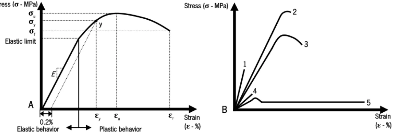
Figure 8. Schematic tensile stress-strain curve. A: Elastic and plastic behavior (zones) until fracture and main mechanical parameters. B: ductile vs brittle materials (1-brittle ceramic; 2- brittle
metal; 3- ductile metal; 4- brittle polymer; 5- ductile polymer) [114][115]. 44
Figure 9. Reduction of MTS compound to a final formazan product (manufacturer’s protocol). 46 D. Results
Figure 10. SEM images of native cBC and gxBC membranes submitted to different drying
procedures, before and after pre-rehydration (Magnification 1.00 K x). 52
Figure 11. SEM images of native cBC membranes soaked in SBF for different time periods
(magnification 1.00 K x). 53
Figure 12. SEM images of native gxBC membranes soaked in SBF for different time periods
(magnification 1.00 K x). 54
Figure 13. SEM images of NaOH modified cBC membranes (30 seconds NaOH 3M, 1 day NaCl free SBF) soaked in SBF for different time periods. Non-modified samples were only subjected
to pre-rehydration and air-drying (magnification 1.00 K x). 55
Figure 14. SEM images of NaOH modified cBC membranes (1 minute NaOH 3M, 1 day NaCl free SBF) soaked in SBF for different time periods. Non-modified samples were only subjected to
rehydration and air-drying (magnification 1.00 K x). 57 Figure 16. SEM images of NaOH modified gxBC membranes (30 seconds NaOH 3M, 1 day NaCl free SBF) soaked in SBF for different time periods. Native samples were only subjected to
pre-rehydration and air-drying (magnification 1.00 K x). 58
Figure 17. SEM images of NaOH modified gxBC membranes (1 minute NaOH 3M, 1 day NaCl free SBF) soaked in SBF for different time periods. Native samples were only subjected to
pre-rehydration and air-drying (magnification 1.00 K x). 59
Figure 18. SEM images of NaOH modified gxBC membranes (3 minute NaOH 3M, 1 day NaCl free SBF) soaked in SBF for different time periods. Native samples were only subjected to
pre-rehydration and air-drying (magnification 1.00 K x). 60
Figure 19. SEM micrographs of BC membranes produced taking into consideration optimized
parameters: cBC and gxBC freeze-dried native, NaOH modified and mineralized. 62
Figure 20. Cryo-SEM cross section images revealing internal structure of produced membranes:
cBC and gxBC freeze-dried native, NaOH modified and mineralized. 63
Figure 21. XRD diffraction pattern of native cBC and gxBC (* identifies BC characteristic peaks). 65 Figure 22. XRD diffraction pattern of NaOH modified cBC and NaOH modified gxBC (* identifies
BC characteristic peaks). 66
Figure 23. XRD diffraction pattern of mineralized cBC and mineralized gxBC (* identifies BC
characteristic peaks and • HA characteristic peaks). 67
Figure 24. Water uptake profile for native cBC, NaOH modified cBC and mineralized cBC over time. Data is represented as mean±SD (n=3) and was analysed by non-parametric Kruskal-Wallis test (P<0.05): (A) denotes significant differences compared with the native cBC, within the same time point; (B) denotes significant differences compared with the NaOH modified cBC, within the same time point; (C) denotes long term significant differences compared with the
native cBC after 1 day of immersion. 68
Figure 25. Water uptake profile for native gxBC, NaOH modified gxBC and mineralized gxBC over time. Data is represented as mean±SD (n=3) and was analysed by non-parametric Kruskal-Wallis test (P<0.05): (A) denotes significant differences compared with the native gxBC, within the same time point; (B) denotes significant differences compared with the NaOH modified
Figure 26. Weight loss profile of native cBC, NaOH modified cBC and mineralized cBC over time. Data is represented as mean±SD (n=3) and was analysed by non-parametric Kruskal-Wallis test (P<0.05): (A) denotes significant differences compared with the native cBC, within the
same time point. 70
Figure 27. Weight loss profile of native gxBC, NaOH modified gxBC and mineralized gxBC over time. Data is represented as mean±SD (n=3) and was analysed by non-parametric
Kruskal-Wallis test (P<0.05). 70
Figure 28. Metabolic activity of 3T3 cells cultured onto native cBC, NaOH modified cBC and mineralized cBC samples. Data is represented as mean±SD (n=15) and was analysed by non-parametric Kruskal-Wallis test (P<0.05): (A) denotes significant differences compared with the native cBC, within the same time point; (B) denotes significant differences compared with the NaOH modified cBC, within the same time point; (C) denotes long term significant differences compared with the native cBC after 1 day of incubation; (D) denotes long term significant differences compared with the NaOH modified cBC after 1 day of incubation; (E) denotes long term significant differences compared with the mineralized cBC after 1 day of incubation. 73 Figure 29. Metabolic activity of 3T3 cells cultured onto native gxBC, NaOH modified gxBC and mineralized gxBC samples. Data is represented as mean±SD (n=15) and was analysed by non-parametric Kruskal-Wallis test (P<0.05): (A) denotes significant differences compared with the native gxBC, within the same time point; (B) denotes long term significant differences compared with the native gxBC after 1 day of incubation; (C) denotes long term significant differences compared with the NaOH modified gxBC after 1 day of incubation; (D) denotes long term significant differences compared with the mineralized gxBC after 1 day of
incubation. 74
Figure 30. Metabolic activity of MC 3T3-E1 cells cultured onto native cBC, NaOH modified cBC and mineralized cBC samples. Data is represented as mean±SD (n=15) and was analysed by non-parametric Kruskal-Wallis test (P<0.05): (A) denotes significant differences compared with the native cBC, within the same time point; (B) denotes long term significant differences compared with the native cBC after 1 day of incubation; (C) denotes long term significant
by non-parametric Kruskal-Wallis test (P<0.05): (A) denotes significant differences compared with the native gxBC, within the same time point; (B) denotes significant differences compared with the NaOH modified gxBC, within the same time point; (C) denotes long term significant differences compared with the native gxBC after 1 day of incubation; (D) denotes long term significant differences compared with the NaOH modified gxBC after 1 day of incubation; (E) denotes long term significant differences compared with the mineralized gxBC after 1 day of
incubation. 76
Figure 32. Fluorescence photographs of live and dead stained 3T3 cells after 1, 3, 7, 14, 21 days of incubation on native, NaOH modified and mineralized cBC. Live cells are stained in green
and dead cells are stained in red. 78
Figure 33. Fluorescence photographs of live and dead stained 3T3 cells after 1, 3, 7, 14, 21 days of incubation on native, NaOH modified and mineralized gxBC. Live cells are stained in green
and dead cells are stained in red. 79
Figure 34. Fluorescence photographs of live and dead stained MC 3T3-E1 cells after 1, 3, 7, 14, 21 days of incubation on native, NaOH modified and mineralized cBC. Live cells are stained
in green and dead cells are stained in red. 80
Figure 35. Fluorescence photographs of live and dead stained MC 3T3-E1 cells after 1, 3, 7, 14, 21 days of incubation on non- native, NaOH modified and mineralized gxBC. Live cells are
stained in green and dead cells are stained in red. 81
E. Discussion
Figure 36. Schematic representation of the mineralization mechanism proposed for BC. Adapted
A. Introduction
Table 1. Features to consider when choosing or designing bone replacement solutions [5][30] 11
Table 2. Cellulose-producing bacteria [57-58] 19
Table 3. Comparison of plant and bacterial cellulose [49] 22
Table 4. Different types of calcium orthophosphates [71-72] 26
C. Materials and methods
Table 5. Reagents for SBF and NaCl-free SBF preparation: order of addition and quantity [71] 41
Table 6. Thickness average values of tested samples 43
D. Results
Table 7. Elemental analysis of produced BC membranes (Wt – mass fraction). Data is represented as mean±SD (n=3) and was analysed by non-parametric Kruskal-Wallis test (P<0.05): (A) denotes significant differences compared with the native cBC; (B) denotes significant differences compared with the native gxBC; (C) denotes significant differences compared with the NaOH modified gxBC. Differences between BC types were analysed by Mann-Whitney U
test (P<0.05) 64
Table 8. Ca and P concentration (% and g/mol) on mineralized BC samples. Ca/P ratio. Data is represented as mean±SD (n=3) and was analysed with Mann-Whitney U test (P>0.05) 65 Table 9. Young modulus (MPa). Data is represented as mean±SD (n=3) and was analysed by
non-parametric Kruskal-Wallis test (P<0.05): (A) denotes significant differences compared with the native cBC tested in wet conditions; (B) denotes significant differences compared with the native gxBC tested in wet conditions. Differences between BC types and testing conditions
were analysed by Mann-Whitney U test (P<0.05) 71
Table 10. Fracture strength(MPa). Data is represented as mean±SD (n=3) and was analysed by
non-parametric Kruskal-Wallis test (P<0.05): (A) denotes significant differences compared with the native gxBC tested in dry conditions; (B) denotes significant differences compared with the native gxBC tested in wet conditions. Differences between BC types and testing
conditions were analysed by Mann-Whitney U test (P<0.05) 72
Table 11. Strain-at-failure (%). Data is represented as mean±SD (n=3) and was analysed by non-parametric Kruskal-Wallis test (P<0.05): (A) denotes significant differences compared with the
native gxBC tested in wet conditions. Differences between BC types and testing conditions
were analysed by Mann-Whitney U test (P<0.05) 72
E. Discussion
Table 12. Mechanical parameters of Biomend Extende®, TBR® and Bio-Gide® commercial
A. Introduction
Introduction
A well-established fact in human biology comprehension is that human beings are not immune to disease nor trauma [1]. In times when average life expectancy was shorter, a common solution to overcome this setbacks was the removal of injured tissues in an attempt to marginally improve quality of life [2]. Nevertheless, since ancient times, Man tries to find solutions to deal with disease, transforming health care in an emerging area that awakens the interest of several scientific domains, including material science. As a result, medical devices where developed for several areas and found application in improving modern society health, now characterized by an high average life expectancy, also due to medical evolution in terms of antiseptics, antibiotics, hygiene and vaccination [2-4].
Biomaterials field emerged from the partnership between materials and biological sciences, outlining a set of “implantable materials that perform their function in contact with living tissues”, as defined by M. Vallet-Regí [3].
Despite no material is fully inert, as all trigger a response by the physiological environment, the first generation of these materials, named bioinert, was intended to restore basic functions of tissues with minimal biological reaction, without toxicity and with a successful outcome [4]. However, these inert solutions are changing to more innovative ones, with desired bioactivity, taking into account the final application [7-9].
With an increasingly elderly population and/or with risk activities on daily routines, bone-related diseases and bone fractures increase and new and attractive solutions are of interest [6].
1.
Bone
Skeletal system is one of the most important structures of vertebrate organisms as it provides support, protection, allows movements in conjunction with muscles, and is a reservoir of minerals and fats and a site of blood cells production. In its constitution is a specialized type of hard endoskeletal connective tissue, bone, associated cartilages, ligaments and joins [15-17].
Bone is the major constituent of this system and the human skeleton is composed of approximately 200 bones, in a total weight of almost 2 Kg [8]. Bone belongs to a large family of biological structures, all having in common mineralized collagen fibers in their structure [15-19]. Some of these are mineralized tendons, dentine and cementum (intern filler of the tooth and layer which connects the teeth roots to jaw, respectively) [9].
It is necessary to keep in mind that a clear distinction must be made between bone as a tissue and bone as an organ. The first, is generally composed of bone cells and mineralized extracellular matrix, while in the second case, in addition to bone tissue, also encompasses cartilage, fibrous tissue, marrow and blood vessels. All these variables relate together yielding four types of bone: long, short, flat and irregular [7][10-11].
1.1. Bone as a tissue
1.1.1. Constitution: bone cells and bone matrix
Bone cells are categorized in four groups, according to their function and origin: osteogenic precursor cells, osteoblasts, osteocytes and osteoclasts [12].
Osteogenic precursor cells, or osteochondral cells, have a broad distribution in bone tissue, being present in all nonresorptive bone surfaces and forming the deep layer of periosteum [12]. Furthermore they are mature and highly metabolic active cells and also the progenitor of osteoblasts [7][12].
These last play a unique role in osteogenesis as they are specialized cells capable of production of both organic and inorganic phase of bone: they are able to produce collagen and proteoglycans and to accumulate calcium and phosphate ions, as well as other enzymes [7][13].
Another type of bone cell are osteocytes that are mature cells, which represent 90-95% of totality of bone cells and are the result of the entrapment of osteoblasts by mineralized collagen matrix [7]. This cell type is less metabolically active when compared with its progenitor, being involved in the balance of calcium and phosphorus concentration, playing an important role in adaptive remodeling behavior in response to local environmental conditions [7][20-22].
Finally, osteoclasts are cells responsible for resorption of bone, having a crucial role in bone reorganisation [7]. This multinucleated cell entity has its origin in mononuclear precursors from red bone marrow, the same stem cells that are progenitor of monocyte (monocyte/macrophage lineage precursors), under strict regulation of osteoblasts, osteocytes and bone marrow stromal cells [7][13].
Bone matrix is responsible for the functional characteristics of bone. It is constituted by an organic and inorganic phases, that in a mature bone represent 35% and 65% of the bone weight, respectively [7]. Some physiological characteristics of this tissue are assigned to each part: organic phase grants flexible bone strength, while the inorganic phase ensures resistance to mechanical forces, hardness, strength and a protective effect on soft tissues [14-16].
The organic portion of bone matrix is essentially composed of collagen fibers and non-collagenous proteins [23-24].
Collagen fibers are the most representative part and are mainly collagen type I. Although little is known about the way these molecules are assembled, their internal organization appears to play an important role in bone mineralization. The arrangement and concentration of these proteins depends on the type of bone, showing higher levels in compact bone. In contrast, it is also in this same type of bone tissue that non-collagenous components appear in minimum concentration [23-24].
Non-collagenous proteins are a wide group comprised of two hundred or more elements, both permanent nature components, like proteoglycans, as well as cellular byproducts like alkaline phosphatase and growth factors, and other molecules normally present in circulating body fluids, including albumin and α2HS-glycoprotein. These components are considered crucial on the induction and regulation of biomineralization [23-24]. In addition, they vary both qualitatively, depending on the type of bone, and quantitatively, depending directly on bone formation rate and indirectly on collagen fibers density [15].
Historically, the description of inorganic phase composition has changed considerably. Nowadays there is a consensual claim that recognises hydroxyapatite as the mineral phase (Ca10(PO4 )6(OH)2), being described by some authors as dahllite (Ca5(PO4, CO3)3(OH)2), with about 5%
carbonate and trace amounts of other ions, like sodium (0.9%) and magnesium (0.5%) [7][15-16]. However, bone hydroxyapatite differs from its nature counterpart and several studies have been carried out in order to unravel its true crystalline nature. Several factors, such as age and inorganic and organic matrix phases’ correlation, influence both composition and structure of the mineral in
the biological environment. Polycrystallinity is considered the true crystalline nature of bone, undergoing changes according to the parameters mentioned above [23-24]. On the other hand, this polycrystalline nature is chiefly associated to the incorporation of the aforementioned ions and to the nonstoichiometry characteristic of this phase [16]. In addition, this biogenic mineral is also classified as a calcium-deficient apatite. In fact the Ca/P molar ratio is a distinctive factor between the two: in bone the ratio varies between 1.57 and 1.71 and in natural hydroxyapatite it is 1.67 [16][23-24].
1.1.2. Types of bone tissue
Besides the different types of mechanisms associated with bone formation (mentioned below), these always involve the formation of a less organized tissue that is replaced by a more mature one [11][14][16].
Primary bone formation is a rapid and unorganized process, resulting in woven bone, also known as immature, embryonic or primary bone. Its fibers are randomly aligned forming bundles of collagen with interfibrilar spaces containing non-collagenous proteins, resulting on calcification nodules or calcospherites formation [11][14-16]. Woven bone is replaced by lamellar bone due to the action of osteoclasts and osteoblasts during secondary bone formation [7][16]. Resultant tissue can also be called mature, secondary, parallel-fibered or osteonic and it is much stronger than the last one presented. Here, collagen fibers are clustered in structures designated as lamellae, in parallel arrangement to each other, alternating in longitudinal and transverse orientations [11][14-16].
Either woven or lamellar bone can be classified as compact and trabecular bone, based on structural organization, mechanical characteristics, amount of bone marrow, cellular function and metabolic activity [14].
Trabecular bone, also named cancellous or spongy bone, is considered, by some authors, as a primary type of bone [12]. It is essentially present in the core of short and flat bones and in epiphyseal ossification centres [7]. Trabeculae, formed by several lamellae, compose the network of this tissue, and its orientation is dependent of stress lines within bone, adaptable to slight changes [7][14].
Compact bone is denser and less porous than its counterpart and it is found in long bone diaphysis and in cortical shell of short and flat bones [7]. In fact, it is characterized by a dense organization where lamellae, in association with osteocytes, are arranged around a central channel called osteon
or haversian system. These structures are parallel to bone axis and contain loose connective tissue, nerves and blood vessels responsible for nutrition and depuration of bone cells. Feeding is performed via Volkmann's canals, which are perpendicular to bone axis [7][14].
1.2. Bone as an organ
As mentioned before, bones can be classified as long, short, flat or irregular [7]. Moreover, in the same bone there are often different types of bone tissue with well-known structural organization and composition characteristics [15-16] .
Long bones, like tibia, are one of the four types of bone, where three distinct parts may be defined: diaphysis or shaft, composed essentially by compact bone with a large space called medullary cavity filled with marrow; epiphysis or end of the bone, composed essentially by cancellous or spongy bone, filled also with marrow; and epiphyseal plate or growth plate, composed by hyaline cartilage, situated between epiphysis and diaphysis and where growth of bone in length takes place, being named of epiphyseal line, a calcificated area, when growth stops [7].
The other three categories of bone are usually devoid of diaphysis or epiphysis, having an interior framework of cancellous bone surrounded by compact bone. The main differences are based on the morphological features: short bones, including finger bones, are nearly cube-shaped or round and medullar cavity is not present; flat bones like skull and ribs for example, are thin, usually curved and with flattened shape; irregular bones, like vertebrae, present shapes diverse from the ones described above.
External and internal surfaces of bone are typically wrapped with periosteum and endosteum membranes, respectively. Periosteum is a connective tissue membrane constituted by two layers: the outer layer is dense and irregular containing essentially blood vessels and nerves, while the inner layer is composed of bone cells. Endosteum has a composition similar to the inner membrane of periosteum [7].
All bones have a complex hierarchical microstructure, where several levels of organization can be distinguished (Figure 1) [10][17].
1.3. Bone dynamics: formation, growth, remodelling and repair 1.3.1. Formation, growth and remodelling
Two distinct developmental processes are involved in bone formation, occurring since embryological development: intramembranous and endochondral ossification [7].
Intramembranous ossification is the process involved in flat bone formation, like the majority of craniofacial complex bones and medial clavicles [7]. In this process, skeletal mesenchymal cells mediate bone formation, without the participation of a cartilage mold [11][18]. Around the fifth week of embryonic development, embryonic mesenchymal cells aggregate and form a highly vascularized membrane of connective tissue [7]. Cells in this membrane become osteochondral progenitor cells, which then differentiate into specialized osteoblasts ready to synthesize bone matrix and develop structures known as ossification centres. Here, connective tissue collagen fibers and cells are entrapped, culminating in the formation of woven bone that will be remodeled to trabecular bone. The process of bone formation continues, leading to trabeculae enlargement, with a decrease in the spacing between other similar structures, resulting in partial closing of vascular connective tissue, which is a precursor of hematopoietic tissue. The tissue becomes organized, forming layers, which are arranged in concentric rings around a blood vessel, known as haversian systems. The formation of the endosteum and periosteum occurs after condensation of outer and inner layers of connective tissue, being areas rich in osteoprogenitor cells [7][26-27].
In endochondral ossification, bone is formed by substitution of a skeletal cartilage template. This process is associated to appendicular skeleton, vertebral column, facial bones and pelvis bone formation [26-27]. This process involves mesenchymal cells differentiation into chondroblasts that are capable of cartilage synthesis. The cartilage template is normally enclosed in a capsule named perichondrium, whose invasion by blood vessels initiates the conversion of osteoprogenitor cells into osteoblasts and synthesis of compact bone by intramembranous ossification on the mold surface, forming the collarbone. At the same time occurs hypertrophy of cartilaginous mold and the hypertrophic chondrocytes (essentially in the middle zone of the structure) produce collagen type X (a hypertrophy indicator) and angiogenic factors such as vascular endothelial cell growth factor (VEGF). These features promote calcification of the template; cells became entrapped and eventually die, forming gaps. These enable the entrance of blood vessels in the calcified mold, carrying osteoprogenitor but also hematopoietic cells and culminating in the formation of trabecular bone on the mold surface and then of cancellous bone on the diaphysis of the structure. As the primary ossification centre is formed, the process of bone matrix deposition continues until all calcified cartilage is replaced by bone [7][18].
Long bones, in particular, present the development of secondary ossification centers in the epiphysis, where the ossification process occurs in a similar way to what happens in primary ossification center. The majority of cartilage in this area is replaced by cancellous bone, with the exception of epiphyseal growth plate and articular cartilage. Long bones and bone projections growth in length occurs in epiphyseal plate, involving interstitial cartilage growth followed by appositional bone formation, thus ensuring bone growth and maintenance of epiphyseal plate thickness. Articular cartilage is the place where epiphyses as well as bones without epiphyseal plate increase in size. The events are similar to those mentioned for epiphyseal line, with the difference that articular cartilage does not ossify. It is important to notice that bone grows both in width and thickness, phenomena associated with periosteum. Summarily appositional bone growth occurs under the periosteum, with varying rates, forming structures named osteons [7][18]. Finally, osteoclasts play an essential role in the diaphysis, resorbing bone in order to form the spinal canal, while the hematopoietic cells will form red bone marrow. Bone is fully formed when all the perichondrium is transformed in periosteum, the epiphyseal plate is mineralized and the process of bone remodelling is finished [7][18].
This last process is continuous, occurring through the entire life cycle. Once the maturity of the structure is reached, it plays a crucial role in the replacement of older bone by new one and in its
response to environmental conditions, like adjustment of bone to stress and regulation of calcium ions in the body [7][19].
In a simple way, bone remodelling occurs through the action of osteoclasts that remove old bone, and of osteoblasts, which deposit new tissue, converting woven bone into lamellar bone [7][19]. Both cortical and trabecular bone remodelling are the two forms of this process. The first occurs within the osteon and it is associated with resorption of old haversian systems and formation of new ones. The last is associated with trabecular endosteal surface, being regarded as a surface process [18].
1.3.2. Repair
As seen before bone is a living and dynamic tissue where a series of biological events occur constantly: resorption, deposition and remodelling [7][11][26-28]. Another important process is bone repair or bone healing [7][20-21]. This complex process is synced in such a way that is effective in repair and restores bone functions. Induced by an immune response triggered by an injury, several entities participate in it, recapturing the processes of bone formation that occur during embryonic development [20].
Bone has the ability of restoring itself without forming a scar and during the last two decades, attention has been focused in understanding the fracture healing mechanism, unveiling the diverse pathways involved in it. Classically the process can be direct and indirect [30-31].
The majority of fractures heal by indirect bone healing, also named of secondary bone healing. This process is characterized by the use of intramedullary and external fixation and conservative treatment using a plaster cast. These solutions allow fracture stabilization, while maintaining a controlled motion between the fractured surfaces subjected to load, thus enhancing the process if the right stability is in fact achieved. This process combines both intramembranous and endochondral ossification in overlapping phases, with the involvement of a cartilaginous template: hematoma formation, cartilage callus formation and ossification and bone remodeling (Figure 2) [29-31].
Direct fracture healing or primary bone healing occurs by direct contact between the cortical ends of fractured bones and depends on the action of functional units called “cutting cones” to re-establish the lamellar bone structure, without the involvement of a cartilage intermediate [11][21]. Furthermore, absolute stability is desired to ensure correct healing of the structure. In this process, a correct anatomic reduction of the fracture fragments is essential and a rigid internal fixation is achieved by compression, which promotes a reduction of interfragmentary force, elimination of any failure within the interfragmentary area and the need for an external callus or vessels and cells for the process to unfold [11][29-31]. The direct route can be divided in contact or gap healing, being the main difference between the two processes the fact that in the second case, bone union and haversian remodeling occurs separately in time [21].
1. Hematoma formation and inflammation
a) The damage of the surrounding tissues leads to the disruption of blood vessels and consequent formation of hematoma by the activation of coagulation mechanisms. This structure encloses the fracture area.
b) Acute inflammatory response: the avascular zones at the end of fractured bone lead to the appearance of apoptotic cells in that area, which produce chemotactic molecules (IL-1, IL-6, IL-11, IL-18 and TNF-α) responsible for the recruitment of other inflammatory cells, as well as mesenchymal cells. This increase in local vascularity is essential to the phagocytosis of necrotic material and the mesenchymal cells will differentiate into osteoprogenitor cells, essential to the repair of this tissue.
2. Cartilage callus formation and ossification
a) The hematoma organizes into granulation tissue, which results in the reduction of in situ strain to <10%, crucial to cartilage or soft callus formation. This phenomenon occurs initially in less stable areas in order to attenuate this factor (between the fracture ends and external to periosteal sites).
b) At the same time, intramembranous ossification occurs at periosteum and an external callus is formed, resulting in a semi-rigid structure able to support weight bearing.
c) The soft callus undergoes endochondral ossification becoming hard callus, essentially constituted by woven bone, a structure increasingly solid, rigid and with better mechanical properties. The formation of this structure can be followed by measurement of markers such as type I procollagen, osteocalcin, alkaline phosphatase and osteonectin.
3. Bone remodeling
a) In order to reach the right mechanical characteristics, woven bone is remodeled into lamellar bone and the vascular supply returns to normal levels. A perfect balance between bone resorption by osteoclasts, bone deposition by osteoblasts, biochemical mediators (IL-1, TNF-α, BMP), adequate blood supply and a gradual increase in mechanical stability is essential.
1.4. Bone defects
There are several pathological scenarios, such as trauma and congenital malformation, fractures, surgery, periodontitis, osteoporose, osteomyelitis or tumors, which promote the destruction of essential components of bone tissue and/or compromise its correct function. In general, bone defects can be classified as critical-sized defects, calvarial defects or long bone or mandible segmental defects. If inappropriate osseous reconstruction occurs, it can result in impaired bone healing with the formation of fibrotic tissue, pseudoarthrosis or artificial joints, with no physiological significance [8][22][32-33].
Diverse factors influence both quantity and quality of bone matrix, including age and sex. In fact, the majority of bone defects can be correctly healed in younger people. Nevertheless and regardless of age, larger defects may need the use of advanced solutions [17][24]. On the other hand, men typically have denser bones than women as a result of their higher body mass and levels of testosterone. However, in both sexes, increasing age has deleterious effects on skeleton and bones become more brittle due to a decrease on collagen production and less dense due to changes in the equilibrium of bone resorption and formation. All this factors are favourable to the development of an osteoporosis scenario, where the probability of fracture is increased [7-8][19]. Another condition where bone becomes more fragile and there is a greater likelihood of fracture is osteomalacia. This results from organ decalcification and it is a case of attention during pregnancy when the need for calcium is higher for fetus development [7].
Bone fractures or defects can be the result of conditions that weakens and decreases the mechanical strength of bone, or of external aggressions, including trauma situations. Fractures are characterized by a break or discontinuity in the bone as a result of an applied force that exceeds the limits of tension or compression that the tissue is able to handle. These fractures may be complete or incomplete, displaced or undisplaced, comminuted or simple, open or closed [25].
Osteomyelitis and periodontitis are another example of conditions that affect bone integrity. Osteomyelitis is a bacterial infection that can lead to complete destruction of bone structure and is
caused, in the majority of cases, by Staphylococcus aureus but also by Mycobacterium
tuberculosis, known as bone tuberculosis [7][22]. Periodontitis is associated with the oral environment and the destruction of periodontal tooth supportive structures. These and other situations of chronical dental disease, either caused by inflammation or infections, may end in bone jaw resorption as well as reduction of jaw bone volume, which complicates implants placement [8][26].
Bone tumors and tooth extraction constitute other examples that can be associated with bone defects [7][26-27]. Although they do not directly originate bone defects, the removal process may lead to the formation of these gaps [26-27].
1.5. Solutions
The search for new advanced solutions for bone defects repair has been a defying goal for the medical community since ancient times. In fact, traces of the use of materials as bone extracts is dated as far as the Neolithic [5].
Presently, the availability of solutions for bone reconstruction include procedures that make use of bone grafts and bone graft substitutes [28]. These solutions must fulfil a set of properties that should be taken into consideration when choosing the most appropriate procedure to be used or in research of new solutions (Table 1) [7-9].
Intended clinical application Availability Ethical issues Biocompatibility
Defect size Desired bioactivity Durability Sterilization
Biomechanical properties Desired resorption rate Associated side effects Cost Chemical composition Handling characteristics
Bone grafts are seen as the traditional solutions employed in bone defect treatment. These comprise autografts, allografts/alloimplants or xenografts/xenoimplants. However, some drawbacks associated to these solutions have fostered the growth of orthopaedic biomaterials market [17][27].
A broad range of materials are currently studied and employed either as bone grafts or for guided bone regeneration (GBR) or tissue engineering approaches. Commonly used materials include polymers, ceramics, metals and composites, either of natural or synthetic origin [17][29]. These are available in several forms, like chips and granules of different sizes, putty or injectable pastes, which is extremely important for surgeons in daily practice [30].
As research on novel solutions is moving from inert to bioactive materials, features like osteoconduction, osteoinduction and/or osteogenesis become crucial [11-14][31]:
Osteoconduction: material's ability to ensure structural support for superficial and/or internal growth of blood capillaries and cells, promoting growth of new bone. This property may be associated with either viable or nonviable material and is heavily dependent on
structural properties of the material, such as porosity, pore size and interconnectivity and surface roughness.
Osteoinduction: material's ability to induce recruitment of cells from the surrounding tissue into the material, thus stimulating bone regeneration. This ability is attributed to a wide range of molecules such as bone morphogenic proteins (BMPs), platelet-derived growth factors (PDGF), insulin-like growth factors I and II (IGF-I and IGF-II), fibroblast growth factors (FGF), epidermal growth factor (EGF), transforming growth factor beta (TGF-β), retinoic acid, sulphated glycosaminoglycans and amino acids.
Osteogenesis: material's ability to induce new bone formation due to the presence of osteoblasts and stem cells in the transplanted/implanted material.
1.5.1. Bone grafts
Autografts consist in transplantation of patient own bone tissue from a site to another within the organism, allowing the utilization of a bone solution with natural osteoconduction, osteoinduction and osteogenic characteristics. Considered the gold standard of bone defects treatment, it offers beyond inherent biocompatibility and minimal ethical issues, rapid healing and complete incorporation, lack of disease transmission and immunogenicity. On the other hand, increased operative time, limited availability of tissue, infection probability, inflammation, compromised biomechanical properties, donor site morbidity and pain are some of the disadvantages associated with this solution [2][8][11-14][17][30].
Donor site morbidity and availability of tissue are some of the drawbacks that may be resolved with the use of allogenic bone grafts and implants. These can be obtained from cadavers or living donors; however, the immunogenic potential and rejection rate associated with fresh, fresh frozen and freeze dried materials has to be considered. In order to try to minimize this, strategies based on demineralized freeze-dried allogenic bone have been proposed. Slow osteointegration, risk of disease transmission and chronic inflammation constitute another set of drawbacks [11-13][30][32].
Xenografs or xenoimplants are another option, which relies on living or non-living tissues from nonhuman species are used, more routinely bovine or porcine. Even when chemically processed, these solutions are far from what is considered ideal, presenting innumerous disadvantages mainly due to the lack of biocompatibility, pathogen transmission, immunogenic rejection, prolonged graft integration, and fracture. However, these solutions are utilized in diverse
situations as an end-line solution, including heart valve replacements (porcine) or bone substitutes (bovine), as ethical considerations have also to be considered [2][28].
1.5.2. Tissue Engineering
“An interdisciplinary field of research that applies the principles of engineering and the life sciences towards the development of biological substitutes that restore, maintain or improve tissue function” is Langer and Vacanti’s definition of tissue engineering [37-38].
There are three main constituents that come into play in a tissue engineering strategy: cells, growth factors or signalling molecules and matrices or scaffolds. The combination of these elements depends on many factors, including patient age, gender, health, systemic conditions, habits and anatomical shape of the implant [37-38].
In the particular case of bone, cells that normally compose the tissue engineered construct should possess osteogenic potential. Periosteal cells, osteoblasts and mesenchymal stem cells (MSCs) are widely used, from which MSCs are the most often applied in bone tissue engineering [29][34].
In order to provide support for cells’ expansion, differentiation and proliferation, but also for the load and subsequent release of growth and differentiation factors, scaffolds constitute an essential component in the overall tissue engineering approach. Pore size, surface and mechanical properties, osteoinductivity, biodegradability, biocompatibility and processability are some of the most important properties of a scaffold. In this sense, the appropriate choice of the materials to be used plays a critical role. However, there is a wide availability of materials that can be employed in tissue engineering research, ranging from metals, ceramics, polymers and composites, from natural and/or synthetic origin, and which can be used in solid preformed structures or injectable form [29][33].
Natural or synthetic hydroxyapatite, calcium phosphate solutions, like β-tricalcium phosphate and bioactive glass, are some of the ceramic materials employed in scaffolds manufacturing. Some advantages of these materials include osteoconductive and osteoinductive properties, being brittleness, low mechanical stability, difficulty to predict degradation/dissolution rates and poor reproducibility due to high variability associated to ceramics, some of the known drawbacks [29][33][35].
Polymers are seen as an attractive alternative to ceramics and can also be divided in natural and synthetic. Natural polymers widely studied for bone tissue engineering comprehend collagen, fibrinogen, chitosan, starch, hyaluronic acid and/or calcium alginate, which exhibit good
biocompatibility, low immunogenic potential, chemical versatility, osteoconductive properties and, in some cases, biodegradability and osteoinductivity [29][33][35]. On another hand, lack of mechanical stability and biochemical changes brought by sterilization techniques are described drawbacks [29][35]. Synthetic polymers are chemically more versatile and different physical and mechanical characteristics and degradation profiles may be obtained. Poly(α-hydroxy acids), poly(ε-caprolactone), poly(propylene fumarates), poly(carbonates), poly(phosfazenes), poly(lactic acid), poly(glycolic acid), poly(doxanonone), poly(anhydrides) associated copolymers are some of the materials included in this group [29][33][35].
Titanium and tantalum are two metallic materials that have been applied in metallic scaffolds’ fabrication and as prosthetic implants coatings, with many different shapes and textures. These materials are biocompatible, resistant to corrosion and durable. Yet, they are bioinert and not biodegradable that together with their high stiffness when compared to bone are the main disadvantages of these metallic solutions [29].
In order to combine advantageous individual characteristics of different types of materials, composites have emerged as a new class. Some interesting combinations include polymeric-ceramic composites and metallic-polymeric-ceramic composites, leading to improved properties as brittleness, drug delivery and mechanical properties in the first case and bioactivity in both [29].
The last main constituents of any tissue engineering approach are growth factors or signalling molecules. Particularly relevant for bone tissue engineering applications are cytokines, specifically BMPs, which are essential in the osteogenic differentiation process [38-39]. Infuse® Bone Graft/LT Cage® fusion device is a specific case of an existing commercial solution based on a bovine collagen matrix loaded with recombinant human BMP-2 (rhBMP-2) for the treatment of degenerative disc disease [35].
1.5.3. Guided Bone regeneration
Developed in 1980, based on the philosophy of guided tissue regeneration, GBR is another procedure applied in bone reconstruction, with particular emphasis on periodontics and implant dentistry [25][32].
The principle of this method relies on preventing the invasion of the defect by soft tissues (mainly composed of non-osteogenic cells), formation of fibrous connective tissue and both establishing and maintaining a propitious environment for recruitment, proliferation, differentiation and
maturation of osteoprogenitor cells on the isolated defect cavity. This is usually accomplished by the use of occlusive or barrier membranes [40-42].
GBR is influenced by a number of factors, including patient-related like smoking habits, diabetes or another limitative pathology; defect-related like morphology, length and angle; and membrane-related, including membrane fixation, passive flap tension, cortical penetration, excessive swelling and membrane composition [32].
A cellulose acetate filter (Millipore) was the first membrane used in this type of approach [32][37]. Nowadays, available solutions have evolved in order to meet the diverse requirements for the expected positive results. These include biocompatibility, space making ability, cell occlusion properties, prevention of bacterial infection integration by the host tissues and clinical manageability, adequate mechanical properties and ability to isolate and stabilize the defect and the fibrin clot or other materials of different origins, such as autogenous bone chips, allografts, xenografts or alloplasts (synthetic) that may be used to occupy the defect and facilitate bone ingrowth [32][41-43].
In general, GBR materials may be characterized as non-resorbable or resorbable materials and their properties are designed to optimize tissue regeneration. These materials may possess antimicrobial properties, be enriched with bioactive molecules or tailored to possess desirable physical or chemical characteristics [39][41-42].
Non-resorbable membranes are mostly synthetic and include expanded and high-density polytetrafluoreethylene (e-PTFE: Gore-tex®; and dPTFE: TefGen-FD®, respectively), titanium mesh (Ti-mesh) and titanium-reinforced ePTFE (Ti-e-PTFE) [32][38]. However, the natural polysaccharide bacterial cellulose, as Gengiflex from BioFill company, have also been studied with promising results [40-41].
Biocompatibility, predictable profile during the healing process, mechanical strength and easy handling are the main advantages that support the use of non-resorbable materials. However, there is the need for a second surgical intervention to remove these membranes, which represents increased costs and patient discomfort [36-37][39][42].
Resorbable solutions partly solve these problems, but immunogenicity, porosity, unpredictable duration of barrier function (degradation and resorption rates) and mechanical performance, when compared to the non-resorbable counterparts, represent some of the disadvantages of these solutions [39][40-41]. Used resorbable membranes may be from natural or synthetic origin. Poly(glycolic acid), poly(lactic acid), poly(ε-caprolactone), poly(urethane), poly(glactin 910),
poly(dioxanon) and their co-polymers represent the synthetic polymeric solutions (Guidor®, Resolute®, Epi-Guide®, Mempol®, Vicryl Periodontal Mesh®, Atrisorb®, Experimental Mempol®). Their natural equivalents include collagen derived from animal skin, tendon or intestines (BioGide®, BioMend®, Avitene®, Collistat®) [32][38-39][42].
2. Bacterial cellulose (BC)
Polymers are ubiquitous in the living world, forming the building blocks of life. On the other hand, polymers are extensively studied and used in diverse industrial areas, ranging from food, textiles, cosmetics, pharmacy and/or medicine, among others [47-48].
By definition a polymer (from the Greek poly – “many”; meres – “parts”) is a macromolecule composed of small repeating units, named monomers, linked by covalent forces. These materials can be classified according to their origin, structure, polymerization mechanism, preparative techniques or thermal behaviour [43].
Focusing on polymer’s nature, they can be classified as natural or synthetic [47-49]. Natural based polymers, or biopolymers, in particular, possess diverse properties that make them suitable to be applied in highly demanding areas such as the medical one. Similarity to biological macromolecules is one of the major advantages of these polymers, as compared to their synthetic counterparts, which enhance biological recognition, cell adhesion and metabolic acceptance and processing. Some disadvantages of this type of materials are related to batch-to-batch consistency; material source, which can lead to undesirable immune reactions due to, for example, the presence of impurities or endotoxins; and to processing technics that can impact polymer’s final properties, including mechanical properties or biodegradability. However, the continuous research and development of novel methods for polymer production, purification and processing will allow an increasing control over material’s properties and enhance its biofunctionality and contributing for the overall reduction of toxicity and chronic inflammation and immunological reactions [48-49]. Natural polymers are instinctively appealing and researchers are investing considerable efforts on their synthesis and production. In this context, biotechnological pathways arise as a strategic alternative, based on microorganisms’ fermentation and enzymatic processes [45]. Considering their chemical structure, polysaccharides, proteins, polyesters, but also inorganic polyanhydrides, enclose the main classes of polymers produced by bacteria [49-50].
Polysaccharides are a group of macromolecules abundant in nature, composed of monosaccharides linked through O-glycosidic linkages. The variety of monosaccharides that can build them up, bonding, shapes and molecular weight greatly contribute for the diversity associated to this class of molecules. Any variation in any of these parameters will consequently result in a different structure with different surface, interfacial and physicochemical properties, including solubility, flow behaviour and gelling potential, and functionality [45-46].
Polysaccharides produced via microorganism’s fermentation can be classified according to their morphological localization in intracellular polysaccharides, found within the cell or as a part of the cytoplasmic membrane (e.g. glycogen); capsular or cell wall polysaccharides, as a structural component of the capsule or cell wall (e.g. K30 antigen); and extracellular polysaccharides, located outside the cell wall, which can be synthesized or secreted by cell wall-anchored enzymes (e.g. xanthan, dextran, cellulose, hyaluronic acid) [50-52].
Cellulose is one of the most abundant biopolymer on earth and can be found in several natural reservoirs (Figure 3) [53-54].
One of the best known and well established industrial sources of cellulose are plants, where cellulose is the major component of the vegetable biomass. Plant-origin cellulose constitutes the primary component of cell walls, naturally associated to other components like acidic polysaccharides, glycoproteins and waxy aromatic substances, including hemicelluloses, pectins and lignins. Cellulose is also naturally present in some fungi and algae, where it plays an important role in structural maintenance [50-52][55-56]. It is also a representative by-product associated with some bacteria, where it acts as an important reserve material, but also as a component responsible for structural protection, particularly relevant under unfavourable environmental conditions (Table 2) [57-58].
Figure 3. Principal pathways of cellulose production and molecular arrangement with identification of β-1→4 bond [55].
β-1→4 bond
Genus Produced molecule Biological Role
Acetobacter Extracellular pellicle composed of ribbons Keep aerobic environment
Achromobacter
Cellulose fibrils
Flocculation in wastewater
Aerobacter Alcaligenes
Pseudomonas No distinct fibrils
Zoogloea Not defined
Agrobacterium Short fibrils Attached to plants Rhizobium
Sarcina Amorphous celulose Unknown
Bacterial cellulose (BC) is an exopolysaccharide produced by several species of bacteria, particularly gram-negative species, such as Gluconacetobacter (formerly known as Acetobacter), Agrobacterium, Aerobacter, Achromobacter, Azotobacter, Rhizobium, Sarcina, Pseudomonas, Alcaligenes and Salmonella, but also by the gram-positive Sarcina ventriculi. The microorganism model used for cellulose studies is Gluconacetobacter xylinus (G. xylinus) due to its ability to produce this polymer in high quantities using a wide range of carbon and nitrogen sources as substrate [50-52][56-57].
Cellulose, with the molecular formula (C6H10O5)n, is a linear homopolysaccharide, composed
of glucose (D-glucopyranose as D-glucose is present as a pyranosyl ring), linked through a β-1→4 glycosidic bond [46][52]. Assembly of the β-1→4 glucan chains will determine the crystallinity of the material (Figure 3) [48].
2.1. BC fermentation and biosynthesis
G. xylinus, also known as Acetobacter xylinus, is a rod-shaped gram-negative α-proteobacteria, found in diverse natural habitats, like vegetables, vinegar, alcoholic beverages, rotting fruits and niches where carbon is fixed in the form of sugar or alcohol [48]. This strictly aerobic and non-photosynthetic microorganism is characterized by its ability to convert several organic substrates, like glucose and glycerol, into cellulose. Despite being a non-flagellated cell, some strains show relative mobility, observed microscopically, during formation of cellulose ribbon [53-55].
BC production is dependent on several conditions, like environmental conditions, culture media and by-products [52-53].
Among different environmental conditions, pH, temperature, dissolved oxygen and culture agitation are the most widely studied for their effect on cellulose synthesis [52].
![Figure 2. Indirect bone healing mechanism [7][29-31] .](https://thumb-eu.123doks.com/thumbv2/123dok_br/17694060.827802/34.892.133.766.106.348/figure-indirect-bone-healing-mechanism.webp)
![Figure 3. Principal pathways of cellulose production and molecular arrangement with identification of β-1→4 bond [55]](https://thumb-eu.123doks.com/thumbv2/123dok_br/17694060.827802/43.892.153.752.399.642/figure-principal-pathways-cellulose-production-molecular-arrangement-identification.webp)
![Figure 4. Mechanism and SEM micrograph of cellulose ribon formation by G. Xylinus [49][54].](https://thumb-eu.123doks.com/thumbv2/123dok_br/17694060.827802/46.892.152.750.109.213/figure-mechanism-sem-micrograph-cellulose-ribon-formation-xylinus.webp)
![Figure 5. Molecular hydrogen bonds (indicated by arrows) established in cellulose [49]](https://thumb-eu.123doks.com/thumbv2/123dok_br/17694060.827802/49.892.248.653.436.688/figure-molecular-hydrogen-bonds-indicated-arrows-established-cellulose.webp)

![Table 5. Reagents for SBF and NaCl-free SBF preparation: order of addition and quantity [71]](https://thumb-eu.123doks.com/thumbv2/123dok_br/17694060.827802/66.892.173.721.138.347/table-reagents-sbf-nacl-preparation-order-addition-quantity.webp)