http://dx.doi.org/10.1590/s2175-97902018000217617
Article
*Correspondence: R. B. Bazotte. Departamento de Farmacologia e Terapêutica, Universidade Estadual de Maringá. Avenida Colombo 5790 - Bloco K68 – sala 101, CEP: 87020-900 - Maringá – PR – Brasil. Tel.: +55-44-3011-5161; Fax: +55-44-3011-5050. E-mail: rbbazotte@gmail.com
# This authors equally contributed to the present work.
Oral lactate intensifies insulin toxicity during severe insulin-induced
hypoglycemia in mice
Vanessa Rodrigues Vilela
1#, Marina Masetto Antunes
1#, Vilma Aparecida Ferreira Godoi
2, Patricia
Batista Travassos
1, Helenir Medri de Souza
3, Roberto Barbosa Bazotte
1*1Department of Pharmacology and Therapeutics, State University of Maringá, Maringá, Paraná, Brazil, 2Department of Physiological Sciences, State University of Maringá, Maringá, Paraná, Brazil, 3Department of Physiological Sciences, State
University of Londrina, Londrina, Paraná, Brazil
We investigated whether oral lactate could prevent seizures and deaths in mice with severe hypoglycemia induced by a high dose of insulin. For this purpose, mice were fasted for 15 h and then given an intraperitoneal injection of regular insulin (5.0 U/kg or 10.0 U/kg). Immediately after insulin injection, the mice received an oral dose of saline (control), glucose (5.5 mmol/kg), or lactate (18.0 mmol/kg). Glucose and lactate levels were measured in the blood and brain before and after the seizures began. Glucose and lactate delayed (p < 0.05) the onset of seizures associated with severe insulin-induced hypoglycemia. Elevated (p < 0.05) brain levels of lactate were associated with an absence of seizures in mice that received glucose or lactate, suggesting that lactate could prevent convulsions associated with severe insulin-induced hypoglycemia. However, the same oral dose of lactate that delayed the onset of convulsions also increased the mortality rate. In contrast, diazepam (3.0 mg/kg) prevented seizures and markedly decreased the frequency of death during severe insulin-induced hypoglycemia. The results demonstrated that in contrast to oral glucose, oral lactate intensifies insulin toxicity.
Keywords: Lactatemia. Oral glucose. Diazepam. Seizure. Anticonvulsant effect.
INTRODUCTION
Insulin-induced hypoglycemia, the major acute adverse effect of insulin therapy (Sanches et al., 2013; Vilela et al., 2014), is clinically relevant because glucose is the main fuel for the brain (Bazzigaluppi et al., 2017).
Because glucose administration has an evanescent effect in treating hypoglycemia, diabetes patients must be advised to eat regular meals to prevent a recurrence of insulin-induced hypoglycemia. However, during sleep, this method is not adequate to treat nocturnal hypoglycemia. In fact, nocturnal hypoglycemia associated with “dead in bed syndrome” is responsible for 5-6% of deaths among patients with type 1 diabetes (Weston, 2012).
Therefore, it is necessary replace glucose with substances that are more potent and have prolonged effects.
In this regard, the effects of glucose have previously been compared with those of glutamine, alanine, lactate, and pyruvate (Galende et al., 2009; Hartman et al., 2010; Nunes Santiago et al., 2013; Rodrigues et al., 2011).
Because glucose inhibits liver gluconeogenesis and does not alleviate hypoglycemia induced by insulin administration (Galende et al., 2009), liver glucose continues to be produced from lactate during periods of hypoglycemia (Hartman et al., 2010; Rodrigues et al., 2011). Moreover, lactate has been proposed as an alternative brain energy fuel (Pellerin, Magistretti, 1994, Wyss et al., 2011). In fact, the lactate produced from glucose in astrocytes is released and used by neurons. Thus, the concept of glucose being the main brain fuel remains unchanged; however, the current idea is that lactate produced from glucose, and not glucose per se, is the main energy fuel for neurons (Pellerin, Magistretti, 1994; Pellerin, Magistretti, 2012).
(Pellerin, Magistretti, 2012; Proia et al., 2016; Smith et al., 2003), suggests the possibility of using lactate rather than glucose to treat insulin induced hypoglycemia (Chan
et al., 2013; De Feyter et al., 2013; Herzog et al., 2013; Oldenbeuving et al., 2014; Rooijackers et al., 2016).
Here, we evaluate the effect of oral lactate on the occurrence of seizures and deaths among mice that received high doses of insulin. To complement this investigation, the blood and brain levels of glucose and lactate, and the ability of diazepam to prevent seizures and deaths were also evaluated.
MATERIAL AND METHODS
Animals
The study protocol was approved by the Animal Ethics Committee (079-PRO 051.2011) of the State University of Maringá. All experimental procedures were performed in accordance with the Brazilian Directives on Research Animal Protection and Experimentation (11.794/2008), and reported in accordance with ARRIVE (Animal Research: Reporting in Vivo Experiments).
Mice (6-weeks of age) were fasted for 15 h, and then given a single intraperitoneal (ip) injection of regular insulin (Humulin®). Immediately after insulin injection, the mice were randomly selected to receive an oral gavage dose of glucose (5.5 mmol/kg), or lactate(18.0 mmol/kg), or a single ip dose of diazepam (3.0 mg/kg).
The doses of insulin (Deprez-Poulain et al.,
2015; Wang, Liao, 2012), glucose (Guo et al., 2015), lactate (Thurston, Hauhart, Schiro, 1983)and diazepam (Keshavars, Showraki, Emamghoreishi, 2013)were selected based on doses used in previous studies.
Dose response curve and seizures
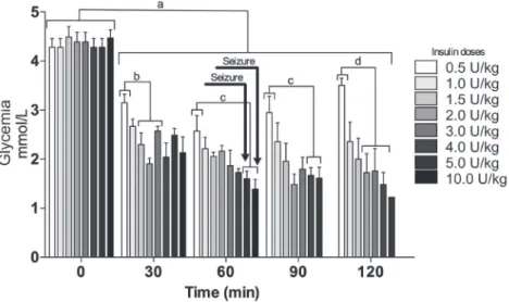
Mice were divided into 8 groups (n = 3-4 per group) which were given increasing ip doses of insulin: (0.5, 1.0, 1.5, 2.0, 3.0, 4.0, 5.0, or 10.0 U/kg). Blood samples were collected from the tail at 0, 30, 60, 90, and 120 min after insulin injection, as previously described (Marques
et al., 2016). Glycemia (mmol/L) was evaluated with a home glucometer (Optium Xceed®, Abbott Laboratories, Chicago, IL, USA), which is known to show good accuracy compared with the gold standard method used to measure plasma glucose (Robinson, Sharp, 2012).
The results of these experiments are summarized in the Figure 1.
Because seizures were observed only after insulin was administered at a dose of 5.0 U/kg or 10.0 U/kg (Figure 1), those doses were chosen to evaluate the effect of oral glucose or lactate administration on seizures and deaths.
The effects of oral glucose or lactate on seizures and deaths among hypoglycemic mice
The effects of oral glucose (5.5 mmol/kg) or oral lactate (18.0 mmol/kg) on seizures and deaths among
FIGURE 1 - Dose response curve and seizures. Blood glucose levels at 0, 30, 60, 90, and 120 min after an intraperitoneal injection
(0.5, 1.0, 1.5, 2.0, 3.0, 4.0, 5.0 or 10.0 U/Kg) of insulin into 15 h fasted mice. The mice were sacrificed when a seizure started. Results represent the mean ± standard error (n = 3-4). Data were analyzed using the Newman-Keuls multiple comparisons test. a
p < 0.05 as compared to time 0; b p < 0.05 as compared to the dose (1.5 U/kg, 2.0 U/kg or 3.0 U/kg); c p < 0.05 as compared to the
mice that received ip insulin (5.0 U/kg or 10.0 U/kg) were evaluated.
Thirty mice that received a 5.0 U/kg dose of insulin and thirty mice that received a 10.0 U/kg dose of insulin were divided into the following 3 groups (n = 10 mice per group): insulin + saline, insulin + lactate, and insulin + glucose. Saline, lactate or glucose was administered immediately after the insulin injection. The mice were them observed for a period of 300 min, which started immediately after an ip dose of insulin followed by an oral dose of saline, lactate orglucose.
To evaluate seizures after insulin injection, each group of mice (10 mice per cage) was kept in a silent room. While in the room, data for the following parameters was recorded for each group of mice: the of onset of the first seizure; time between seizures; number of seizures; death rate; time of death. As previously described by Zhao et al. (2003), seizure was defined as when the mouse
showed rhythmic jerks (clonic) and continuous tension or contraction of muscles (tonic).
Blood and brain levels of glucose and lactate
The blood and brain levels of glucose and lactate in mice that received ip insulin (10.0 U/kg) + oral saline (Ins group), ip insulin (10.0 U/kg) + oral lactate (Ins + Lac group) or ip insulin (10.0 U/kg) + oral glucose (Ins + Glc group) were quantified. A normoglycemic Control group that received ip saline and oral saline (Control group) was also included.
All mice (n = 6 per group) were sacrificed by decapitation. This method ensures a quick blood and brain collection without interference from anesthetics in the brain.
After decapitation, the blood was collected, centrifuged (10 min at 5 °C), and the plasma was kept on ice for subsequent evaluations of blood lactate and glucose levels. Lactate concentrations were measured using enzymatic techniques (Gutmann, Wahlefeld, 1974). Glucose was measured by using commercial kits.
For evaluations of brain lactate and glucose levels, the brains were quickly removed and stored in liquid nitrogen for several minutes. The brains were then homogenized (20 seconds) with perchloric acid containing EDTA (1 mM) in a van Potter-Elvehjem homogenizer; after which, the homogenates were placed in an ice bath and centrifuged at 10000 g (for 20 min at 5 °C.) The supernatant fractions were neutralized with KOH (5 mmol) containing 50 mmol triethanolamine, and then maintained on ice for 15 min before being centrifuged again (Kepler, Decker, 1974). After centrifugation, the supernatant lactate and glucose levels were measured
using enzymatic techniques (Gutmann, Wahlefeld, 1974) and commercial kits, respectively.
The following studies were performed as two sets of experiments.
In one set of experiments, the mice (n = 6 per group) were sacrificed when a seizure started in the Ins group but not in the other groups. This experimental approach allowed us to compare mice with a seizure (Ins group) and without a seizure. In the other set of experiments, each mouse was sacrificed when it began to convulse (n = 4 per group). This experimental approach allowed for comparisons of all mice which experienced a seizure.
The effects of diazepam on seizures and deaths among mice that received ip insulin
Mice that received ip insulin (10.0 U/kg) + ip saline (n =9 per group) were compared with mice that received
ip insulin (10.0 U/kg per group) + ip diazepam (3.0 mg/kg) (n=10 per group). The 300 min observation period started immediately after injection of either ip insulin + ip saline or ip insulin + ip diazepam.
Statistical analyses
All data were analyzed by ANOVA (Newman-Keuls post hoc test) using Graph-Pad Prism Version 5.0 software. Results are reported as the mean ± standard error (SE) of the mean. P-values < 0.05 were considered statistically significant.
RESULTS
As shown in Table I, 90%, 100%, and 80% of mice that received ip insulin (5.0 U/kg) plus oral saline, ip
insulin (5.0 U/kg) plus oral lactate or ip insulin (5.0 U/kg) plus oral glucose, respectively, had seizures.
The number of seizures in the group that received ip
insulin (5.0 U/kg) plus oral glucose was higher (p < 0.05) than those in the other groups. Moreover, the group that received ip insulin (5.0 U/kg) plus oral lactate had a higher death rate (Table I).
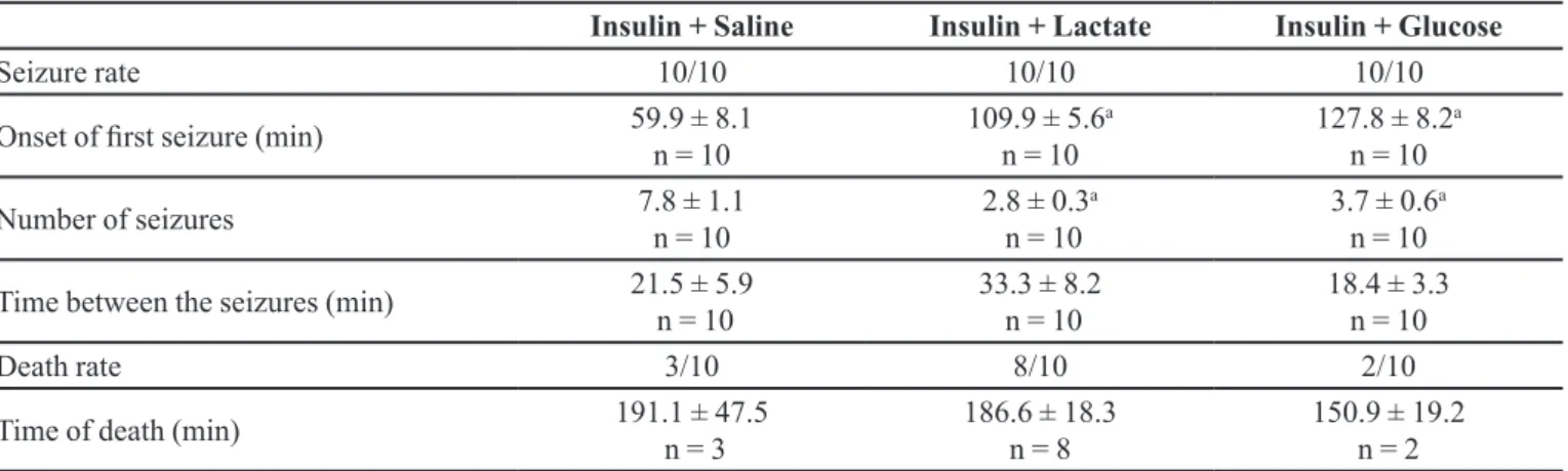
As shown in Table II, all mice that received ip insulin (10.0 U/kg) had seizures.
Furthermore, the seizures started earlier (p < 0.05) in the group that received ip insulin (10.0 U/kg) + oral saline than in the other groups. Moreover, the group that received
ip insulin (10.0 U/kg) + oral lactate had a higher death rate. Seizure rate, onset of the first seizure, number of seizures, time between seizures, death rate, and time of death during the 300 min observation period following ip administration of insulin. Oral saline, lactate or glucose was administered immediately after insulin injection. Results are reported as the mean ± standard error or a proportion (n/n). Data were analyzed using the Newman-Keuls multiple comparisons test. a p < 0.05 as compared to the Insulin + saline group.
Because all mice given 10.0 U/kg of insulin had seizures, that dose was chosen to evaluate the glucose and lactate levels in the blood and brain.
The data shown in Figure 2A-D, compare the blood
and brain glucose and lactate levels of mice with a seizure (Ins group) and without a seizure (Ins + Glc group and Ins + Lac group).
The glycemia values of all groups that received ip
insulin were lower (p < 0.05) than those in the Control group (Figure 2A). Additionally, the Ins + Glc group had higher (p <0.05) glycemia values than the Ins group (Figure 2A). In contrast, the brain glucose levels were similar in all groups (Figure 2B).
The lactatemia values were greater (p < 0.05) in the Ins + Lac group than in the other groups. In contrast, the Control, Ins, and, Ins + Glc groups had similar blood lactate levels (Figure 2C).
Moreover, the Ins group had lower (p < 0.05) brain lactate levels than the other groups (Figure 2D).
The results presented in Figure 2E-H compare the blood and brain glucose and lactate levels of all animals after seizure onset in each animal; thus the levels were measured at variable intervals after receiving insulin.
TABLE I - Effect of oral glucose (5.5 mmol/kg) or lactate (18.0 mmol/kg) on seizures and deaths among hypoglycemic mice that
received intraperitoneal insulin (5.0 U/kg)
Insulin + Saline Insulin + Lactate Insulin + Glucose
Seizure rate 9/10 10/10 8/10
Onset of first seizure (min) 71.7 ± 11.7 n = 9
100.4 ± 9.0 n = 10
83.3 ± 6.3 n = 8
Number of seizures 4.8 ± 1.5
n = 9
2.5 ± 0.8 n = 10
8.5 ± 1.1a,b
n = 8
Time between the seizures (min) 14.5 ± 1.3 n = 5
17.5 ± 6.9 n = 6
21.4 ± 11.1 n = 8
Death rate 1/10 7/10 1/10
Time of death (min) 192.8
n = 1
139.0 ± 10.0 n = 7
170.0 n = 1
TABLE II - Effect of oral glucose (5.5 mmol/kg) or lactate (18.0 mmol/kg) on seizures and deaths among hypoglycemic mice that
received intraperitoneal insulin (10.0 U/kg)
Insulin + Saline Insulin + Lactate Insulin + Glucose
Seizure rate 10/10 10/10 10/10
Onset of first seizure (min) 59.9 ± 8.1 n = 10
109.9 ± 5.6a
n = 10
127.8 ± 8.2a
n = 10
Number of seizures 7.8 ± 1.1
n = 10
2.8 ± 0.3a
n = 10
3.7 ± 0.6a
n = 10
Time between the seizures (min) 21.5 ± 5.9 n = 10
33.3 ± 8.2 n = 10
18.4 ± 3.3 n = 10
Death rate 3/10 8/10 2/10
Time of death (min) 191.1 ± 47.5
n = 3
186.6 ± 18.3 n = 8
The glycemia (Figure 2E) and brain lactate levels (Figure 2H) were lower (p < 0.05) in all groups that received ip insulin than in the Control group. However, the Ins, Ins + Lac, and Ins + Glc groups showed no difference in their glycemia (Figure 2E), blood lactate (Figure 2G), brain glucose (Figure 2F) and brain lactate (Figure 2H) levels.
The numbers of seizures and deaths among mice that received ip insulin (10.0 U/kg) and mice that received ip
insulin (10.0 U/kg) plus ip diazepam (3.0 mg/kg) were compared. As shown in Table III the ip administration of diazepam prevented seizures and decreased the death rate among mice that received ip insulin (10.0 U/kg).
DISCUSSION
Nocturnal hypoglycemia, especially common in type 1 diabetic patients, is a danger for any patient being treated
FIGURE 2 - Blood and brain levels of glucose and lactate in mice with () and without () seizures. The Control group received
intraperitoneal (ip) saline + oral saline. The experimental groups received ip insulin + oral saline (Ins group), ip insulin + oral lactate (Ins and Lac group) orip insulin + oral glucose (Ins and Glc group). Oral saline, lactate (18.0 mmol/kg) or glucose (5.5 mmol/kg) was administered immediately after ip saline (Control group) or insulin (10.0 U/kg). In one set of experiments (A, B, C, D; n = 6), the mice were sacrificed when the first seizure started in the Ins group, and while the other groups did not have seizures. In the other set of experiments, each mouse was sacrificed when it began seizing (E, F, G, H; n = 4). Data are reported as the mean ± standard error. Data were analyzed using the Newman-Keuls multiple comparison test, a p < 0.05 as compared to the Control group; b p <
0.05 as compared to the Ins group; c P < 0.05 as compared to the Ins + Lac group; d p < 0.05 as compared to the Ins + Glc group.
TABLE III - Effect of intraperitoneal (ip) diazepam (3.0 mg/kg) on seizures and the death rate among mice that received ip insulin
(10.0 U/kg)
Seizure rate
Onset of first
seizure (min)
Number of seizures
Time between the seizures (min)
Death Rate
Time of death (min)
Insulin + Saline 9/9 67.9 ± 8.9
n = 9
7.4 ± 1.2 n = 9
19.5 ± 4.4
n = 9 5/9
174.9 ± 27.6 n = 5
Insulin + Diazepam 0/10 NA
n = 10
NA n = 10
NA
n = 10 1/10
by insulin because the patient is not awake and cannot take appropriate measures or receive help.
In view of the vulnerability of the brain and the absence of suitable antidotes for nocturnal hypoglycemia, the possibility of using lactate to treat or prevent prolonged hypoglycemia must be considered.
The main reason for this proposition is based on the fact that not only glucose (Bazzigaluppi et al., 2017; Vilela
et al., 2014), but also lactate (Proia et al., 2016; Wyss et al., 2011) are important fuels for the brain.
Because, it remains to be clarified whether lactate can be used to treat hypoglycemia, we evaluated the effects of the oral lactate on the occurrence of seizures and deaths during a period of five hours, among mice that received high doses of insulin.
Higher (p < 0.05) brain levels of lactate (Figure 2D: Control, Ins + Lac or Ins + Glc groups versus Ins group) were associated with the absence of seizures. In addition, the role of brain lactate to prevent seizures was reinforced by the fact that when seizures began, the brain levels of lactate in the Ins, Ins + Lac, or Ins + Glc groups were lower (p < 0.05) than those in the control group (Figure 2H). In agreement with these results, Wiegers et al. (2016) demonstrated that in type 1 diabetes patients, symptoms of
insulin-induced hypoglycemia are related to the decreased availability of lactate in the brain.
Interestingly, hypoglycemic mice that received oral glucose (Ins + Glc group) or lactate (Ins + Lac group) had similar brain lactate levels (Figure 2D), confirming that oral glucose is an important source of brain lactate (Bazzigaluppi et al., 2017).
Despite the fact that glucose prolonged the time until the first seizure, mice that received glucose (Ins + glc group) or saline (Ins + saline group) had similar death rates (Tables I and II). Those results can be attributed to the fact that the dose of glucose was sufficient to prolong the time until the first seizure induced by the high doses (5 U/kg and 10.0 U/kg) of insulin but was not enough to prevent severe hypoglycemia.
The same dose of oral lactate that delayed the onset of first convulsion also increased hypoglycemia-induced mortality (Table II and Table III). One possible explanation for this finding is that lactate has a low Km value, i.e., about 2.5 mM (Shulman, Hyder, Rothman, 2001), which is near its physiological level in blood. Therefore, there is a limitation in the transport of lactate through the blood-brain barrier and the elevated blood lactate levels cannot supply the brain lactate deficit due to hypoglycemia. As
FIGURE 3 - Scheme based on the model proposed by Pellerin and Magistretti (2012). Our hypothesis is that during severe
consequence of this limitation, the elevation in blood lactate concentration after lactate administration (Figure 2C) was not followed by an elevation of brain lactate (Figure 2D). Moreover, the decrease in brain lactate levels which occurred during severe insulin induced hypoglycemia (Figure 2H) may promote neuronal death.
Taken together, the results of the present study, when combined with recent reports (Newman, Korol, Gold, 2011; Falkowska et al., 2015; Boury-Jamot et al., 2016) concerning the roles of glucose, lactate, and glutamate in brain metabolism, permit us formulate the hypothesis illustrated in Figure 3. Our hypothesis, based on the the astrocyte-neuron lactate shuttle model (Pellerin, Magistretti, 2012, Shen et al., 2014), states that during insulin-induced hypoglycemia, there is a sharp decrease in the availability of glucose to the astrocytes promoting decreased glycosysis in these cells and reduced release of lactate to the neurons. In other words, there is a disruption of the harmonic interaction between astrocytes (glycolysis, lactate release, and glutamate uptake) and neurons (lactate uptake and its use as a fuel source in oxidative metabolism). But, it must be emphasized that the interactions between astrocytes and neurons involve not only glucose/lactate metabolism, but also neuroprotective mechanisms similar than that triggered during ischemia (Gouix et al., 2014), chronic hyperglycemia (Rivera-Aponte et al., 2015), and traumatic brain injury (Crupi et al., 2013).
Diazepam increases GABAergic inhibition of glutamatergic neurons (Lason, Chelebicka, Rejdak, 2013; Malhi et al., 2014) promote an anticonvulsant effect (Table
III). Therefore, we suggest that an elevated availability of excitatory neurotransmitters could have triggered seizures and deaths (Sulkowski, Dabrowska-Bouta, Struzynsk, 2013).Consistent with this hypothesis, it was previously proposed that excitatory amino acid pathways may mediate the insulin-induced hypoglycemic seizures in Swiss mice (Anuradha, Hota, Pandhi, 2004).
Finally, our results did not change the current view that lactate is the main energetic fuel to the neurons. However, in contrast with glucose, oral lactate intensifies insulin toxicity.
ACKNOWLEDGMENTS
This research was supported by PRONEX/CNPq/ Fundação Araucária.
REFERENCES
Anuradha K, Hota D, Pandhi P. Investigation of central mechanism of insulin induced hypoglycemic convulsions in mice. Indian J Exp Biol. 2004;42(2):368-72.
Bazzigaluppi P, Ebrahim Amini A, Weisspapir I, Stefanovic B, Carlen PL. Hungry neurons: metabolic insights on seizure dynamics. Int J Mol Sci. 2017;18(11):pii:E2269.
Boury-Jamot B, Carrard A, Martin JL, Halfon O, Magistretti PJ, Boutrel B. Disrupting astrocyte-neuron lactate transfer persistently reduces conditioned responses to cocaine. Mol Psychiatry. 2016;21(8):1070-6.
Chan O, Paranjape AS, Horblitt A, Zhu W, Sherwin RS. Lactate-induced release of GABA in the ventromedial hypothalamus contributes to counterregulatory failure in recurrent hypoglycemia and diabetes. Diabetes. 2013;62(12):4239-46.
Crupi R, Paterniti I, Campolo M, Di Paola R, Cuzzocrea S, Esposito E. Exogenous T3 administration provides neuroprotection in a murine model of traumatic brain injury. Pharmacol Res. 2013;70(1):80-9.
De Feyter HM, Manson GF, Shulman GI, Rothman DL, Petersen KF. Increased brain lactate concentrations without increased lactate oxidation during hypoglycemia in type 1 diabetic individuals. Diabetes. 2013;62(9):3075-80.
Deprez-Poulain R, Hennuyer N, Bosc D, Liang WG, Enée E, Marechal X, et al. Catalytic site inhibition of insulin-degrading enzyme by a small molecule induces glucose intolerance in mice. Nat Commun. 2015;6:8250.
Falkowska A, Gutowska I, Goshorska M, Nowacki P, Chulubek D, Baranowska-Bosiacka I. Energy metabolism of the brain, including the cooperation between astrocytes and neurons, especially in the context of glycogen metabolism. Int J Mol Sci. 2015;16(11):25959-81.
Galende SB, Neto OCO, Santos LF, Peicher MV, Souza HM, Bazotte RB. Glucose administration inhibits the hepatic activation of gluconeogenesis promoted by insulin-induced hypoglycemia. Braz Arch Biol Technol. 2009;52(4):849-54.
Guo A, Daniels NA, Thuma J, Mccall KD, Malgor R, Schawartz F L. Diet is critical for prolonged glycemic control after short-term insulin treatment in high-fat diet-induced type 2 diabetic male mice. PLoS One. 2015;10(1):e0117556.
Gutmann I, Wahlefeld AW. L-(+)-Lactate. Determination with lactate dehydrogenase and NAD. Meth Enzym Anal. 1974;1464-72.
Hartmann EM, Garcia RF, Gazola VAFG, Barrena HC, Bazotte RB. Investigation of glycemia recovery with oral administration of glycerol, pyruvate, and L-lactate during long-term, insulin-induced hypoglycemia. J Diabetes Complications. 2010;24(5):301-5.
Herzog RI, Jiang L, Herman P, Zhao C, Sanganahalli BG, Blondeau N et al. Lactate preserves neuronal metabolism and function following antecedent recurrent hypoglycemia. J Clin Invest. 2013;123(5):1988-98.
K e p l e r D , D e c k e r K . G l y c o g e n d e t e r m i n a t i o n w i t h amyloglucosidase. Meth Enzym Anal. 1974;1127-31.
Keshavarz M, Showraki A, Emamghoreishi M. Anticonvulsant effect of guaifenesin against pentylenetetrazol-induced seizure in mice. Iran J Med Sci. 2013;38(2):116-121.
Lason W, Chlebicka M, Rejdak K. Research advances in basic mechanisms of seizures and antiepileptic drug action. Pharmacol Rep. 2013;65(4):787-801.
Malhi M, Jawed H, Hanif F, Ashraf N, Zubair F, Siddiqui BS, et al. Modulation of c-Fos and BDNF protein expression in pentylenetetrazol ekindled mice following the treatment with novel antilepileptic compound HHL-6. Biomed Res Int. 2014;2014:876712.
Marques ACR, Schiavon FPM, Travassos PB, Eik VF, Godoy G, Schamber CR, Bazotte RB. Evaluation of the impact of orally administered carbohydrates on postprandial blood glucose levels in different pre-clinical models. Braz J Pharm Sci. 2016;52(4):761-69.
Newman LA, Korol DL, Gold PE. Lactate produced by glycogenolysis in astrocytes regulates memory processing. PLoS One. 2011;6(12):28427.
Nunes Santiago A, de Godoi-Gazola VA, Fachin Milani M, de Campos VC, Rodrigues Vilela V, Diaz Pedrosa MM, Bazotte RB. Oral glutamine is superior than oral glucose to promote glycemia recovery in mice submitted to insulin-induced hypoglycemia. Int J Endocrinol. 2013;2013:841514.
Oldenbeuving G, Mcdonald JR, Goodwin ML, Sayilir R, Reijngoud DJ, Gladden LB et al. A patient with acute liver failure and extreme hypoglycaemia with lactic acidosis who was not in a coma: causes and consequences of lactate-protected hypoglycaemia. Anaesth Intensive Care. 2014;42(4):507-11.
Pellerin L, Magistretti PJ. Glutamate uptake into astrocytes stimulates aerobic glycolysis: a mechanism coupling neuronal activity to glucose utilization. Proc Nat Acad Sci. 1994;91(22):10625-9.
Pellerin L, Magistretti, PJ. Sweet sixteen for ANLS. J Cereb Blood Flow Metab. 2012;32(7):1152-66.
Proia P, Di Liegro CM, Schiera G, Fricano A, Di Liegro I. Lactate as a metabolite and a regulator in the central nervous system. Int J Mol Sci. 2016;17(9):pii:E1450.
Rivera-Aponte DE, Mendez-Gonzalez MP, Rivera-Pagan AF, Kucheryavikh YV, Kucheryavykh LY, Skatchkov SN, et al. Hyperglycemia reduces functional expression of astrocytic Kir4.1 channels and glial glutamate uptake. Neuroscience. 2015;310:216-23.
Robinson CS, Sharp P. Tighter accuracy standards within point-of-care blood glucose monitoring: how six commonly used systems compare. J Diabetes Sci Technol. 2012;6(3):547-54.
Rodrigues R, Feitosa KP, Felisberto-Junior AM, Barrena HC, Curi R, Bazotte RB. Comparative effects of short-term and long-term insulin-induced hypoglycemia on glucose production in the perfused livers of weaned rats.Pharmacol Rep. 2011;63(5):1252-7.
Rooijackers HMM, Wiegers EC, Tack CJ, Van Der Graaf M, De Galan BE. Brain glucose metabolism during hypoglycemia in type 1 diabetes: insights from functional and metabolic neuroimaging studies. Cell Mol Life Sci. 2016;73(4): 705-22.
Oral lactate intensifies insulin toxicity during severe insulin-induced hypoglycemia in mice
Shulman RG, Hyder F, Rothman D. Cerebral energetics and the glycogen shunt: Neurochemical basis of functional imaging. Proc Natl Acad Sci USA. 2001;98(11):6417-22.
Shen Y, Tian Y, Shi X, Yang J, Ouyang L, Gao, J et al. Exposure to high glutamate concentration activates aerobic glycolysis but inhibits ATP-linked respiration in cultured cortical astrocytes. Cell Biochem Funct. 2014;32(6):530-7.
Smith D, Pernet A, Hallett WA, Bingham E, Marsden PK, Amiel SA. Lactate: a preferred fuel for human brain metabolism in vivo. J Cereb Blood Flow Metab. 2003;23(6):658-64.
Sulkowski G, Dabrowska-Bouta B, Struzynska L. Modulation of neurological deficits and expression of glutamate receptors during experimental autoimmune encephalomyelits after treatment with selected antagonists of glutamate receptors. Biomed Res Inter. 2013;2013:186068.
Thurston JH, Hauhart RE, Schiro JA. Lactate reverses insulin-induced hypoglycemic stupor in suckling-weanling mice: biochemical correlates in blood, liver, and brain. J Cereb Blood Flow Metab.1983;3(4):498-506.
Vilela VR, Marques ACR, Schamber CR, Bazotte RB. Hypoglycemia induced by insulin as a triggering factor of cognitive deficit in diabetic children. Sci World J. 2014;2014: 616534.
Wang C, Liao JK. A mouse model of diet-induced obesity and insulin resistance. Methods Mol Biol. 2012;821:421-33
Weston PJ. The dead in bed syndrome revisited: a review of the evidence. Diabetes Manag. 2012;2(3):233-41.
Wiegers EC, Rooijackers HM, Tack CJ, Heerschap A, De Galan BE, Van Der Graaf M. Brain lactate concentration falls in response to hypoglycemia in patients with type 1 diabetes and impaired awareness of hypoglycemia. Diabetes. 2016;65(6):1601-5.
Wyss MT, Jolivet R, Buck A, Magistretti PJ, Weber B. In vivo evidence for lactate as a neuronal energy source. J Neuroscences. 2011;31(20):7477-85.
Zhao WJ, Ma YH, Fei J, Mei ZT, Guo LH. Increase in drug-induced seizure susceptibility of transgenic mice overexpressing GABA transporter-1. Acta Pharmacol Sin. 2003;24(10):991-95.
Received for publication on 20th September 2017