RESUMO: A doença de Petri é complexa, ataca plantas jovens de videira e é difícil de ser controlada. O fungo Phaeomoniella chlamydospora é o principal agente causal dessa doença. Os obje-tivos deste estudo foram: avaliar o local prevalente dos fungos da doença de Petri, em diferentes partes de plantas de videira; ava-liar a suscetibilidade de porta-enxertos de videira para o fungo
P. chlamydospora; avaliar o efeito da solarização e da biofumiga-ção seguido de tratamento com água quente sobre a desinfecbiofumiga-ção de estacas do porta-enxerto IAC 766 infectadas com o fungo
P. chlamydospora; avaliar o efeito da solarização e da biofumigação seguido de tratamento com água quente sobre o enraizamento de estacas do porta-enxerto IAC 766. Para o teste de colonização, as espécies de fungos detectadas e identificadas em Niagara Rosada enxertada em dois porta-enxertos diferentes foram P. chlamydospora
e Phialemoniopsis ocularis. Este é o primeiro relato de P. ocularis
em parrerais jovens de videira no Brasil. Ambos os fungos, em particular P. chlamydospora, colonizaram somente a parte basal das plantas, destacando-se os porta-enxertos como foco para medidas de controle. Medidas das estrias escuras no sistema vascular revela-ram que Golia foi o porta-enxerto menos suscetível, e o IAC 572 foi o mais suscetíveis para P. chlamydospora. Além disso, a biofu-migação ou a temperatura de 37ºC aplicadas por 7 e 14 dias segui-das de tratamento com água quente eliminaram P. chlamydospora
em estacas do porta-enxerto IAC 766 sem afetar o enraizamento. No entanto, novos estudos são necessários ainda para validar a eficiência dessas técnicas de desinfecção.
PALAVRAS-CHAVE: doença de Petri; material de propagação; controle; resistência; Vitis L.
ABSTRACT: Petri disease is complex, attacks young vine plants and it is difficult to be controlled. The fungus
Phaeomoniella chlamydospora (Phc) has been identified as the main causative agent of this disease. This study aimed to evaluate the prevalent colonization of the Petri disease fungi in different portions of vine plants; to assess the susceptibility of grapevine rootstocks to the fungus P. chlamydospora; to assess the effect of solarization and biofumigation, followed by hot-water treatment (HWT), on the disinfection of cuttings of the rootstock IAC 766 infected with P. chlamydospora, and assess the effect of solarization and biofumigation, followed by HWT, on the rooting of cuttings of the rootstock IAC 766. For the prevalent colonization test, the fungus species detected and identified in ‘Niagara Rosada’ grafted on two rootstocks different were Phc and Phialemoniopsis ocularis. This is the first report of P. ocularis in a young vineyard in Brazil. Both fungi, in particular Phc, colonized only the plant’s basal part, drawing attention to the rootstock as target for control measures. Measurement of the dark streaks in the vascular system revealed that Golia was the least susceptible rootstock, and IAC 572 was the most susceptible to Phc. Moreover, biofumigation or temperature of 37°C applied for 7 and 14 days, both followed by HWT, suppressed Phc in cuttings of the rootstock IAC 766 without hampering their rooting. Meanwhile, new studies are needed to validate the efficiency of these disinfection techniques.
KEYWORDS: Petri disease; propagating material; control; resistance; Vitis L.
Colonization of vines by Petri disease fungi,
susceptibility of rootstocks to
Phaeomoniella
chlamydospora
and their disinfection
Colonização de videiras pelos fungos da doença de Petri, suscetibilidade de
porta-enxertos ao fungo Phaeomoniella chlamydospora e sua desinfecção
Ana Beatriz Monteiro Ferreira1, Luís Garrigós Leite1, José Luiz Hernandes2, Ricardo Harakava3,
Carlos Roberto Padovani4, César Junior Bueno1*
1Instituto Biológico – Campinas (SP), Brazil 2Instituto Agronômico – Jundiaí (SP), Brazil 3Instituto Biológico – São Paulo (SP), Brazil
4Universidade Estadual Paulista “Julio de Mesquita Filho”, Instituto de Biociências, Campus de Botucatu – Botucatu (SP), Brazil *Corresponding author: cjbueno@biologico.sp.gov.br
Received on: 10/23/2017. Accepted on: 04/19/2018
INTRODUCTION
Petri disease causes decline and dieback of grapevines, mainly in young vines. It is a complex disease, and therefore difficult to be controlled. This disease is caused by combination of
Phaeomoniella chlamydospora (W. Gams, Crous, M. J. Wingf & L. Mugnai Crous & W. Gams) and several species of
Phaeoacremonium, and also by Cadophora lutea- olivaceae (F. H. Beyma) T.C. Harrington & McNew. However, P. chlamydospora
is more often associated with typical symptoms of Petri disease, causing the largest lesions and being more frequently re-isolated compared to other fungi related to this disease (GRAMAJE et al. 2011; HALLEEN et al., 2007; MOSTERT et al., 2006; MUGNAI et al., 1999). Other genera of fungi that might be associated to the decline in nurseries or in young vineyards are
Acremonium (A. charticola and A. ochraceum)and Phialemoniopsis curvata (= Phialemonium curvatum) (HALLEEN et al., 2007; PERDOMO et al., 2013).
External symptoms of the Petri disease show late bud-break, stunted shoot growth, reduced vegetative vigor, short-ened internodes, lower stem diameter, interveinal chlorosis, foliage with necrotic margins, premature defoliation, wilting, and dieback. Internal symptoms (xylem vessel) of the trunk show black spots and black streaking, tyloses and black gums (AROCA; RAPOSO, 2009; GRAMAJE; ARMENGOL, 2011; MOSTERT et al., 2006; MUGNAI et al., 1999).
Petri disease has been reported in different parts of the world where grapevine is cultivated (ABREO et al., 2011; CROUS; GAMS, 2000). In Brazil, Petri disease pathogens were found in the state of Rio Grande do Sul (GARRIDO et al., 2004) as well as in the Northeast region of the country (CORREIA et al., 2013).
LORENA et al. (2001) inoculated P. chlamydospora in the root of Paulsen 1103 rootstock and observed that the fun-guscolonization differed between vine plant portions, being greatest at the root collar level and at the base of the stem, becoming less frequent and disappearing above the 7th/8th
inter-node. Thus, the Petri disease pathogens will colonize mostly the rootstocks in grafted vine plants.
Rootstocks and scions of grapevines are susceptible to the Petri disease pathogens (GRAMAJE; ARMENGOL, 2011); however, some grapevine rootstocks inoculated with
C. luteo-olivacea, Phaeoacremonium spp. and P. chlamydospora
have shown to be less susceptible in field conditions (GRAMAJE et al., 2010), suggesting the necessity of new studies to verify the susceptibility of different rootstocks to the Petri disease pathogens.
The main sources of inoculum of the Petri disease fungi are infected propagation material, processes for propagation of grapevine plants, infected mother vines, infested soils, and aerial inoculum (AROCA et al., 2010; MOSTERT et al., 2006; MUGNAI et al., 1999). To avoid the dissemination of Petri disease fungi in the field, new studies should be carried
out with the purpose of disinfecting rootstock cuttings for the production of healthy mother vines and, consequently, to obtain healthy planting material.
A controversial measure to control Petri disease fungi in grapevine dormant cutting is the hot-water treatment (HWT) (GRAMAJE et al., 2009; ROONEY; GUBLER, 2001). Another measure with potential to disinfect grapevine dormant cutting is biofumigation, a sustainable method for disinfesting the soil. Biofumigation generates anaerobic conditions, toxic volatile compounds (isothiocynates), high temperature (GOPI et al., 2016), and all these factors can weaken or eliminate soilborne phytopathogenic fungi and their resistant structures. Solarization and biofumigation can be simulated in glass flasks kept in a growth chamber, providing an environment that is called microcosm (BUENO et al., 2004). So far, there is no study assessing the effect of biofumigation and solarization in a microcosm, tested solely or complemented with HWT, for the control of Petri disease fungi in rootstock cuttings.
Thus, this study aimed to:
1. evaluate the prevalent colonization of the Petri disease fungi in different portions of vine plant;
2. assess the susceptibility of grapevine rootstocks to the fungus P. chlamydospora;
3. assess the effect of solarization and biofumigation in microcosm, followed by hot-water treatment (HWT), on the disinfection of cuttings of the rootstock IAC 766 infected with P. chlamydospora, and
4. assess the effect of solarization and biofumigation in a microcosm, followed by HWT, on the rooting of cut-tings of the rootstock IAC 766.
MATERIAL AND METHODS
Colonization of vines
by Petri disease fungi
This study aimed to assess the portions with the most preva-lence for colonization of Petri disease fungi in vine plants: the basal part (rootstock), the stem part (40 cm above the basal part), or the branch part (40 cm far from stem part). To this end, three entire plants of ‘Niagara Rosada’ grapevine (Vitis labrusca L. x Vitis vinifera L.) grafted on the rootstock Ripária do Traviú (3 years old) and three plants grafted on the root-stock IAC 766 (2 years old) were collected randomly in a com-mercial vineyard infected with Petri disease, in the municipal-ity of Jundiaí, São Paulo state (SP), Brazil (23°07’83.0”S and 46°56’85.3”W). Thus, three treatments and three replications were established, represented by the three parts of each plant, and by the three plants of each variety, respectively.
them for 30 seconds in 70% alcohol and 1 min in a 1.5% solution of sodium hypochlorite, followed by immersion in sterile distilled water. Then, the samples were dried on ster-ile filter paper and cut into smaller fragments (0.5 cm) with a sterile scalp. The samples were distributed in Petri dishes, using 4 fragments per dish and 15 dishes per each plant part.
The Petri dishes were incubated in a growth chamber under 23°C for a 12-hour photoperiod for 21 days. The incidence of the Petri disease pathogens was expressed as the percentage of infected fragments per each plant part.
Identification of
the Petri disease fungi
The isolates of the Petri disease that grew in the medium were processed according to CORREIA et al. (2013) for molecular identification. After this, DNA was extracted by following the CTAB method described by DOYLE; DOYLE (1987). The polymerase chain reaction (PCR) was performed to amplify the ITS-5.8S region of the rDNA with the oligonucle-otide primers ITS1 (5’-TCCGTAGGTGAACCTGCGG-3’) and ITS4 (5’-TCCTCCGCTTATTGATATGC-3’) (WHITE et al., 1990). Fragments of the beta tubulin gene (β Tubulin) were amplified with the pair of primers Bt2a (5’-GGTAACCAAATCGGTGCTGCTTTC-3’) and Bt2b (5’-ACCCTCAGTGTAGTGACCCTTGGC-3’) o f G L A S S ; D O N A L D S O N ( 1 9 9 5 ) a n d B t u b - F : A A G G G H C AY TAY A C Y G A R G G a n d B t u b - R : CATGTTGGACTCDGCCTC of LAZAROTTO et al. (2014). The reactions were performed in a PTC100 ther-mocycler (MJ Research) according to the following protocol: initial denaturation at 94°C/2 min, 40 cycles of 94°C/30 s – 54°C/30 s – 72°C/60 s, and final extension at 72°C/4 min. The PCR products were purified by precipitation with polyethylene glycol, according to a protocol described by SCHMITZ; RIESNER (2006). Sequencing was performed by the chain termination method with the reagent BigDye 3.1 (Applied Biosystems) and ABI3500 automatic sequencer (Applied Biosystems).
Nucleotide alignment was carried out by MUSCLE pro-gram (EDGAR; MUSCLE, 2004). Phylogenetic trees were built by the Neighbor Joining method using the MEGA 6.0 pro-gram, with evaluation of the topology’s reproducibility through bootstrap with 1,000 repetitions. The support value obtained for each branch of the tree is shown in Figure 1, not adopting a minimum value to separate the branches.
Susceptibility of rootstocks
to the fungus
P. chlamydospora
This study aimed to assess the susceptibility of nine rootstocks to the fungus P. chlamydospora. The rootstocks used were: • IAC 313 “Tropical” – Golia x Vitis cinerea;
• IAC 572 “Jales” –V. caribaea x 101-14 Mgt (V. riparia x
V. rupestris);
• IAC 571-6 “Jundiaí” – V. vinifera (Pirovano 57) x
V. caribaea;
• IAC 766 “Campinas” – Ripária do Traviú (106-8 Mgt) x V. caribaea;
• Ripária do Traviú (106-8 Mgt) – V. riparia x (V. cordifolia
x V. rupestris);
• Ripária Gloire de Montpellier – seedling of V. riparia; • Golia – cross of Castel 156-12 (V. vinifera x V. riparia)
x V. rupestris;
• SO4 – V. berlandieri x V. riparia;
• Paulsen 1103 – V. berlandieri x V. rupestris.
The experiment was performed in randomized blocks with six replications. Each replication consisted of five pots contain-ing 4.0 kg of soil and a plant inoculated or not inoculated with the fungus P. chlamydospora of each rootstock. The sanity of the cuttings of each rootstock was attested before installing the test.
The fungus P. chlamydospora (IBVD 01) was isolated from the ‘Niagara Rosada’ plant grafted on the rootstock Ripária do Traviú from a commercial vineyard in the municipality of Jundiaí, SP (23°07’83.0”S and 46°56’85.3”W). The inocu-lum of the fungus was produced in Petri dishes containing a PDA medium, and incubated in a growth chamber under 23°C for a 12-hour photoperiod for 30 days.
The methodology to inoculate P. chlamydospora in root-stocks was adapted from ESKALEN et al. (2001). The base of the stem (2 cm above soil level) was injured with a metal-lic cork borer (0.5 cm) and, then, a plug of PDA medium (0.5 cm) colonized by the fungus was inserted into a circular wound and fixed by sealing the wound with gauze soaked in sterile distilled water and parafilm.
Four months after inoculation, the length of dark streaks caused by P. chlamydospora in the vascular system of each root-stock was measured.
Effect of technique combinations on
disinfection and rooting of rootstock
Disinfection
Eight treatments were established, consisting of the follow-ing four techniques (BUENO et al. 2004), complemented or not with hot-water treatment set to 51°C for 30 min (HWT) (GRAMAJE et al., 2009):
1. soil plus kale plants at 37°C (biofumigation); 2. soil without kale plants at 37°C (solarization); 3. without soil and without kale plants at 37°C, and 4. without soil and without kale plants at 23°C (control).
Continue...
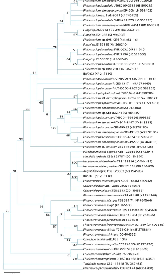
Figure 1. Phylogenetic trees showing the relationship between the isolates of fungi detected in ‘Niagara Rosada’ vine in comparison with the genera and species of fungi isolated from vines and deposited in GenBank-NCBI. The trees were constructed on the basis of
the following sequences: (A) ITS-5.8S region – showing condensed tree with 50% cut-off value; and (B and C) parts of the beta tubulin
gene. The isolates Pleurostoma richardsiae (= Pleurostomophora richardsiae) and Conioachaeta lignicola (= Lecythophora lignicola) were used as the outgroup. The accession number of the sequences of the isolates in GenBank-NCBI is given parenthetically.
Phialemonium dimorphosporum E7620j (HM 992502) 81
51
61
57
96
91
84 65
61
57
60
98
51 51
100
65
92
100
78
72
98
56
57 83
88 78
78
96 60 94 93 99
100
100 100 66 74
Phialemonium aff. dimorphosporum II-056.3b (AY 188371)
Paraphaeomoniella capensis CBS 123535 (FJ 372391) Philaemoniopsis oculares FMR 6632 (NR111515)
Phialemonium cf. curvatum CBS 115998 (EF 042105)
Phaeoacremonium angustius CBS 249.95 (AB 278178) Philaemoniopsis ocularis CNRMA 12.278 (HG 933293)
Philaemoniopsis curvata UTHSC 04-956 (HE 599295)
Celerioriella prunicola STEU:6343 (GQ 154588) Phialemonium sp. BRO-2013 (KF 367530)
Celerioriella dura CBS:120882 (GQ 154597)
Togininella acerosa CBS 113648 (EU 367453) Phialemonium dimorphosporum E9430h (JN 559402)
Phialemonium dimorphosporum (AJ 012300)
Aequabiliella effusa CBS:120883 (GQ 154598) Fungal sp. E15807B (KM 266242)
Neophaeomoniella zymoides CBS:121168 (GQ 154600)
Phialemonium inflatum BK239 (KU 702692) Fungal sp. AM2013 167 JMp (KC 506319)
Philaemoniopsis curvata CBS 490.82 (AB 278180)
Phaeoacremonium australiense CBS 113589 (KF 764560) Philaemoniopsis cornearis UTHSC 06-1820 (NR 111516)
Phaeoacremonium scolyti (KC 166688) Phialemonium sp. 695 ICIPE (KM 463116)
Philaemoniopsis curvata UTHSC 06-4324 (HE 599288)
Phaeoacremonium viticola Y271-03-1d (JF 275864) Philaemoniopsis cornearis UTHSC 06-1465 (HE 599285)
Phaeoacremonium fraxinopennsylvanicum UCR389 (JN 693515) Philaemoniopsis ocularis UTHSC 09-2358 (HE 599282)
Philaemoniopsis pluriloculosa UTHSC 09-3589 (HE 599287)
Neophaeomoniella niveniae CBS 131316 (JQ 044435) Philaemoniopsis oculares FMR 7190 (HE 599280)
Minutiella tardicola CBS: 121757 (GQ 154599)
Phialemonium obovatum CBS 279.76 (HE 610365) Phialemonium dimorphosporum NRRL 44611 (HM 060271)
Phialemonium curvatum UTHSC R-3447 (AY 818323)
Phaeoacremonium inflatipes CBS 391.71 (KF 764564) IBVD 02 (KP 213119)
Phaeoacremonium venezuelense CBS 651.85 (KF 764568)
Pleurostomophora richardsiae CBS723.74 (AB364700) Phialemonium sp. 1 AE-2013 (KF 746155)
Phialemonium sp. CBS 832.71 (AY 464130)
Phaeomoniella chlamydospora AQ04 18S (FJ 530942) Philaemoniopsis oculares UTHSC 05-2527 (HE 599281)
IBVD 01 (KP 213118)
Phialemonium atrogriseum UTHSC 03-986 (HE 610359) Fungal sp. E2128B (KT 996028)
Phialemonium dimorphosporum CBS 491.82 (AB 278185)
Phaeoacremonium parasiticum JQ 665454) Philaemoniopsis cornearis CBS 131711 (KJ 573445)
Phaeoacremonium subulatum CBS 113584 (KF 764565) Fungal sp. E15718E (KM 266210)
Phialemonium dimorphosporum CBS 492.82 (AY 464128)
Calosphaeria minima (EU 851104)
Philaemoniopsis pluriloculosa UTHSC 04-7 (HE 599286)
Phaeoacremonium minimum (DQ 404355)
0.05
P. chlamydospora) per treatment. Each replication consisted of eight rootstocks (IAC 766) infected with P. chlamydospora. To obtain the infected plants, rootstocks (sanity attested) planted in bags with solarized soil were inoculated with the
P. chlamydospora isolate according to the methodologies of inoc-ulum preparation and inoculation as described for the previous assay (Resistance of rootstocks to the fungus P. chlamydospora). Four months after inoculation, 96 plants were removed from the soil, their root system washed, and eight rootstocks were grouped together and then placed inside one of the two glass
bottles that comprise the microcosm (BUENO et al., 2004). All treatments were carried out inside the microcosms and incubated within growth chambers under 37±2°C and 23±2°C for a 12-hour photoperiod for 7, 14, and 21 days (BUENO et al., 2004). After each period, eight rootstocks per treatment were removed from each microcosm and four of them were submitted to additional HWT.
Three identical and independent tests were performed, using an independent microcosm for each period mentioned. The soil used in the experiment (Table 1) was moistened with distilled
Figure 1. Continuation.
IBVD 02 (MH 142383)
Phialemonium inflatum CBS 259.39 (HE 599347) Phialemoniopsis ocularis FMR 6632 (HE 599296)
Phialemoniopsis pluriloculosa UTHSC 04-7 (HE 599303)
Preussia terricola CBS 527.84 (GQ 203687)
Phialemoniopsis ocularis CNRMA 12.278 (HG 933290)
Phialemoniopsis curvata CBS 490.82 (HE 599307)
Phialemonium obovatum CBS 730.97 (HE 599335) Phialemoniopsis ocularis UTHSC 09-2358 (HE599299)
Phialemoniopsis cornearis CBS 131711 (KJ 573460) Phialemoniopsis ocularis CBS 110031 (KJ 573459)
Phialemoniopsis endophytica ACCC:38980 (KT 799563)
Phialemonium atrogriseum UTHSC 03-986 (HE 599343) Phialemoniopsis ocularis UTHSC 05-2527 (HE 599298)
Phialemoniopsis pluriloculosa UTHSC 09-3589 (HE 599304)
Coniochaeta lignicola CBS 267.33 (HE 610353)
Phialemoniopsis ocularis FMR 7190 (HE 599297)
Phialemoniopsis curvata CBS 491.82 (HE 599306)
Preussia isomera CBS 388.78 (GQ 203686) Phialemoniopsis cornearis UTHSC 06-1465 (HE 599302)
Phialemoniopsis cornearis UTHSC 06-1820 (HE 599301)
0.01
50 50 45 46
60 77
9
22 4
4
23 28
14
21
32
100 19
37
IBVD 02 (KP 213109)
Phaeoacremonium parasiticum CBS 101007 (AF 246804)
Phaeomoniella clamydospora Pach-55 (JX 679883)
Phaeoacremonium australiense STE-U 5960 (EU 128069) Togniniella acerosa CBS 113648 (KT 716486)
Phaeoacremonium scolyti (KC 013294)
Phaeoacremonium fraxinopennsylvanicum KER-U-PMOAP2 (KF 467605)
Phaeoacremonium angustius CBS 101739 (AF 246816)
Phialemonium sp. BRO-2013 (KF 428848)
Phaeoacremonium inflatipes CBS 391.71 (AF 246805) Phaeoacremonium venezuelense P4 (HQ 605012)
Pleurostoma richardsiae CBS 270.33 (AY 579334)
IBVD 01 (KP 213108)
Phaeoacremonium subulatum STE-U 6094 (EU 128092) Phaeoacremonium minimum P49 (HQ 605024)
Phaeoacremonium viticola Y271-03-1d (HQ 700718)
0.05
100
100
100 75
99
45
80
50 57
99 99 76 30
B
water at a proportion of 20% (w/v). For biofumigation, 60 g of kale (Brassica oleracea var. acephala L.) (Table 2) were crushed and mixed into 3 kg of soil, providing a 15-cm thick layer of growing medium in the microcosm (BUENO et al. 2004).
For evaluation, after each period of exposition to the treat-ments, fragments from vascular system of the basal part of each plant (replication) were removed and disinfested superficially, similarly to the colonization test. The fragments were cut into smaller pieces (0.5 cm) with a sterile scalp, and distributed in a Petri dish containing a PDA medium, with four pieces per dish and 5 dishes per plant. The dishes were incubated in a growth chamber under 23°C for a 12-hour photoperiod for 21 days. Fragments infected with the P. chlamydospora fungus were counted and converted into percentages of infected frag-ments to compare the efficiency of the treatfrag-ments.
Rooting
To assess the effect of the technique combinations on the rooting of the rootstock, eight healthy cuttings contain-ing 2-3 buds (replications) were grouped together and then placed into one of the two glass bottles that comprised the microcosm. The treatments and the methodology to expose the cuttings to the technique combinations were the same as described before (effect of technique combinations on disin-fection of rootstock).
After the different exposition periods, the cuttings were planted in a flowerbed (4×1.5 m), with a spacing of 20 cm between rows and 12 cm between plants. After planting, the soil was moistened, and then a mulching was spread on the soil, around the cuttings, in order to maintain appropri-ate moisture levels. After germination of the first buds, the mulching was removed.
The formation of the radicular system in the cuttings was evaluated 40 days after planting.
Data Analysis
Colonization data were analyzed by the non-parametric anal-ysis of variance for repeated measurements in independent
groups, complemented with the Dunn multiple comparison test (ZAR, 2009), with 5% significance.
Data from the screening test of rootstocks were sub-jected to analysis of variance for the experiment in random-ized blocks with replicates inside the blocks, complemented with the T test at 1% significance (ZAR, 2009).
The data from the disinfection and from the rooting tests were analyzed by the Goodman association test involv-ing contrasts between and within multinomial populations (GOODMAN, 1964; 1965), at 5% significance.
RESULTS
Colonization of vines
by Petri disease fungi
‘Niagara Rosada’ grapevine plants collected from young commercial vineyard were infected by two different fungal species, which were molecularly identified as
Phaeomoniella chlamydospora (IBVD01) and Phialemoniopsis ocularis (IBVD02)(Fig. 1).
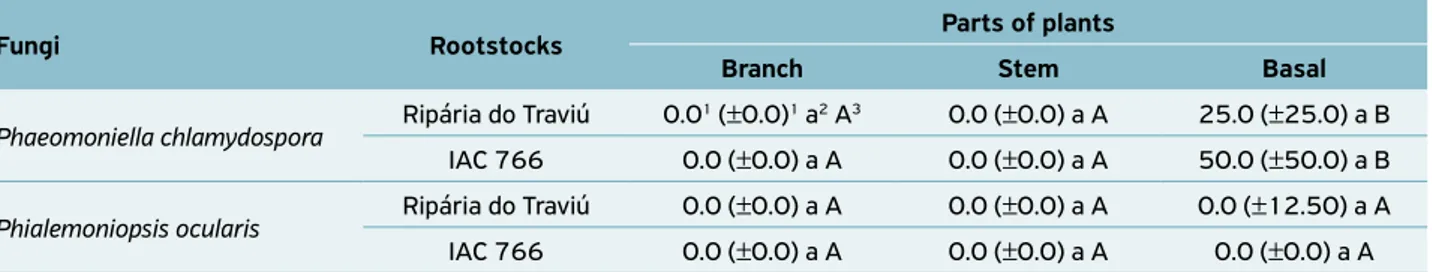
‘Niagara Rosada’ plants grafted on rootstock Ripária do Traviú were infected by two pathogens that colonized only the basal part, with significant more colonization by P. chlamydospora
than by P. ocularis. The plants grafted on rootstock IAC 766 were infected just by P. chlamydospora, whichalso colonized only the plant’s basal part (Table 3).
Susceptibility of rootstocks
to the fungus
P. chlamydospora
Control treatment showed no dark streaks; thus, the data on the control were not included in the analyses and in Figure 2.
Golia rootstock was the least susceptible to P. chlamydospora, whereas Ripária Glorie, Ripária do Traviú, IAC 766, SO4 and Paulsen 1103 were moderately susceptible, and IAC 572 was the most susceptible (Fig. 2).
Table 2. Mineral composition (macro and micronutrients) of the kale plants used.
N P K Ca Mg S C Fe Mn Cu Zn B Al Humidity
%
Macronutrients (g/Kg) Micronutrients (mg/Kg)
30.6 3.3 34.3 32.5 5.8 2.4 396.3 290.5 218.5 11.3 155.6 40.9 387.3 89.2
pH CaCl2
O.M. g/dm3
Presin mg/dm3
Al3+ H+Al K Ca Mg SB CTC
V% S B Cu Fe Mn Zn
mmolc/dm3 mg/dm3
4.3 10 12 5 28 1.0 2 2 5 33 15 7 0.14 0.3 9 2.2 0.2
Fungi Rootstocks Parts of plants
Branch Stem Basal
Phaeomoniella chlamydospora Ripária do Traviú 0.0
1 (±0.0)1 a2 A3 0.0 (±0.0) a A 25.0 (±25.0) a B
IAC 766 0.0 (±0.0) a A 0.0 (±0.0) a A 50.0 (±50.0) a B
Phialemoniopsis ocularis Ripária do Traviú 0.0 (±0.0) a A 0.0 (±0.0) a A 0.0 (±12.50) a A
IAC 766 0.0 (±0.0) a A 0.0 (±0.0) a A 0.0 (±0.0) a A
Table 3. Median values and quartile semi-amplitude of the percentage of incidence of Phaeomoniella chlamydospora and
Phialemoniopsis ocularis fungi in samples removed from the vascular system of different parts of ‘Niagara Rosada’ vines grafted on
two different rootstocks.
1Median and quartiles semi-amplitude of 60 fragments analyzed; 2Lowercase letters compare the rootstocks by fungus in the parts of the plants
sampled, according to the Dunn multiple comparison test (ZAR, 2009) at 5% significance; 3Uppercase letters compare the different parts of the
plants of the rootstock by fungus, according to the Dunn multiple comparison test (ZAR, 2009) at 5% significance.
Figure 2. Susceptibility of vine rootstocks to the fungus
Phaeomoniella chlamydospora.
*Letters compare the different rootstocks for their susceptibility,
according to the T test at 1% probability (ZAR, 2009).
Effect of technique combinations on
disinfection and rooting of rootstock
The treatments without hot-water treatment (HWT) did not kill the fungus in the vascular system of the cuttings of the rootstock IAC 766 (Table 4).
The only treatments that suppressed the P. chlamydospora
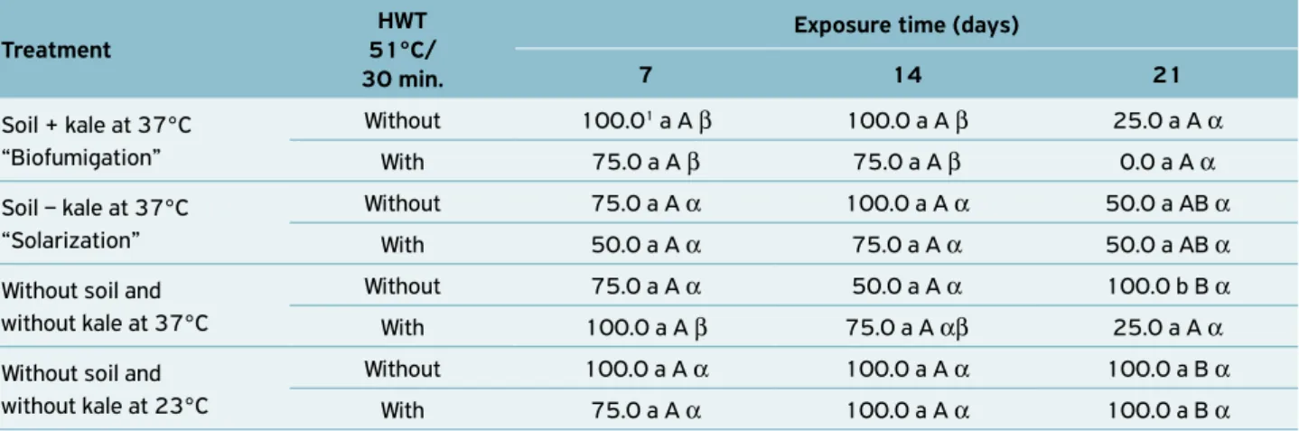
fungus in the vascular tissues of the cuttings of the rootstock IAC 766, in all exposure times tested, were biofumigation and temperature of 37°C, complemented with HWT. However, the biofumigation and temperature of 37ºC applied for 14 days and complemented with HWT suppressed the fungus and allowed rooting on 75% of the rootstock cuttings (Tables 4 and 5). This rate of rootstock rooting is acceptable for later planting and production of healthy rootstock mother plants.
The HWT treatment (51°C for 30 min) tested solely (23°C complemented with HWT) did not affect the rootstock cuttings
Treatment
HWT 51°C/ 30 min.
Exposure time to treatment
(days)
Incidence (%)
Soil + kale at 37°C “Biofumigation”
Without
7 25.01 b B β
14 6.3 a AB α
21 7.5 ab A α
Soil - kale at 37°C “Solarization”
7 20.0 a B β
14 1.3 a A α
21 45.0 b B β
Without soil and without kale at 37°C
7 2.5 a A α
14 16.3 a B β
21 15.0 a A β
Without soil and without kale at 23°C
7 0.0 a A α
14 13.8 b AB α
21 2.5 ab A α
Soil + kale at 37°C “Biofumigation”
With
7 0.0 a A α
14 0.0 a A α
21 0.0 a A α
Soil - kale at 37°C “Solarization”
7 0.0 a A α
14 0.0 a A α
21 1.3 a A α
Without soil and without kale at 37°C
7 0.0 a A α
14 0.0 a A α
21 0.0 a A α
Without soil and without kale at 23°C
7 2.5 a A α
14 2.5 a A α
21 1.3 a A α
Lowercase letters: comparison of different periods by fixing the thermotherapy and treatment; Uppercase letters: comparison of treatments by fixing the thermotherapy and the period; Greek letters: comparison of thermotherapy by fixing the treatment and the
period. The comparisons were done in accordance with the Goodman
association test (GOODMAN, 1964; 1965) at 5% significance
1Medium of 80 fragments analyzed.
Table 4. Percentage of incidence of Phaeomoniella chlamydospora
in samples of the vascular system of the basal part of the rootstock
IAC 766, submitted to different treatments for different periods,
with and without added hot-water treatment (HWT).
Lenght (cm) – dark str
eacks
in the vascular system
Ripária Gloir
e
Ripária do T
raviú
IAC 766 IAC 572
IAC 571-6 IAC 313
S04
P
aulsen 1103
Golia
Rootstocks 45.0
40.0 35.0 30.0 25.0 20.0 15.0 10.0 5.0 0.0
d d d
c b
for root formation, but also did not eliminate P. chlamydospora
fromplants (Tables 4 and 5).
The biofumigation for 21 days followed or not by HWT, and the temperature of 37ºC followed by HWT treatments hampered the root formation in the rootstock cuttings, result-ing in 0%, 25% and 25% of root formation for these treat-ments (Table 5). Thus, the application of these treattreat-ments for periods over 14 days, complemented with HWT, negatively affected the root formation in the cuttings.
DISCUSSION
This is the first report of the Phialemoniopsis ocularis fungus in young vineyards in Brazil. The P. chlamydospora fungus that was found infecting ‘Niagara Rosada’ grafted on two differ-ent rootstocks is often the main responsible for Petri disease (HALLEEN et al., 2007; MUGNAI et al., 1999). This fun-gus had already been found in Brazil, infecting the rootstock SO4 (CORREIA et al., 2013).
HALLEEN et al. (2007) isolated Phialemoniopsis curvata
(=Phialemonium curvatum) from vascular tissues from an asymptomatic vines nursery, and stated that this fungus may cause decline in nurseries or young vineyards. According to the phylogenetic tree of the present study (Fig. 1), the fungus
Phialemonium curvatum (EF042105) found by HALLEEN et al. (2007) should belong to the Phialemoniopsis cornearis species.
The genus Phialemoniopsis was created by PERDOMO et al. (2013) to accommodate the Phialemoniopsis curvata
(=Phialemonium curvatum) and Phialemoniopsis ocularis
(=Sarcopodium oculorum) fungi, and two new species,
Phialemoniopsis cornearis and Phialemoniopsis pluriloculosa. Interestingly, the GenBank sequences available for the P. ocularis
species are related to specimens that cause opportunistic infections in humans and in other animals. A review about
Phaeoacremonium species involved in Petri disease and Esca shows that some species of Phaeoacremonium are able to infect Vitis vinifera and humans (MOSTERT et al., 2006). According to HALLEEN et al. (2007), the relative importance of P. cornearis
and P. ocularis to the decline in grapevines should be confirmed by assessing the frequency of incidence of these fungi on diseased grapevines with different ages, which grew in different places.
P. chlamydospora was found predominantly in the basal part of ‘Niagara Rosada’ vines (rootstocks Ripária do Traviú and IAC 766), confirming results obtained by ABREO et al. (2011) and LORENA et al. (2001). According to ABREO et al. (2011), the P. chlamydospora fungus may also be found in other parts of grapevine such as in the apical part. However, the rootstock is the main target for Petri disease fungi. Thus, stud-ies are still necessary to find rootstocks with high level of resis-tance to Petri disease or techniques that ensure the total dis-infection of the rootstock cuttings.
Reviewing on Phaeoacremonium species and on plant sus-ceptibility to the Petri and Esca diseases, MOSTERT et al. (2006) emphasized the absence of rootstocks or scions with immunity or with a high level of resistance. In agreement with MOSTERT et al. (2006), none of the rootstocks tested in our study were immune or showed high level of resistance to P. chlamydospora.
The rootstocks Paulsen 1103 and Richter 110, artificially inoculated with P. chlamydospora, were more susceptible to the fungus compared to the V. vinifera cultivars, Chardonnay and Aglianico (ZANZOTTO et al., 2008). Similar results were obtained in Australia, where seven rootstocks (Ramsey, 99 Richter, Schwarzmann, Kober 5BB, P 1103, 101-14 Millardet and SO4) were more susceptible to P. chlamydospora
than five cultivarsof V. vinifera (Merlot, Cabernet Sauvignon,
Treatment
HWT 51°C/ 30 min.
Exposure time (days)
7 14 21
Soil + kale at 37°C “Biofumigation”
Without 100.01 a A β 100.0 a A β 25.0 a A α
With 75.0 a A β 75.0 a A β 0.0 a A α
Soil – kale at 37°C “Solarization”
Without 75.0 a A α 100.0 a A α 50.0 a AB α
With 50.0 a A α 75.0 a A α 50.0 a AB α
Without soil and without kale at 37°C
Without 75.0 a A α 50.0 a A α 100.0 b B α
With 100.0 a A β 75.0 a A αβ 25.0 a A α
Without soil and without kale at 23°C
Without 100.0 a A α 100.0 a A α 100.0 a B α
With 75.0 a A α 100.0 a A α 100.0 a B α
Table 5. Percentage of root formation in the rootstock IAC 766 cuttings submitted to disinfection in different periods, with and
without added hot-water treatment (HWT).
1Medium of four cuttings analyzed. Lowercase letters: comparison of thermotherapy (with and without) by fixing the treatment and the period;
Pinot Noir, Shiraz PT10 and Shiraz PT23) (WALLACE et al., 2004). These studies show that V. vinifera materials are indeed less susceptible to the P. chlamydospora fungus compared to the rootstocks. Furthermore, GRAMAJE et al. (2010) suggested that grapevine rootstock crosses of V. riparia x V. berlandieri
could be less susceptible to the patogens C. luteo-olivacea,
Phaeoacremonium spp. and P. chlamydospora, which are involved with Petri disease in young vines.
In the present study, the rootstocks with low and moderate susceptibility come from Vitis riparia, V. rupestris, V. berlandieri
and V. vinifera, which suggests that their genetic backgrounds from species of Vitis can be responsible for the least suscepti-bility to P. chlamydospora. New breeding studies are needed to obtain a rootstock with a satisfactory resistance level to Petri disease pathogens.
In agreement with ROONEY; GLUBER (2001), the appli-cation of HWT at 51°C for 30 min solely as a curative mea-sure is not sufficient to control P. chlamydospora and P. inflatipes
from dormant material. On the other hand, GRAMAJE et al. (2009), when applying HWT with temperatures above 50°C for different periods, managed to eliminate P. chlamydospora
from vine materials, but the process affected the sprouting and the weight of branches of the combination Temp – scion and 161-49 C – rootstock. In the present study, HWT (51°C for 30 min) tested as sole treatment (23°C followed by HWT) did not affect the rootstock cuttings for root formation, but also did not eliminate P. chlamydospora from theplants.
The biofumigation or temperature of 37°C treatments should be applied for 14 days, followed by the complementary treatment of HWT at 51°C for 30 min in order to eliminate
P. chlamydospora fromrootstock cuttings without affect the rooting. The interaction among different treatments such as biofumigation plus HWT can be more efficient to control the Petri disease fungi in rootstock cuttings compared to HWT tested as a sole technique.
Once the efficiency of these new techniques is validated, the healthy and normal cuttings can be used by growers to obtain healthy rootstock mother plants and, consequently,
produce healthy nursery plants, eliminating the following problems reported by other authors:
• transmission of the Petri disease fungi in nurseries through the use of infected mother plants (MOSTERT et al., 2006), and
• use of HWT with temperatures above 50°C for long peri-ods, which eradicates the fungus P. chlamydospora, but that can damage the plants (GRAMAJE et al., 2009).
CONCLUSION
In conclusion, P. chlamydospora and P. ocularis colonized preva-lently the basal part of ‘Niagara Rosada’ plants, denoting colo-nization of the rootstock. P. ocularis was detected by the first time in young vineyards in Brazil. Golia was the least suscep-tible rootstock to P. chlamydospora, and IAC 572 was the most susceptible. Biofumigation or temperature of 37°C applied for 7-14 days and followed by HWT at 51°C for 30 min, when used as treatments, suppressed P. chlamydospora in the root-stock cuttings without hampering the rooting. Meanwhile, new studies are necessary to validate the efficiency these dis-infection techniques.
ACKNOWLEDGEMENTS
The authors would like to thank Coordenação de Aperfeiçoamento de Pessoal de Nível Superior - Brazil (CAPES) for partial financial support received - Finance Code 001. The authors also would like to thank Mr. Daniel Fernando Miqueletto (Agricultural Engineer of City Council of Louveira, São Paulo state, Brazil) for the financial support received (FUNDAG – IB – Porta Enxertos 1018), and Mr. Fernando Perez, owner of the vine-yard (Jundiaí city, São Paulo state, Brazil), for authorization to collect ‘Niagara Rosada’ grapevines with Petri disease.
REFERENCES
ABREO, E.; MARTÍNEZ, S.; BETTUCCI, L.; LUPO, S. Phaeomoniella chlamydospora and Phaeoacremonium spp. in grapevines from Uruguay. Phytopathologia Mediterranea, Bologna,
v.50, p.577-585, 2011. http://dx.doi.org/10.14601/
Phytopathol_Mediterr-8682
AROCA, A.; RAPOSO, R. Pathogenicity of Phaeoacremonium species
on grapevines. Journal of Phytopathology, Berlin, v.157, p.413-419,
2009. http://dx.doi.org/10.1111/j.1439-0434.2008.01513.x
AROCA, Á.; GRAMAJE, D.; ARMENGOL, J.; GARCÍA-JIMÉNEZ, J.; RAPOSO, R. Evaluation of the grapevine nursey propagation
process as a source of Phaeoacremonium spp. and Phaeomoniella chlamydospora and occurrence of trunk disease pathogens in rootstock mother vines in Spain. European Journal of Plant Pathology, Dordrecht, v.126, p.165-174, 2010. http://dx.doi.
org/10.1007/s10658-009-9530-3
BUENO, C.J.; AMBRÓSIO, M.M.Q.; SOUZA, N.L.; CERESINI,
P.C. Controle de Fusarium oxysporum f.sp. lycopersici raça 2,
CORREIA, K.C.; CÂMARA, M.P.S.; BARBOSA, M.A.G.; SALES JR.; R., AGUSTÍ-BRISACH, C.; GRAMAJE, D.; LÉON, M.; GARCÍA-JIMÉNEZ, J.; ABAD-CAMPOS, P.; ARMENGOL, J.; MICHEREFF, S.J. Fungal
trunk pathogens associated with table grapes decline in North-eastern Brazil. Phytopathologia Mediterranea, Bologna, v.52, n.2, p.380-387, 2013.
DOYLE, J.J.; DOYLE, J.L. A rapid DNA isolation procedure for
small quantities of fresh leaf tissue. Phytochemical Bulletin,
Sussex, v.19, n.1, p.11-15, 1987.
EDGAR, R.C. Muscle: multiple sequence alignment with high accuracy and high throughput. Nucleic Acids Research,Oxford, v.32, n.5,
p.1792-1797, 2004. http://dx.doi.org/10.1093/nar/gkh340
ESKALEN, A.; GLUBER, W.D.; KHAN, A. Rootstock susceptibility
to Phaeomoniella chlamydospora and Phaeoacremonium spp.
Phytopathologia Mediterranea, Bologna, v.40, p.S433-S438, 2001.
http://dx.doi.org/10.14601/Phytopathol_Mediterr-1636
GARRIDO, L.R.; SÔNEGO, O.R.; GOMES, V.N. Fungos Associados
com o Declínio e Morte de Videiras no Estado do Rio Grande do Sul. Fitopatologia Brasileira, Brasília, v.29, n.3, p.322-324, 2004.
GLASS, N.L.; DONALDSON, G.C. Development of primer sets
designed for use with the PCR to amplify conserved genes from
filamentous ascomycetes. Applied and Environmental Microbiology, Washington, v.61, n.4, p.1323–1330, 1995.
GOODMAN, L. A. Simultaneous confidence intervals for contrasts
among multinomial populations. Annals of Mathematical Statistics, Ann Arbor, v.35, p.716-725, 1964.
GOODMAN, L.A. On simultaneous confidence intervals for multinomial proportions. Technometrics, Washington, v.7, n.2,
p.247-254, 1965. http://dx.doi.org/10.1080/00401706.1
965.10490252
GOPI, R.; AVASTH, R.K.; YADAV, A.; KALITA, H. Biological soil
disinfestation and biofumigation alternatives for chemical fumigation in organic farming. Advances in Plants & Agriculture Research., v.4, n.2, p.00135, 2016. http://dx.doi.org/10.15406/
apar.2016.04.00135
GRAMAJE, D.; ARMENGOL, J.; SALAZAR, D.; LÓPEZ-CORTÉS, I.; GARCÍA-JIMÉNEZ, J. Effect of hot-water treatments above 50ºC
on grapevine viability and survival of Petri disease pathogens.
Crop Protection, Guildford, v.28, n.3, p.280-285, 2009. http://
dx.doi.org/10.1016/j.cropro.2008.11.002
GRAMAJE, D.; GARCÍA-JIMÉNEZ, J.; ARMENGOL, J. Field evaluation
of Grapevine Rootstocks inoculated with Fungi associated with Petri disease and Esca. American Journal of Enology and Viticulture, Davis, v.61, n.4, p.512-520, 2010.
GRAMAJE, D.; ARMENGOL, J. Fungal trunk pathogens in the
grapevine propagation process: potential inoculums sources, detection, identification and management strategies. Plant Disease, Saint Paul, v.95, n.9, p.1040-1055, 2011. http://
dx.doi.org/10.1094/PDIS-01-11-0025
GRAMAJE, D.; MOSTERT, L.; ARMENGOL, J. Characterization of
Cadophora luteo-olivacea and C. melinii isolates obtained from grapevines and environmental samples from grapevine nurseries
in Spain. Phytopathologia Mediterranea, Bologna, v.50, p.S112– S126, 2011.
HALLEEN, F.; MOSTERT, L.; CROUS, P.W. Pathogenicity testing
of lesser-known vascular fungi of grapevines. Australasian Plant Pathology, Clayton, v.36, p.277-285, 2007. https://doi. org/10.1071/AP07019
LAZAROTTO, M.; BOVOLINI, M.P.; MUNIZ, M.F.B.; HARAKAVA, R.; REINIGER, L. R.S.; SANTOS, A.F. Identification and characterization
of pathogenic Pestalotiopsis species to pecan tree in Brazil. Pesquisa Agropecuária Brasileira, Brasília, v.49, p.440-448, 2014.
http://dx.doi.org/10.1590/S0100-204X2014000600005
LORENA, T.; CALAMASSI, R.; MORI, B.; MUGNAI, L.; SURICO, G.
Phaemoniella chlamydospora-grapevine interaction: histochemical reactions to fungal infection. Phytopathologia Mediterranea, Bologna, v.40, n.3, p.S400-S406, 2001.
MOSTERT, L.; HALLEEN, F.; FOURIE, P.; CROUS, P.W. A review of
Phaeoacremonium species involved in Petri disease and esca of grapevines. Phytopathologia Mediterranea, Bologna, v.45, p.S12-S29, 2006.
MUGNAI, L.; GRANITI, A.; SURICO, G. Esca (black measles) and
brown woodstreaking: two old and elusive diseases of grapevine.
Plant Disease, Saint Paul, v.83, n.5, p.404-418, 1999. https:// doi.org/10.1094/PDIS.1999.83.5.404
PERDOMO, H.; GARCÍA, D.; GENÉ CANO, J.J.; SUTTON, D.A.; SUMMERBELL, R.; GUARRO, J. Phialemoniopsis, a new genus
of Sordariomycetes, and new species of Phialemonium and Lecythophora. Mycologia, New York, v.105, n.2, p.398-421, 2013. https://doi.org/10.3852/12-137
ROONEY, S.; GUBLER, W.D. Effect of hot water treatments on
eradication of Phaeomoniella chlamydospora and Phaeoacremonium inflatipes from dormant grapevine wood. Phytopathologia Mediterranea, Bologna, v.40, n.3, p.S467-S472, 2001. http://
dx.doi.org/10.14601/Phytopathol_Mediterr-1617
SCHMITZ, A.; RIESNER, D. Purification of nucleic acids by
selective precipitation with polyethylene glycol 6000. Analytical Biochemistry, New York, v.354, n.2, p.311-313, 2006. http://
dx.doi.org/10.1016/j.ab.2006.03.014
WALLACE, J.; EDWARDS, J.; PASCOE, I.G.; MAY. P. Phaeomoniella chlamydospora inhibits callus formation by grapevine rootstock and scion cultivars. Phytopathologia Mediterranea, Bologna, v.43, p.151-152, 2004.
WHITE, T.J.; BRUNS, T.; LEE, S.; TAYLOR, J. Amplification and direct
sequencing of fungal ribosomal RNA genes for phylogenetics. In:
INNIS, M.A.; GELFAND, D.H.; SHINSKY, J.J.; WHITE, T.J. (Eds.).
PCR protocols: a guide to methods and applications. San Diego: USA. Academic., 1990. p.315-322.
ZANZOTTO, A.; GARDIMAN, M.; LOVAT, L. Effect of Phaeomoniella chlamydospora and Phaeoacremonium sp. on in vitro grapevine plants. Scientia Horticulturae, Amsterdam, v.116, n.4, p.404-408,
2008. http://dx.doi.org/10.1016/j.scienta.2008.03.002
ZAR, J.H. (Ed.). Biostatistical analysis. 5 ed. New Jeresey: Practice-Hall, 2009. 960p.