Abstract: Polysaccharides from fungal wastes were partially characterized and evaluated for their protective effects against bacterial spot caused by Xanthomonas gardneri on four tomato cultivars: Santa Cruz Kada, Natália, BRS Sena and Forty. The polysaccharides were extracted from spent mushroom substrate of Pleurotus ostreatus, residual brewery yeast (Saccharomyces
cerevisiae), and basidiocarps discarded from Lentinula edodes production. These polysaccharides were characterized for total carbohydrates, phenolics and proteins content, pH, scatter intensity, conductivity, Zeta potential, DPPH scavenging assay and infrared spectroscopy. The effects of time interval between treatment and inoculation (4 or 7 days) and polysaccharide concentrations (0.5 or 1.5 mg.mL–1) were assessed for disease severity using a susceptible
tomato cultivar. The polysaccharide action mode was investigated by determining the activity of peroxidases and phenylalanine ammonia-lyase and by quantifying flavonoids and total phenolics in the plants
PLANT PROTECTION - Article
Residual polysaccharides from fungi reduce
the bacterial spot in tomato plants
Tarsis Aguiar, Caroline Luiz, Argus Cezar Rocha Neto, Robson Marcelo Di Piero* Federal University of Santa Catarina - Plant Sciences - Florianópolis (SC), Brazil.
*Corresponding author: robson.piero@ufsc.br Received: Dec. 13, 2016 – Accepted: May 29, 2017
treated and challenged with X. gardneri. The polysaccharides obtained from Lentinula edodes (PSHII), Saccharomyces cerevisiae (PRC) and Pleurotus ostreatus (PSPO) (1.5 mg.mL-1) reduced bacterial
spot severity by 50% on tomato cotyledons, leaflets and five-leaf plants. Furthermore, PRC and PSHII (1.5 mg.mL–1) could decrease
disease severity in all tested cultivars. PSHII, the most effective, did not cause change in phenylalanine ammonia-lyase activity or flavonoid content on the cultivars Kada and Natália. However, an increase in peroxidase activity and total phenol content on cv. Kada was noted. The polysaccharides obtained from food industry wastes could provide protection against bacterial spot on tomato cultivars by inducing defense mechanisms and can be useful in formulating products with phytosanitary potential.
Key words: induced resistance, Lentinula edodes, Pleurotus ostreatus,
INTRODUCTION
Tomato (Solanum lycopersicon L.) is one of the eighth-most cultivated crops in the world, contributing significantly to the economy of many countries (Faostat 2013). Bacterial spot caused by Xanthomonas spp. is one of the main diseases that can cause severe to moderate damages to tomato crops, especially in tropics and in temperate regions (Mansfield et al. 2012). The disease has a potential to result in a substantial damage to crops due to the lack of efficient chemical control, the absence of resistant cultivars, and being seed borne (Quezado-Duval et al. 2004). Xanthomonas axonopodispv. vesicatoria,
X. vesicatoria, or X. gardneri, can cause the disease; therefore, attempts were made to identify the pathogen species present in this region. A total of 215 strains were obtained from 10 commercial areas in 1997, 1998, and 2000. The strains were characterized using pulsed-field gel electrophoresis (PFGE). In the tomato-producing region of Alto Vale do Rio do Peixe, Santa Catarina state, Brazil, the BOX-PCR technique was employed to analyze the causative agents of the bacterial spot, and almost 80% of the isolates associated were identified as
Xanthomonas gardneri, 11% as Xanthomonas perforans
and 9% as Xanthomonas vesicatoria (Costa et al. 2012). The main strategy to control plant pathogens is the application of pesticides. However, they not only increase production costs (Carrer Filho et al. 2008), but also contaminate the soil, groundwater, harm human health, the ecosystem, and eventually result in the selection of resistant pathogens (Pacumbaba et al. 1999). One alternative to the commercial pesticides commonly used against pathogens could be the use of fungi that secrete enzymes or antibiotics, compete by nutrient or induce host plant defenses (Punja and Utkhede 2003). For the last case, as a result of the interaction between plant and pathogen, defense elicitors stimulate the synthesis of phytoalexins and pathogenesis-related proteins (PR-proteins) eventually after binding to the receptor proteins of plant cell wall (Di Piero et al. 2006).
At present, there is an increasing demand for environmentally acceptable alternatives instead of traditional crop protection methods (Burketova et al. 2015). A new approach for this purpose is to use the food residual substances effectively in agriculture (Hamasaki et al. 2014), which also avoid the loss of valuable raw material
(Israilides and Philippoussis 2003). The biomass generated from brewing and growing mushroom industries is around 3 billion tons of residual yeast per year at the global level (Faostat 2013; Cardoso et al. 2015) and the same is 50 million tons for mushrooms substrate (Williams et al. 2001; Faostat 2013). This biomasses can be used for the extraction of the byproducts such as polysaccharides. Recently, Osińska-Jaroszuk et al. (2015) have demonstrated that the fungal polysaccharides obtained through ethanol precipitation have the potential for the environmental and agricultural applications as bio-fertilization, soil/water bioremediation, and plant bio-protection. Further, the carbohydrates are also known to elicit defense responses in plants and reduce disease symptoms (Trouvelot et al. 2014).
The ability of polysaccharides obtained from mushrooms, crustaceans, plants, algae and microbial cultures to protect plants against phytopathogens has been well documented (Di Piero et al. 2006; Coqueiro et al. 2011; Luiz et al. 2015; Delgado et al. 2013; Hahn and Albersheim 1978). Some of the polysaccharides, such as chitosan and that obtained from aloe (Aloe barbadensis
Miller), reduced tomato bacterial spot acting on the phenylpropanoid metabolism and increasing peroxidase activity on treated leaves (Coqueiro et al. 2011; Luiz et al. 2015). Moreover, a neutral polysaccharide extracted from the fruiting body of Lentinula edodes (Berk.) Pegler, called lentinan, inhibited the Tobacco mosaic virus multiplication (Wang et al. 2013). The extract from residual brewer’s yeast containing β-glucans and mannoproteins induced the expression of defense genes in Arabidopsis thaliana, which first activated the jasmonate/ethylene signaling pathways and, subsequently, the salicylic acid pathway (Narusaka et al. 2015). Therefore, the absence of studies related to the resistance inducers obtained from the fungal biomass discarded during food production processes justifies the importance of the present study.
Thus, the aim of this study was to evaluate the effect of different polysaccharides obtained from solid wastes of residual brewery yeast (Saccharomyces cerevisiae Meyen ex Hansen), spent mushroom substrate (SMS) from
MATERIAL AND METHODS
Plant pathogen
Xanthomonas gardneri (ex Šutič 1957) Jones et al.2006; Xan 166 (Group D) was isolated from the tomato plants showing the disease symptoms and grown in Águas Mornas (Santa Catarina, Brazil). The initial inoculum was provided by Sakata Seed Sudamerica and identified by BOX-PCR at Embrapa Hortaliças (Brasília, DF, Brazil). The bacterium was stored in phosphate buffer at pH 7 (8.6 mM K2HPO4; 7.4 mM KH2PO4) and 25 °C until use (Coqueiro and Di Piero 2011). Prior to the experiments, the bacteria culture was grown on nutrient agar medium (NA, 28 g.L–1, HIMEDIA®) and
incubated at 25 °C for 48 h. Then, bacterial suspensions were prepared by adding distilled water to the colonies formed on growth medium. Finally, the bacterial suspension density was optically adjusted with the aid of a spectrophotometer (U-1800 Spectrophotometer) at 600 nm (Luiz et al. 2015) to 0.3 or 0.6 absorbance units corresponding to 0.9 × 10⁸ and
1.9 × 10⁸ CFU.ml–1, respectively.
Crude extracts
The spent mushroom substrate (SMS) from P. ostreatus
was provided by Cogumelos da Gula Company (Garopaba, Santa Catarina, Brazil). The substrate was removed from theplastic bags and separated by hand. Then, 500 g of fresh residual substrate were mixed with 1.5 L of distilled water and autoclaved (30 min, 120 °C). After cooling, the product was filtered and an aqueous extract of substrate (ESPO) was made (Parada et al. 2012).
The residual basidiocarps from Lentinula edodes (shiitake mushroom), provided by Dr. Márcio José Rossi (Department of Microbiology and Parasitology, Federal University of Santa Catarina, Brazil), were sliced and mixed with water in a proportion of 1:3 (w/v). The samples were autoclaved as described previously for ESPO and an aqueous extract from shiitake fruiting bodies (ESHII) was obtained.
A sample of biomass rich with S. cerevisiae cells (Safale US-05 DRY ALE YEAST, Lesaffre International R&D), collected after beer brewing, was donated from Cerveja Amanita (Rancho Queimado, Santa Catarina, Brazil). The yeast slurry was centrifuged (7,500 rpm, 5 min, 25 °C), and the precipitate oven dried (60 °C). The samples, after attaining a constant weight, were ground to powder in an analytical mill and stored at –20 °C. An aqueous extract from
brewer residue (ERC) was obtained from the mixture of 200 g of dry biomass suspended to 1 L of distilled water, being stirred in Ultra Turrax (model T-25 Basic IKA® WERKE) at
13,500 rpm for 3 min and autoclaved for 2 h at 121 °C (Zanardo et al. 2009). After cooling, the material was centrifuged (7,500 rpm, 5 min, 25 °C) and the supernatant considered as ERC.
Polysaccharides fractions
The aqueous extracts of ERC, ESPO, and ESHII were precipitated with ethanol (80%), using an ethanol-extract proportion of 1:1 (v/v) (Hahn e Albersheim 1978), and the solutions were maintained at –20 °C for 48 h (Delgado et al. 2013). After precipitation, the supernatants were discarded and the precipitate of each material was collected (PRC, PSPO and PSHII, respectively). The fractions were oven dried (45 °C) until constant weight, ground in an analytical mill, and stored at –20 °C till further analysis. Prior to in situ
experiments, the polysaccharide fractions were solubilized in distilled water under constant stirring with an Ultra Turrax homogenizer for 3 min at 13,500 rpm.
Polysaccharides characterization
The polysaccharides from fungal residues PRC, PSPO and PSHII were characterized for total carbohydrates, phenolics and proteins content, pH, scatter intensity, conductivity, Zeta potential, DPPH scavenging assay, and infrared spectroscopy type IV (FTIR). For each fraction of polysaccharides, a mixture of three different extractions was used, and each analysis was carried out in triplicate.
Determination of carbohydrate, total phenolic and protein contents
The total carbohydrate content of PRC, PSPO and PSHII was determined by the phenol-sulfuric acid method (DuBois et al. 1956) adapted by Masuko et al. (2005) with some modifications. The fractions were diluted in distilled water (1.5 mg.mL–1), from which 50 µL was transferred into
Testplate 96F, Switzerland) and the absorbance was measured at 490 nm using a microplate reader (Molecular Devices, Spectra Max® Paradigm Multi-Mode Detection Platform,
Austria). The total carbohydrate content of each sample was calculated using a standard curve of glucose ranging from 0 to 5 mg.mL–1 (y = 0,288x + 0,055; R2 = 0.989), and expressed
in milligrams of glucose.
The phenolic compounds in polysaccharide suspensions (1.5 mg.mL–1) were quantified according to Popova et al.
(2007) with some modifications. An aliquot (3.2 mL of each suspension) was transferred to test tubes containing 200 µL of Folin-Ciocalteu reagent and 600 µL of 20% sodium carbonate solution (w/v). The samples were incubated in the dark for 2 h at room temperature and the absorbance was measured at 760 nm. The phenolic content was calculated based on a standard curve (0-25ng.ml–1; y = 0.019 + 0.076x; R2 = 0.993)
of gallic acid. The analysis was done in triplicate and the data were transformed to log (x) for statistical analysis. The concentration of phenolic compounds for each sample was expressed as nanograms of gallic acid equivalents (ng GAE).
The total protein content of the samples was determined by the Bradford method (1976) with some modifications. To 240 µL of a polysaccharide suspension (1.5 mg.mL–1), 60 µL
of concentrated Bradford reagent were added. Then, after 10 minutes the absorbance was read at 595 nm using a micro plate reader (Spectramax®). The total protein content of
each sample was calculated using a standard curve of bovine serum albumin (BSA) (0 to 60 µg.ml–1) and expressed in
milligrams of total proteins.
pH, scatter intensity, conductivity, Zeta potential, DPPH scavenging assay and infrared spectral analysis. The pH of each polysaccharide suspension (1.5 mg.mL–1) was measured
on a pH meter (Tec-3MP, Technical) in combination with electrode style Mettler Toledo InLab Easy BNC.
The polysaccharide suspensions (PRC, PSPO and PSHII) at 1.5 mg.mL–1 were pipetted into a capillary cell
(model DTS1070) and Zeta potential, scatter intensity, and conductivity were measured (nine readings per sample) using Zetasizer Nano ZS Malvern 90 UK.
The free radical scavenging activity of PRC, PSGPO and PSHII was evaluated against DPPH (1,1-dipheny-l,2-picrylhydrazyl) (Sigma-Aldrich, St. Louise, MO) according to the method adapted from Wang et al. (2015). The reaction solution containing 1 mL of ethyl alcohol and 0.2 mM DPPH was added to 1 mL of the polysaccharide suspensions (1.5 mg.mL–1). The resultant solution was incubated at 25 °C,
in the dark, 30 min, and the absorbance of polysaccharides was measured at 517 nm. Absolute ethanol was used as the blank. The antioxidant activity of polysaccharides was evaluated according to:
Sequestration Rate (%) = [1 − (Aj − Ai/Ac) × 100%] where Ac is the absorbance of 1 mL of DPPH and 1 mL absolute ethanol, while Ai and Aj are the absorbance values of 1 ml of the sample and 1 mL of DPPH or absolute ethanol, respectively.
The total polysaccharide fractions (PRC, PSPO and PSHII) were subjected to the infrared spectroscopy (Agilent Technologies – Series FTIR Spectrometer Cary 600). The polysaccharides were mixed with KBr powder, ground and pressed to form pellets (1 mm). The FTIR spectra were recorded in the range of 4000 – 400 cm–1.
Evaluation of bacterial spot severity
The protective effect of polysaccharides was evaluated on tomato plants at different phenological stages and environmental conditions. Initially, the polysaccharides were applied on the seedlings under two environmental conditions (greenhouse and growth room). In a greenhouse, the polysaccharides were applied on tomato cultivars of different groups.
Seedlings bioassays in greenhouse and growth room.
Firstly, the efficiency of the polysaccharides in controlling bacterial spot was evaluated using tomatoes seedling (cv. Santa Cruz Kada, Paulista). For the bioassays conducted in the greenhouse, tomato plants were grown in 128-cell Styrofoam trays containing Tropstrato HT Hortaliças® substrate, at a
temperature of 24.3 ± 4.7 °C. Whereas, inside the growth room, the seedlings were grown on the same substrate, described above, in plastic trays (20 cm length, 10 cm wide; without divisions), at a temperature of 25 °C and a photoperiod of 12 h. The seedlings, after attaining a height of 8 cm(with two primary leaves and about two weeks after sowing), were sprayed with PRC, PSPO and PSHII at 1.5 mg.mL–1 or distilled
Four replicates per treatment were used, and an experimental plot consisting of eight seedlings in each bioassay was employed. In the greenhouse conditions, the disease severity on leaflets was evaluated at 14 days after inoculation, while in the growth room, the severity per cotyledon was recorded after 30 days of inoculation. Two leaflets or cotyledon per plant were collected, photographed, and the obtained images were processed using QUANT software to obtain the severity values (Vale et al. 2002).
Protection assays for tomato plants at five-leaf stage in a greenhouse
The effect on a highly susceptible cultivar.
Tomato seeds (cv. Santa Cruz Kada Paulista, acquired at an agricultural supply store) were sown in Styrofoam trays containing substrate Tropstrato HT Hortaliças®. Fifteen days
after planting, two seedlings were transplanted into 2 L pots, containing soil and substrate Tropstrato HT (4:1, v/v). The experiments were conducted in a greenhouse, at a temperature of 24.3 ± 4.7 °C and a photoperiod of 12 h. At the five-leaf stage of the plants, 15 mL of each polysaccharide fractions, PRC, PSPO and PSHII, at 0.5 or 1.5 mg.mL–1 concentration
was sprayed on the plants. For the control plants, distilled water was used for spraying. The spray (polysaccharide or water) were applied onto abaxial and adaxial leaf surfaces with the aid of a paint spray gun coupled to an air compressor, at four or seven days before inoculation.
Protection and genetic resistance of tomato cultivars.
Two polysaccharides and a commercial inducer were selected for this analysis.The ability of PRC and PSHII to protect tomato plants was evaluated in cultivarswith different levels of susceptibility to X. gardneri. The seeds of cultivars, viz., Natália, BRS Sena and Forty were kindly provided by Sakata Seed Sudamerica, Eagle Flores Frutas & Hortaliças and Syngenta, respectively, and grown under greenhouse conditions previously described. The polysaccharides PRC and PSHII (1.5 mg.mL–1) were applied to the plants of the
cultivars, Santa Cruz Kada, Forty, Natalia and BRS Sena, at five-leaf stage, four days before inoculation. Distilled water and ASM (acibenzolar-S-methyl), an inducer of systemic acquired resistance, at 25 mg.L–1 concentration, served as
negative and positive controls, respectively.
In both experiments with plants at the five-leaf stage,
X. gardneri inoculum was adjusted spectrophotometrically
(0.6 OD at 600 nm) and, after inoculation, the plants were kept in a humid chamber (48 h). The experiments were randomized with five replicates per treatment and the experimental unit was represented by a plot with two plants. The disease severity was visually estimated at 10, 20 and 30 days post-inoculation with the aid of a diagrammatic scale (Mello et al. 1997) composed by five percentage levels of the infected foliar area (1%, 5%, 15%, 25% and 50%). The third and fourth true leaves were analyzed in a total of four leaves per experimental unit.
Biochemical defense mechanisms
Two cultivars with indeterminate growth habit were used for this analysis. Plants from Santa Cruz Kada (susceptible) and Natália (moderately resistant) at the five-leaf stage were sprayed with distilled water (negative control) or PSHII (1.5 mg.mL–1) and were inoculated with X. gardneri (0.6 OD;
600 nm) or left uninoculated at four days after treatment. The leaf samples (the third and fourth leaves, from base to apex) were collected at 0, 4, and 7 days after treatment (dat). Four leaves for each replication were collected and four replications were used. The collected samples were wrapped in aluminum foil and immediately frozen in liquid nitrogen. The samples were stored at –80 °C until biochemical analysis.
Peroxidase and phenylalanine ammonia-lyase (PAL) activity.
For peroxidase (POD) and phenylalanine ammonia-lyase (PAL) activities, the samples (100 mg fresh weight) were ground in liquid nitrogen and homogenized using 1 mL of extraction buffer (0.1 M sodium phosphate, pH 7.5, containing 1 mM ethylenediamine tetraacetic acid (EDTA) and 1% polyvinylpyrrolidone (PVP)). The homogenate was centrifuged (20,000 × g, 30 min, 4 °C) and the supernatant (protein extract) was recovered for enzymatic analysis (Coqueiro et al. 2011). Total protein content in each sample was determined by the Bradford method (1976).
and 90 mM hydrogen peroxide. Then, the absorbance was read at 470 nm for 4 min at a 30-s internal and at 30 °C. The results were expressed as optical density units at 470 nm per mg protein per minute (OD 470 nm.min–1.mg–1 protein).
PAL activity was determined according to Falcón et al. (2008), with some modifications. In this case, phenylalanine was used at 50 mM concentration as a substrate in 0.1 M sodium borate buffer (pH 8.8). Of the protein extract, 50 µL was added to 450 μL of the substrate and the mixture incubated at 40 °C for 1 h. The reaction was stopped by the addition of 200 μL of 5 N HCl and allowed to cool in ice for 5 min. Afterward, 300 μL of water was added and the absorbance recorded at 290 nm. The results were expressed as nanomoles of trans-cinnamic acid formed per minute per milligram of protein (nmol trans-cinnamic acid min–1.mg–1 protein).
Total phenolic compounds and flavonoids content.
For total phenolic compounds (PHENOL) and flavonoids (FLAVO) quantification, 100 mg of leaf tissues was ground in liquid nitrogen and homogenized with 3 mL of 80% acidified methanol (methanol: HCl = 80: 1, v/v). The extract was incubated in dark for 1 h at room temperature (Luiz et al. 2015) and centrifuged (3,500 rpm, 5 min).
The phenolic compounds were quantified according to McCue et al. (2000) and Coqueiro et al. (2011), with some modifications. The extract (0.5 mL) was mixed with 0.5 mL of 95% methanol (v/v), 1 mL of 95% ethanol, 1 mL of distilled water and 0.5 mL of Folin-Ciocalteu reagent. After 5 min, 1 mL of Na2CO3 5% (w/v) was added to each sample, followed by incubation in the dark for 1 h at room temperature. The absorbance was measured at 725 nm and the phenolic quantification was calculated using the standard curve (0.5 – 150 μg; y = 0.0012 x − 0.224; R2 = 0.978) of
gallic acid. The results are expressed in micrograms of gallic acid equivalents per gram of fresh weight (μg GAE. g–1 FW).
The flavonoids content was determined accordingly to Woisky and Salatino (1998), with some modifications. To the obtained extract (0.5 mL), 2.5 mL of 99% ethanol and 0.5 mL of 2% aluminum chloride solution in methanol were added, followed by incubation in the dark for 1 h at room temperature. The absorbance was read at 420 nm and the flavonoid content was determined using a quercetin standard curve (0–200 μg; y = 0.0063 x − 0.0015; R2 = 0.994). The total phenolic content was expressed as
the micrograms of quercetin equivalent per gram of fresh weight (μg QE.g–1 FW).
Statistical analysis
Analysis of variance (one way or factorial ANOVA) and Tukey’s post-hoc test (multiple comparisons) at 5% probability were used to detect the differences between treatments. All analyses were performed using the statistical software Statistica 8.0.
RESULTS AND DISCUSSION
The polysaccharides extracted from fungal residual biomass improved tomato plant resistance against the infection of X. gardneri. Thus, this study ascertains the use of waste biomass in the control of tomato bacterial spot. The polysaccharides from L. edodes (PSHII) were the most efficient and significantlyreduced the disease severity in different developmental stages and host genotypes.
The polysaccharides of Ascomycetes and Basidiomycetes show efficient antioxidant, antitumor and antimicrobial properties (Osińska-Jaroszuk et al. 2015). There has been an increase in the interest toward carbohydrates for their roles in plant immunity. They are defense elicitors of plants and can act as signaling molecules in a manner similar to plant hormones (Trouvelot et al. 2014).
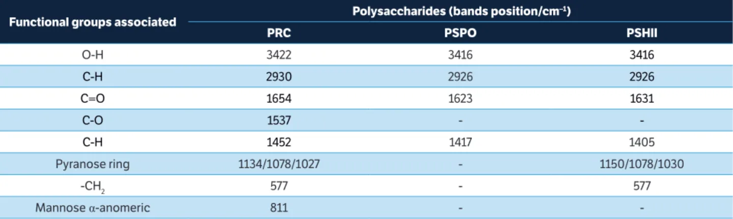
In the present study, fungal polysaccharides were successfully extracted and characterized. The main vibrational characteristics of functional groups associated with the surface of polysaccharides (PRC, PSPO, and PSHII) are given in Table 1. The IR type IV spectra of PRC, PSPO and PSHII were associated with stretches, peaks, and patterns similar to those shown by purified polysaccharides described in the literature by Chen et al. (2011) and Wang et al. (2015). FTIR analysis is a well-established technique for analysis of glucans from yeast (Thanardkit et al. 2002). The glucans present in fungal cell wall can activate plant immune system (Klarzynski et al. 2000; Di Piero et al. 2006; Nars et al. 2013). The bands characteristic of glucans extracted from Pleurotus spp. (Gutiérrez et al. 1996) for regions between 1000 and 1150 cm–1 were found during
the FTIR analysis carried out in our study. The FTIR analysis of PSHII demonstrated a band at 1150 cm–1
effects could be attributed to lentinan, a beta-glucan extracted from L. edodes mushroom, which also shows antitumor, antibacterial, antiviral, and anticlotting activities (Zanardo et al. 2015).
Total carbohydrate content was higher in PRC (1.1 mg.mL–1) compared to other fractions (Table 2). This
fact was related to the large amount of malt sugar used in brewing process. However, no differences were found between PSHII (0.4 mg.mL–1) and PSPO (0.5 mg.mL–1) regarding
total carbohydrate content. The suspensions differed in total protein content (PRC showed a higher amount of protein than PSPO and PSHII, 52.1% and 17.3%, respectively). This higher content of proteins detected in PRC can be related to the longer autoclaving time used to obtain the initial extract (later used in ethanolic precipitation). However, with a smaller and similar autoclaving period for PSHII and PSPO, the differences between them could be related to the characteristics of the residue used for extraction. Basidiocarps (material for obtaining PSHII) have naturally a higher protein content (m/v) in relation to spent mushroom substrate (material for obtaining PSPO), composed basically of straw and mycelium.
No differences were found between the dispersion intensity and pH values. Thus, it could be suggested that biopolymers constituting the three distinct treatments were homogeneously dispersed on plants and consequently resulted in the reduction of disease severity. The pH values were derived from the polysaccharide suspensions without any need of adjustment and it poses a low risk of damage to the plants.
Zeta potential (mV) was lower in PSHII, compared to PRC and PSPO (Table 2). The stability of polysaccharide
suspensions was evaluated according to the values of zeta potential and pH. Among the assessed polysaccharides, PSHII showed better tendency to stability than PRIC and PSPO (Zeta-Meter, Inc., 2005). Moreover, the conductivity of the three polysaccharides differed significantly. The highest value was observed for PSPO, while PRC had the lowest value. The total phenolic content also differed significantly, it was observed to be the highest for PSHII (89.8 ng GAE). PSHII also had the highest values of DPPH scavenging rate (89%), when compared to PRC and PSPO, which had similar values (Table 2).
The effect of PSHII against tomato bacterial spot could be ascribed to its higher phenolic content and antioxidant activity (DPPH Scavenging Rate). This correlation between the high antioxidant activity and the amount of phenolic compounds was detected in L. edodes and Volvariella volvacea mushroom extracts (Cheung et al. 2003). Phenolic compounds are chemical constituents mainly responsible for beneficial biological activities, especially antimicrobial and antioxidant properties (Popova et al. 2007).
The elicitors extracted from fungal residues could be used for the sustainable management of plant disease. In the present study, the polysaccharides, viz., PRC, PSPO and PSHII (1.5 mg.mL–1) significantly reduced the bacterial spot severity
on cotyledons (44.8%) in growth room bioassays, when compared to the control plants. In greenhouse conditions, PSHII, PRC and PSPO reduced severity (49, 30 and 33%, respectively) of the disease on young plants (Table 3). The unequal periods of disease evaluation (14 or 30 days for greenhouse and growth room, respectively) could be related to the higher temperature amplitude and a faster development of the plant inside a greenhouse.
Functional groups associated Polysaccharides (bands position/cm
–1)
PRC PSPO PSHII
O-H 3422 3416 3416
C-H 2930 2926 2926
C=O 1654 1623 1631
C-O 1537 -
-C-H 1452 1417 1405
Pyranose ring 1134/1078/1027 - 1150/1078/1030
-CH2 577 - 577
Mannose α-anomeric 811 -
The effect of SMS to protect plants against pathogens has been well documented. For example, Joshi et al. (2009) observed that the severity of bean angular leaf spot, caused by
Phaeoisariopsis griseola, was limited after the use of SMS and its extracts in treatments employed by amendments in soil and through foliar sprays. In another study, Parada et al. (2011) observed that the aqueous extracts from Lyophyllum decastes
SMS protected cucumber plants against the anthracnose caused by C. orbiculare, leading to an increased defense gene expression (chitinase and b-1,3-glucanase).
On five-leaf plants (cv. Kada), all polysaccharides (PRC, PSPO and PSHII) and doses (0.5 and 1.5 mg.mL–1) were
effective in reducing the severity of bacterial spot by about 55%, compared to the control plants, when applied four days prior to X. gardneri inoculation. In the interval of seven days between the treatment and inoculation, the level of protection was similar (Table 4). This indicates that the polysaccharides could be acting over an extended period of time. Coqueiro and Di Piero (2011) studying the same pathosystem (tomato – X. gardneri) observed that the polysaccharide chitosan (3 mg.mL–1) was not efficient to control disease symptoms with
an interval of six days between treatment and inoculation. The authors reported that when used at shorter intervals (24, 48 and 72 h before inoculation), chitosan conferred protection to tomato (70%) against the bacterial spot.
Doses of 0.5 and 1.5 mg.mL–1 provided the same level
of protection at both time intervals (Table 4). The dose of 1.5 mg.mL–1 was used in the next tests to allow a better
comparison with other polysaccharides (chitosan and
Aloe polysaccharides) that have been evaluated to control bacterial spot.
Table 2. Characterization of polysaccharides extracted from residual brewery yeast Saccharomyces cerevisiae (PRC), spent mushroom substrate of Pleurotus ostreatus (PSPO) and basidiocarps of Lentinula edodes (PSHII): protein content, total carbohydrates, scattering intensity (Kcps), pH, Zeta potential (mV) and conductivity (mS/cm).
.
Means followed by the same letter in line does not differ significantly by Tukey test (p < 0.05). SD; Standard Deviation.
Characteristic Polysaccharide (1.5 mg.mL
–1) ± SD
PRC PSPO PSHII
Total Protein (mg.mL–1) 0.023 ± 0.001 A 0.011 ± 0.0005 C 0.019 ± 0.001 B
Total Carbohydrate (mg.mL–1) 1.109 ± 0.109 A 0.563 ± 0.016 B 0.462 ± 0.110 B
Scattering Intensity (Kcps) 125.33 ± 19.91 A 180.53 ± 51.33 A 175.30 ± 15.56A
pH 5.5 ± 0.202 A 5.7 ± 0.156 A 5.4 ± 0.041 A
Zeta Potential (mV) –10.9 ± 0.700 A –9.7 ± 0.493 A –22.9 ± 0.519 B
Conductivity (mS/cm) 0.066 ± 0.001 C 0.467 ± 0.007 A 0.212 ± 0.001 B
Total Phenolics (ng GAE) 24.9 ± 0.4 B 23.0 ± 0.1 C 89.8 ± 3.7 A
DPPH Scavenging Rate (%) 45.2 ± 3.7 B 45.8 ± 2.3 B 89.5 ± 0.2 A
Table 3. Severity (%) of bacterial spot caused by X. gardneri on tomato cotyledons and young plants (cv. Santa Cruz Kada) treated
with polysaccharides (1,5 mg.mL–1)extracted from residual brewery
yeast Saccharomyces cerevisiae (PRC), spent mushroom substrate of Pleurotus ostreatus (PSPO) and basidiocarps of Lentinula edodes (PSHII) compared to control (distilled water).
Means followed by the same letters in the column indicate no significant difference at the level of 5% probability by Tukey’s test. SD; Standard deviation.*Evaluation performed at 30 and 14 days after inoculation on cotyledons and young plants, respectively.
Treatment Severity (%)±SD Cotyledons* Young plants*
Water 3.3 ± 0.7 a 24.8 ± 2.1 a
PRC 1.7 ± 0.3 b 17.3 ± 2.4 b
PSPO 1.7 ± 0.2 b 16.5 ± 2.2 b
PSHII 1.6 ± 0.3 b 12.6 ± 2.3 b
The data related to the severity of bacterial spot caused by X. gardneri on four tomato cultivars indicated significant differences for treatments and cultivars. PSHII reduced the bacterial spot severity in other three tomato cultivars, besides the cultivar Kada, during the evaluation performed 20 days after inoculation (Table 5). At 30 days of inoculation, both PSHII and PRC (1.5 mg.mL–1) significantly reduced
disease severity by 57.2% and 49.6% in all cultivars (Santa Cruz Kada, Forty, Natália, and BRS Sena) compared to the plants sprayed with water. This observation demonstrated that polysaccharides have a broad range of action, protecting cultivars with different resistance levels (Table 5).
in cv. BRS Sena could be ascribed to the process of genetic selection conducted by Embrapa Hortaliças, DF. This cultivar is considered the first Brazilian tomato industrial hybrid
.
Severity was evaluated at 20 days after inoculation. Means followed by the same letter do not differ significantly by Tukey’s test (p < .05). Evaluation performed 20 days after inoculation. SD: standard deviation.
Table 4. Severity (%) of bacterial spot caused by X. gardneri on tomato plants cv. Santa Cruz Kada treated with polysaccharides extracted from residual brewery yeast Saccharomyces cerevisiae (PRC), spent mushroom substrate of Pleurotus ostreatus (PSPO) and basidiocarps
of Lentinula edodes (PSHII), at 0.5 and 1.5 mg.mL–1, with the intervals
between treatment and inoculation of 4 or 7 days.
Treatment (mg.mL–1)
Severity (%)
Mean ± SD 4 days between treatment and
inoculation
PRC PSPO PSHII
0 14.3 ± 4.8 14.3 ± 4.8 14.3 ± 4.8 14.3 ± 4.8 a
0.5 9.7 ± 2.3 5.8 ± 3.2 7.2 ± 3.2 7.5 ± 3.2 b
1.5 8.0 ± 2.7 9.9 ± 4.5 8.4 ± 3.7 8.7 ± 3.6 b
Treatment (mg.mL–1)
Severity (%)
Mean ± SD 7 days between treatment and
inoculation
PRC PSPO PSHII
0 17.3 ± 5.0 17.3 ± 5.0 17.3 ± 5.0 17.3 ± 5.0 a
0.5 6.9 ± 2.4 9.3 ± 3.8 8.2 ± 1.6 8.1 ± 2.7 b
1.5 9.0 ± 4.2 9.8 ± 8.5 7.6 ± 3.9 8.7 ± 5.5 b
Cultivar/ Treatment
Severity (%)
Mean ± SD 20 days after inoculation
WATER PRC PSHII ASM
BRS Sena 3.1 ± 2.9 3.5 ± 1.9 2.0 ± 0.8 1.1 ± 0.4 2.4 ± 1.9 b
Natália 6.0 ± 3.0 5.0 ± 2.4 2.0 ± 1.1 1.1 ± 0.6 3.5 ± 2.7 ab
Forty 8.7 ± 3.3 3.1 ± 1.8 4.2 ± 3.9 1.8 ± 1.5 4.5 ± 3.7 ab
Kada 9.7 ± 6.4 5.1 ± 4.0 6.6 ± 3.1 1.0 ± 0.3 5.6 ± 4.9 a
Mean ± SD 6.9 ± 4.6 A 4.1 ± 2.9 AB 3.7 ± 2.9 BC 1.2 ± 0.8 C
Cultivar/ Treatment
Severity (%)
Mean ± SD 30 days after inoculation
WATER PRC PSHII ASM
BRS Sena 7.6 ± 3.8 4.6 ± 1.7 2.5 ± 1.7 1.5 ± 0.5 4.0 ± 3.1 b
Natália 13.2 ± 7.0 8.1 ± 3.1 4.6 ± 2.1 2.3 ± 1.7 7.0 ± 5.6 b
Forty 14.1 ± 4.4 6.0 ± 2.7 5.5 ± 4.2 2.3 ± 1.4 7.0 ± 5.4 b
Kada 23.1 ± 8.9 10.7 ± 4.9 12.5 ± 3.5 4.2 ± 1.8 12.6 ± 8.5 a
Mean ± SD 14.5 ± 8.1 A 7.3 ± 3.8 B 6.2 ± 4.7 B 2.6 ± 1.6 C
Table 5. Severity (%) of bacterial spot caused by X. gardneri on tomato cultivar Santa Cruz Kada, Forty, Natália and BRS Sena after spraying
distilled water, ASM (25 mg.L–1), polysaccharides (1.5 mg.mL–1) extracted from residual brewery yeast Saccharomyces cerevisiae (PRC) and
basidiocarps discarded from Lentinula edodes production (PSHII).
.
Means followed by the same letter (uppercase between treatments and lowercase among cultivars) did not differ significantly by Tukey’s test (p < .05), one way ANOVA. SD; Standard Deviation.
with higher level of resistance to bacterial spot caused by
Xanthomonas spp. (Quezado-Duval et al. 2014).
The commercial inducer of resistance (ASM) resulted in the lowest levels of disease (2.6%), regardless of the cultivar (Table 5). ASM is a chemical resistance-inducer, which may result in a physiological cost reducing some parameters related to yield, plant height, and fresh and dry weights of shoot (Barbosa et al. 2008). Louws et al. (2001) suggested that the concentration of ASM and the number of its application needed to be optimized. Recently, Pontes et al. (2016) observed a reduction in the yield of tomato plants after ten ASM applications. The polysaccharide utilization could be an important method to control plant diseases in organic production. However, further studies need to be performed to evaluate physiological costs related to the polysaccharide application in plants.
In relation to the mode of action, the polysaccharides from
total phenolic compounds (18.3%, independently of inoculation) compared to the control plants (Figure 2). These changes occurred in plants treated with PSHII, just before inoculation and also some days after contact with the pathogen, and can be associated with the defense mechanisms of the plants to stop bacterial infection or colonization. According to Soylu et al. (2003), during the incompatible interactions between plant and microbe or the treatments with elicitors, an increase in POD activity can often be found associated with the progressive incorporation of phenolic compounds to the cell wall. The plant cell wall enhancement increases the plant resistance against degrading enzymes/toxins produced by pathogens and acts as a physical barrier, reducing the severity of symptoms.
Chitosan and Aloe polysaccharides also promoted the reduction of bacterial spot severity by an increase in peroxidases activity and phenols contents. The protection levels exerted by these polysaccharides when used at concentrations near 1.5 mg.mL–1 (i.e., chitosan at 1−3 mg.mL–1 or aloe
polysaccharides at 0.75 and 1.5 mg.mL–1) were between
56 and 76% (Coqueiro and Di Piero 2011; Coqueiro et al. 2011; Luiz et al. 2012; Luiz et al. 2015). These findings corroborate with the results presented herein in terms of efficiency in bacterial spot control (57%) after residual polysaccharides application (PSHII; 1.5 mg.mL–1).
Similarly, peroxidases (POD) in tomato plants were significantly increased after treatment with L. edodes extract against R. solanacearum (Silva et al. 2007). Further, Di Piero
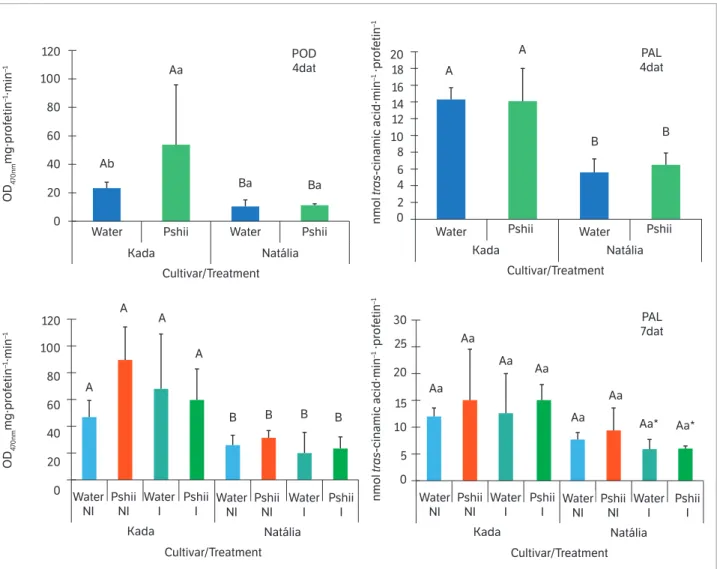
Figure 1. Peroxidase (POD) and phenylalanine ammonia-lyase (PAL) activities in tomato leaves (cv. Santa Cruz Kada and Natália) sprayed with
water or polysaccharides extracted from basidiocarps of Lentinula edodes (Pshii 1.5 mg.mL–1) and inoculated (I) or not (NI) with X. gardneri
(OD 0.6; 600 nm). Mean values recorded at 4 days after treatment (4 dat) and 7 dat (3 days after inoculation). Error bars indicate standard deviation. Means followed by the same letter (uppercase between cultivars; lowercase between treatments within the cultivar) did not differ significantly by Tukey’s test (p < 0.05). * Effect of inoculation relative to the respective non-inoculated control. Bars indicate standard deviation.
Water Pshii Pshii
120 100 80 60 40 20 0 120 100 80 60 40 20 0 2 0 0 5 10 15 20 25 30 6 4 10 8 14 12 16 20 18 Ba Ba Aa Aa Aa Aa Aa Aa Aa Aa* Aa* Ab A A A A A A B
B B B B
B Kada Natália Cultivar/Treatment Pshii Pshii Kada Kada Natália Cultivar/Treatment Natália Cultivar/Treatment POD 4dat PAL 4dat PAL 7dat OD 4 70nm mg·pr of etin –1·min –1 OD 4 70nm mg·pr of etin –1·min –1 nmol tra s -cinamic acid·min
–1 ·pr
of etin –1 nmol tra s -cinamic acid·min
–1 ·pr
et al. (2006) found that the inducers from partially purified
L. edodes fruiting bodies exert a protective effect on cucumber plants against C. lagenarium by increasing POD activity. In the present study, we assume that the values observed for POD in treated leaves with PSHII contribute to the reduction of bacterial disease symptoms.
Increases in peroxidase activity (POD) and phenolic contents (PHENOL) due to PSHII treatment were evidenced only in the highly susceptible cultivar (Santa Cruz Kada). In the moderately resistant cultivar (Natália), no changes in POD and FAL activities or PHENOL and FLAVO contents were detected after treatment with PSHII (1.5 mg.mL–1)
during the evaluated periods (Figures 1 and 2).
The induced resistance conferred by PSHII may act differently on the evaluated cultivars (Santa Cruz Kada and Natália). According to Sharma et al. (2010), BABA (DL–3-amino butyric acid) can induce resistance at different levels against Phytophthora infestans, depending on the tomato genotype used, and the level of induction generally decreases with the increase of leaf age. Additionally, these authors showed that the level of induction does not always relate to the resistance level of the tomato accessions and can be significantly affected by the pathogen isolate used for challenged inoculation. Probably in cv. Natália, the biochemical alterations promoted by PSHII may have occurred earlier and were not detected in the sampled periods. Thus, after contact with the elicitor, the material
Water Pshii Pshii
0 200 400 600 800 1,000 1,200 Kada Natália Cultivar/Treatment Pshii Pshii Kada Kada Natália Cultivar/Treatment Natália Cultivar/Treatment PHENOL 4dat PHENOL 7dat FLAVO 4dat FLAVO 7dat μg QE·g
–1 FW
0 200 400 600 800 1,000 1,200 μg QE·g
–1 FW
0 200 400 600 800 1,000 1,200 μg G AE·g
–1 FW
0 200 400 600 800 1,000 1,200 μg G AE·g
–1 FW
Water Water Water NI Pshii NI Water I Water NI Pshii NI Water I Pshii I Pshii I Kada Natália Cultivar/Treatment Water NI Pshii NI Water I Water NI Pshii NI Water I Pshii I Pshii I Water A A A A A A* A* A B B B B Aa Aa Aa Aa Aa Bb Bb Ba Ba Aa Aa Aa
Figure 2. Total phenolic compounds (PHENOL) and flavonoid contents (FLAVO) in tomato leaves (cv. Santa Cruz Kada and Natália) sprayed
with water or polysaccharides extracted from basidiocarps of Lentinula edodes (Pshii 1.5 mg.mL–1) and inoculated (I) or not (NI) with X. gardneri
responsiveness with intermediate resistance could be faster than in a highly susceptible material.
Considering the differences between cultivars, the highest PHENOL content was recorded in moderately resistant plants, being probably responsible for the protection against bacterial leaf spot compared to the most susceptible plants (Figure 2). In addition, Mandal et al. (2011) showed that the total phenolic content and lignin deposition were significantly higher in tomato plants resistant to R. solanacearum. On the other hand, some biochemical defense mechanism was elicited at a less intensity in cv. Natália, especially the peroxidase activity, in both the analyzed time points (4 and 7 dat) and PAL activity four days after treatments (Figure 1). Therefore, cv. Natália’s partial resistance may involve structural mechanisms or even biochemical mechanisms not evaluated here.
While assessing the effect of inoculation, cv. Natália showed reductions in total phenolic content (Figure 2) and PAL activity (Figure 1). A hypothesis that could explain this finding is that bacterial proliferation in plant tissues causes cell destruction and inhibits or reduces defense compounds production (Coqueiro et al. 2011). Moreover, Kavitha and Umesha (2008) revealed that PAL activity (the first key regulatory enzyme in the phenylpropanoid pathway leading to the production of phenolic substances) was maximum at 21 h after pathogen inoculation, when compared to the control and other samples of different time intervals assayed. Whereas, two tomato cultivars (Golden and Leadbeter) showed a decrease in PAL activity at 21 h after X. axonopodis pv. vesicatory inoculation, in a manner similar to what was observed with cv. Natália in the PAL activity analysis performed 72 h after of inoculation. Thus, depending on innate characteristics of the plant variety analyzed, different results were obtained.
CONCLUSION
The application of polysaccharides suspensions was found as an effective alternative to control bacterial
spot in tomato, probably owing to their ability to induce resistance. Besides, different development stages and host genotypes showed differences in the response to the polysaccharide molecules derived from fungi wastes (PRIC, PSPO, and PSHII). The polysaccharides from L. edodes increased peroxidase activity and total phenolic compounds in tomato plants. These findings support the decrease in plant symptoms and reduction in the severity of tomato bacterial spot. However, large-scale assays would be needed in order to evaluate the cost-effectiveness of the polysaccharide extraction and its application at a commercial level and under in situ
growing conditions.
ACKNOWLEDGEMENTS
The authors acknowledge Prof. Dr. Márcio José Rossi for providing the residual basidiocarps of Shiitake mushroom production. Also, we thank Mrs. Juliane of Cerveja Amanita for the S. cerevisiae biomass and Mr. Gustavo of Cogumelos da Gula for the P. ostreatus SMS. Our thanks to Leticia Mazzarino for performing Zeta potential measurements. We thank the companies Eagle Flores Frutas & Hortaliças, Sakata and Syngenta for seeds kindly provided. The financial support of Coordination for the Improvement of Higher Education Personnel (CAPES) is also acknowledged.
ORCID IDs
T. Aguiar
https://orcid.org/0000-0003-4237-2374 C. Luiz
https://orcid.org/0000-0003-3401-6679 A. C. R. Neto
https://orcid.org/0000-0002-9777-3151 R. M. Di Piero
https://orcid.org/0000-0003-3742-5582
REFERENCES
Barbosa, M. A., Laranjeira, D. and Coelho, R. (2008). Physiological cost of induced resistance in cotton plants at different nitrogen
levels. Summa Phytopathologica, 34, 338-342. http://dx.doi.
org/10.1590/S0100-54052008000400007.
Burketova, L., Trda, L., Ott, P. G. and Valentova, O. (2015). Bio-based resistance inducers for sustainable plant protection against pathogens. Biotechnology Advances, 33, 994-1004.
http://doi.org/10.1016/j.biotechadv.2015.01.004.
Cardoso, M. L., Conrad, R. W., Luz, M. L. G. S., Luz, C. A. S., Gadotti, G. I. and Gomes, M. C. (2015). Análise econômica dos processos de produção para ampliação de uma microcervejaria em Canela-RS. Revista Técnico-Científica do CREA-PR, 1, 1-14.
Carrer Filho, R., Romeiro, R. S. and Garcia, F. A. O. (2008) Biocontrole
de doenças de parte aérea do tomateiro por Nocardioides
thermolilacinus. Tropical Plant Pathology, 33, 457-460. http://doi.
org/10.1590/S1982-56762008000600010.
Chen, Y., Mao, W., Tao, H., Zhu, W., Qi, X., Chen, Y., Li, H., Zhao, C., Yang, Y., Hou, Y., Wang, C. and Li, N. (2011). Structural characterization and antioxidant properties of an exopolysaccharide produced by the mangrove endophytic fungus Aspergillus sp. Y16. Bioresource Technology, 102, 8179-8184. http://doi.org/10.1016/j. biortech.2011.06.048.
Cheung, L. M., Cheung, P. C. K. and Ooi, V. E. C. (2003). Antioxidant activity and total phenolics of edible mushroom extracts. Food Chemistry, 81, 249-255. http://doi.org/10.1016/ S0308-8146(02)00419-3.
Coqueiro, D. S. O. and Di Piero, R. M. (2011) Antibiotic activity against Xanthomonas gardneri and protection of tomato plants by chitosan. Journal of Plant Pathology, 93, 337-344. http://dx.doi. org/10.4454/jpp.v93i2.1188.
Coqueiro, D. S. O., Maraschin, M. and Di Piero, R. M. (2011). Chitosan reduces bacterial spot severity and acts in phenylpropanoid metabolism in tomato plants. Journal of Phytopathology, 159, 488-494. http://doi.org/10.1111/j.1439-0434.2011.01791.x.
Costa, J. R., Araújo, E. R., Becker, W. F., Ferreira, M. A. S. V. and Quezado-Duval, A. M. (2012). Ocorrência e caracterização do complexo de espécies causadoras da mancha bacteriana do tomateiro no Alto Vale do Rio do Peixe, SC. Tropical
Plant Pathology, 37, 149-154. http://doi.org/10.1590/
S1982-56762012000200009.
Delgado, D. Z., Freitas, M. B. and Stadnik, M. J. (2013). Effectiveness of saccharin and ulvan as resistance inducers against rust and angular leaf spot in bean plants (Phaseolus vulgaris). Crop Protection, 47, 67-73. http://doi.org/10.1016/j.cropro.2013.01.003.
Di Piero, R. M., Wulff, N. A. and Pascholati, S. F. (2006). Partial purification of elicitors from Lentinula edodes basidiocarps protecting cucumber seedlings against Colletotrichum lagenarium. Brazilian Journal of Microbiology, 37, 175-180. http://doi.org/10.1590/ S1517-83822006000200015.
DuBois, M., Gilles, K. A., Hamilton, J. K., Rebers, P. A. and Smith, F. (1956). Colorimetric method for determination of sugars and related substances. Analytical Chemistry, 28, 350-356. http://doi. org/10.1021/ac60111a017.
Falcón, A. B., Cabrera, J. C., Costales, D., Ramírez, M. A., Cabrera, G., Toledo, V. and Martínez-Téllez, M. A. (2008). The effect of size and acetylation degree of chitosan derivatives on tobacco plant protection against Phytophthora parasitica nicotianae. World Journal of Microbiology and Biotechnology, 24, 103-112. http:// doi.org/10.1007/s11274-007-9445-0.
Food and Agriculture Organization of the United Nations. (2013). Available at: http://faostat.fao.org/site/339/default.aspx. Accessed on May 20, 2014
Gutierrez, A., Bocchini, P., Galletti, G. C., Martinez, A. T. (1996). Analysis of lignin-polysaccharide complexes formed during grass lignin degradation by cultures of Pleurotus species. Applied and Environmental Microbiology, 62 (6), 1928-1934.
Hahn, M. G. and Albersheim, P. (1978). Host-Pathogen Interactions. Plant Physiology, 62, 107-111.
Hamasaki, T., Kitagawa, T. and Yasuhara, T. (2014). Efficacy of yeast cell wall extract, a byproduct of beer brewing, in tomato
(Solanum lycopersicum) culture. 2nd International Conference on
Environment, Energy and Biotechnology, 76, 21-25. http://dx.doi. org/10.7763/IPCBEE.2014.V76.5.
Hammerschmidt, R., Nuckles, E. M. and Kuc´, J. (1982). Association of enhanced peroxidase activity with induced systemic resistance of cucumber to Colletotrichum lagenarium. Physiological Plant Pathology,
20, 73-82. http://dx.doi.org/10.1016/0048-4059(82)90025-X.
Joshi, D., Hooda, K. S., Bhatt, J. C., Mina, B. L. and Gupta, H. S. (2009). Suppressive effects of composts on soil-borne and foliar diseases of French bean in the field in the western Indian Himalayas. Crop Protection, 28, 608-615. http://dx.doi.org/10.1016/j.cropro.2009.03.009.
Jones, J., Lacy, G., Bouzar, H., Stall, R. and Schaad, N. (2006). Reclassification of the xanthomonads associated with bacterial spot disease of tomato and pepper. Systematic and Applied Microbiology, 29, 85-86. https://doi.org/10.1078/0723202042369884.
Kavitha, R. and Umesha, S. (2008). Regulation of defense-related enzymes associated with bacterial spot resistance in tomato. Phytoparasitica, 36, 144-159. http://dx.doi.org/10.1007/BF02981327.
Klarzynski, O., Plesse, B., Joubert, J. M., Yvin, J. C., Kopp, M., Kloareg, B. and Fritig, B. (2000). Linear beta-1,3 glucans are elicitors of defense responses in tobacco. Plant Physiology, 124, 1027-1038. http://dx.doi. org/10.1104/pp.124.3.1027.
Louws, F. J., Wilson, M., Campbell, H. L., Cuppels, D. A., Jones, J. B., Shoemaker, P. B. Sahin, F. and Miller, S. A. (2001). Field control of bacterial spot and bacterial speck of tomato using a plant activator. Plant Disease, 85, 481-488. http://dx.doi.org/10.1094/PDIS.2001.85.5.481.
Luiz, C., Felipini, R. B., Costa, M. E. B.; Di Piero, R. M. (2012). Polysaccharides from Aloe barbadensis reduce the severity of bacterial spot and activate disease-related proteins in tomato. Journal of Plant Pathology, 94 (2), 387-393. http://dx.doi.org/10.4454/JPP.FA.2012.046.
Luiz, C., Rocha Neto, A. C. and Di Piero, R. M. (2015). Resistance to
Xanthomonas gardneri in tomato leaves induced by polysaccharides from plant or microbial origin. Journal of Plant Pathology, 97, 119-127.
http://dx.doi.org/10.4454/JPP.V97I1.029.
Mandal, S., Das, R. K. and Mishra, S. (2011). Differential occurrence of oxidative burst and antioxidative mechanism in compatible and incompatible interactions of Solanum lycopersicum and Ralstonia
solanacearum. Plant Physiology and Biochemistry, 49, 117-123. http://
dx.doi.org/10.1016/j.plaphy.2010.10.006.
Mansfield, J., Genin, S., Magori, S., Citovsky, V., Sriariyanum, M., Ronald, P., Dow, M., Verdier, V., Beer, S. V., Machado, M. A., Toth, I., Salmond, G. and Foster, G. D. (2012). Top 10 plant pathogenic bacteria in molecular plant pathology. Molecular Plant Pathology, 13, 614-629. http://dx.doi. org/10.1111/j.1364-3703.2012.00804.x.
Masuko, T., Minami, A., Iwasaki, N., Majima, T., Nishimura, S. I. and Lee, Y. C. (2005). Carbohydrate analysis by a phenol-sulfuric acid method in microplate format. Analytical Biochemistry, 339, 69-72. http://dx.doi. org/10.1016/j.ab.2004.12.001.
McCue, P., Zheng, Z., Pinkham, J. L. and Shetty, K. (2000). A model for enhanced pea seedling vigour following low pH and salicylic acid treatments. Process Biochemistry, 35, 603-613. http://dx.doi. org/10.1016/S0032-9592(99)00111-9.
Mello S. C., Takatsu A. and Lopes C. A. (1997). Escala diagramática para avaliação da mancha-bacteriana do tomateiro. Fitopatologia Brasileira, 22, 447-448.
Nars, A., Lafitte, C., Chabaud, M., Drouillard, S., Mélida, H., Danoun, S., Le Costaouec, T., Rey, T., Benedetti, J., Bulone, V., Barker, D. G., Bono, J. J., Dumas, B., Jacquet, C., Heux, L., Fliegmann, J. and Bottin, A. (2013). Aphanomyces euteiches cell wall fractions containing novel glucan-chitosaccharides induce defense genes
and nuclear calcium oscillations in the plant host Medicago
truncatula. PLoS One 8, 1-13. http://dx.doi.org/10.1371/journal.
pone.0075039.
Narusaka, M., Minami, T., Iwabuchi, C., Hamasaki, T., Takasaki, S., Kawamura, K. and Narusaka, Y. (2015). Yeast cell wall extract induces disease resistance against bacterial and fungal pathogens in Arabidopsis thaliana and Brassica crop. PLoS ONE, 10 (1), 114.
http://dx.doi.org/10.1371/journal.pone.0115864.
Osińska-Jaroszuk, M., Jarosz-Wilkołazka, A., Jaroszuk-Ściseł, J., Szałapata, K., Nowak, A., Jaszek, M., Ozimek, E. and Majewska, M. (2015). Extracellular polysaccharides from Ascomycota and Basidiomycota: production conditions, biochemical characteristics, and biological properties. World Journal of Microbiology and Biotechnology, 31, 1823-1844. http://dx.doi. org/10.1007/s11274-015-1937-8.
Pacumbaba, R. P., Beyl, C. A. and Pacumbaba, R. O. (1999). Shiitake mycelial leachate suppresses growth of some bacterial species and symptoms of bacterial wilt of tomato and lima bean in vitro. Plant Disease, 83, 20-23. http://dx.doi.org/10.1094/PDIS.1999.83.1.20.
Parada, R. Y., Murakami, S., Shimomura, N., Egusa, M. and Otani, H. (2011). Autoclaved spent substrate of hatakeshimeji mushroom
(Lyophyllum decastes Sing.) and its water extract protect cucumber
from anthracnose. Crop Protection, 30, 443-450. http://dx.doi. org/10.1016/j.cropro.2010.11.021.
Parada, R. Y., Murakami, S., Shimomura, N. and Otani, H. (2012). Suppression of fungal and bacterial diseases of cucumber plants by using the spent mushroom substrate of Lyophyllum decastes
and Pleurotus eryngii. Journal of Phytopathology, 160, 390-396.
Pontes, N. C, Nascimento, A. R., Golynski, A., Maffia, L. A., Oliveira, J. R. and Quezado-Duval, A. M. (2016). Intervals and number of applications of acibenzolar-s-methyl for the control of bacterial spot on processing tomatoes. Plant Disease, 100, 2126-2133. http://dx.doi. org/10.1094/PDIS-11-15-1286-RE.
Popova, M. P., S.Bankova, V., Bogdanov, S., Tsvetkovac, I., Naydenskic, C., Marcazzand, G. L. and Sabatini, A. G. (2007). Chemical characteristics of poplar type propolis of di ff erent geographic origin. Apidologie, 38, 306-311. http://dx.doi.org/10.1051/apido:2007013.
Punja, Z. K. and Utkhede, R. S. (2003). Using fungi and yeasts to manage vegetable crop diseases. Trends in Biotechnology, 21, 400-407. http://dx.doi.org/10.1016/S0167-7799(03)00193-8.
Quezado-Duval, A. M., Leite, R. P., Truffi, D. and Camargo, L. E. A. (2004). Outbreaks of Bacterial spot caused by Xanthomonas gardneri
on processing tomato in central-west Brazil. Plant Disease, 88, 157-161. http://dx.doi.org/10.1094/PDIS.2004.88.2.157.
Quezado-Duval, A.M., Nascimento, A. R., Pontes, N. de C., Moita, A.W., Assunção, A., Golynski, A., Inoue-Nagata, A. K., Oliveira, R. T., Castro, Y. O. and Melo, B. J. (2014). Desempenho de híbridos de tomate para processamento industrial em pressão de begomovirose e de mancha-bacteriana. Horticultura Brasileira, 32, 446-452. http:// dx.doi.org/10.1590/S0102-053620140000400012.
Sharma, K., Butz, A.F. and Finckh, M. R. (2010). Effects of host and pathogen genotypes on inducibility of resistance in tomato (Solanum
lycopersicum) to Phytophthora infestans. Plant Pathology, 59,
1062-1071. http://dx.doi.org/10.1111/j.1365-3059.2010.02341.x.
Silva, R. F., Pascholati, S. F. and Bedendo, I. P. (2007). Indução de resistência em tomateiro por extratos aquosos de Lentinula edodes e
Agaricus blazei contra Ralstonia solanacearum. Fitopatologia Brasileira, 32, 189-196. http://dx.doi.org/10.1590/S0100-41582007000300002.
Soylu, S., Baysal, Ö. and Soylu E. M. (2003). Induction of disease resistance by the plant activator, acibenzolar-S-methyl (ASM), against bacterial canker (Clavibacter michiganensis subsp. mchiganensis) in tomato seedlings. Plant Science, 165, 1069-1075. http://dx.doi. org/10.1016/S0168-9452(03)00302-9.
Thanardkit, P., Khunrae, P., Suphantharika, M. and Verduyn, C. (2002). Glucan from spent brewer’s yeast: Preparation, analysis and use as a potential immunostimulant in shrimp feed. World Journal of Microbiology and Biotechnology, 18, 527-539. http://dx.doi. org/10.1023/A:1016322227535.
Trouvelot, S., Héloir, M. C., Poinssot, B., Gauthier, A., Paris, F., Guillier, C., Combier, M., Trdá, L., Daire, X. and Adrian, M. (2014). Carbohydrates in plant immunity and plant protection: roles and potential application as foliar sprays. Frontiers in Plant Science, 5, 592. http://dx.doi.org/10.3389/fpls.2014.00592.
Vale, F. X. R., Fernandes Filho, E. I. and Liberato, J. R. (2002). QUANT: image processing software. Viçosa: Universidade Federal de Viçosa (UFV), Versão 1.0.2.
Wang, J., Wang, H. Y., Xia, X. M., Li, P. P. and Wang, K. Y. (2013). Inhibitory effect of sulfated lentinan and lentinan against tobacco mosaic virus (TMV) in tobacco seedlings. International Journal of Biological Macromolecules, 61, 264-269. http://dx.doi.org/10.1016/j. ijbiomac.2013.07.005.
Wang, L., Wang, C., Gao, X., Xu, N., Lin, L., Zhao, H., Jia, S. and Jia, L. (2015). Purification, characterization and anti-aging capacity of mycelia zinc polysaccharide by Lentinus edodes SD-08. BMC Complementary and Alternative Medicine, 15, 111. http://dx.doi. org/10.1186/s12906-015-0630-7.
Williams, B. C., Mc Mullan, J. T. and Mc Cahey, S. (2001). An initial assessment of spent mushroom compost as a potential energy feedstock. Bioresource Technology, 79, 227-230.
Woisky, R.G. and Salatino, A. (1998). Analysis of propolis : some parameters and procedures for chemical quality control. Journal of Apicultural Research, 37, 99-105. http://dx.doi.org/10.1080/0 0218839.1998.11100961.
Zanardo, N. M. T., Pascholati, S. F. and Fialho, M. B. (2009). Resistência de plântulas de pepineiro a Colletotrichum lagenarium
induzida por frações de extrato de Saccharomyces cerevisiae. Pesquisa Agropecuaria Brasileira, 44, 1499-1503. http://dx.doi. org/10.1590/S0100-204X2009001100018.
Zanardo, N. M. T., Pascholati, S. F. and Di Piero, R. M. (2015). Atividade antimicrobiana in vitro de extratos aquosos de isolados de Lentinula edodes contra Colletotrichum sublineolum e Xanthomonas
axonopodis pv. Passiflorae. Summa Phytopathologica, 41 (1),
13-20. https://dx.doi.org/10.1590/0100-5405/1995.