Description of immature stages and natural history of Stigmella schinivora
(Lepidoptera: Nepticulidae), a leaf-miner associated with the Brazilian
peppertree
Cristiano M. Pereira
1, Denis S. Silva
2, Héctor A. Vargas
3, Gilson R.P. Moreira
21Programa de Pós-graduação em Biologia Animal, Departamento de Zoologia, Instituto de Biociências, Universidade Federal do Rio Grande do Sul. Avenida Bento Gonçalves 9500, 91501-970 Porto Alegre, RS, Brazil.
2Departamento de Zoologia, Instituto de Biociências, Universidade Federal do Rio Grande do Sul. Avenida Bento Gonçalves 9500, 91501-970 Porto Alegre, RS, Brazil.
3Departamento de Recursos Ambientales, Facultad de Ciencias Agronómicas, Universidad de Tarapacá. Casilla 6-D, Arica, Chile.
Corresponding author: Gilson R.P. Moreira (gilson.moreira@ufrgs.br)
http://zoobank.org/CCF52BCB-A2C2-44DB-9B64-903BB1233748
ABSTRACT. Stigmella schinivora van Nieukerken, 2016 was described from Cataratas de Iguazú, Misiones, Argentina, based on adults reared from Schinus terebinthifolius Raddi (Anacardiaceae) leaf mines. The aim of this study is to describe for the first time the external morphology of the immature stages of S. schinivora with the aid of light and scanning electron microscopy, based on mines collected on the same host plant, but in Laranjeiras do Sul, Paraná, Brazil. Data on natural history, including histology of the mines, are also provided. The larva passes through four instars, all endophytic, having chewing mouth parts and feeding on the palisade parenchyma. The first three instars are apodous and have a subcylindrical body, bearing only one pair of setae on the tenth abdominal segment; the fourth instar is eruciform, with well-developed ambulatory calli on thorax and abdomen and setae on all tagmata. A serpentine mine is constructed on the adaxial surface, progressively increasing in width during larval development. With the exception of the widened, terminal section, the mine is left filled with larval feces. The fully developed larva of last instar exits through a slit made at the distal end of the mine, building a silk cocoon on the leaf abaxial surface where pupation occurs. This is the first record of S. schinivora from Brazil, which was only known from the type locality in Argentina. KEY WORDS. Atlantic forest, leaf-mining moths, microlepidoptera, nepticulids, Schinus terebinthifolius.
INTRODUCTION
The Nepticulidae is one of the most ancient lineages of Lepidoptera, with a global distribution and approximately 884 described species that are divided into 29 genera (van Nieukerken et al. 2016a, van Nieukerken 2018). They are among the smallest extant lepidopterans, having predominantly leaf miner habits and being associated with several plant families (Braun 1917, van Nieukerken et al. 2016a). Their minute size and the scarcity of the material available in collections has led their taxonomy to be mainly based on the morphology of adults, the immature stages rarely being taken into account (e.g., van Nieukerken et al. 2004). Studies that include the general appearance of their mines are not uncommon (e.g., Braun 1917, Stonis et al. 2013, 2014, van Nieukerken et al. 2016b), but specialization of larval feeding on tissues, if any, is largely unknown.
The worldwide distributed genus Stigmella Schrank, 1802 currently with ca. 420 species, is the largest genus of Nepticuli-dae (van Nieukerken 2018). Species identification in this genus can be difficult, since species complexes are common whose adults have similarities in external appearance; in these cases, only subtle differences in the genitalia morphology can be detected among species (Stonis and Remeikis 2016). There are at least 61 species of Stigmella recognized for the Neotropical Region (van Nieukerken et al. 2016a); however, this genus is still little studied in this region; thus, this number may not reflect its real diversity in the Neotropics (Puplesis and Rob-inson 2000, Šimkevičiūtė et al. 2009, Stonis et al. 2014, van Nieukerken et al. 2016a, Stonis and Remeikis 2017). This aspect is even more relevant in Brazil, where there are no records of Stigmella yet. This is unexpected since this is a megadiverse country, including biomes such as the Atlantic Forest, known
RESEARCH ARTICLE
ZOOLOGIA 35: e24485 ISSN 1984-4689 (online)
for the great diversity of plants and animals and high ende-mism indexes (Myers et al. 2000).
Stigmella schinivora van Nieukerken, 2016 was recently described as a leaf miner of the Brazilian peppertree, Schinus ter-ebinthifolius Raddi (Anacardiaceae) from the region of Misiones, Argentina (van Nieukerken et al. 2016b). Its description relied on morphology of the male and female genitalia. In the present study, using material collected in southwest Paraná state, Brazil, we present a detailed description of the immature stages of S. schinivora, based on light and scanning electron microscopy. We also provide additional information about its natural history, including the histology of its mines on leaves of S. terebinthifolius.
MATERIAL AND METHODS
Specimens used in this study came from leaf mines of S. ter-ebinthifolius collected in Laranjeiras do Sul municipality, Paraná, Brazil, in 2016 and 2017. They were brought to the Laboratório de Morfologia e Comportamento de Insetos (LMCI), Zoology Department of Federal University of Rio Grande do Sul (UFRGS), Porto Alegre city, and then they were either dissected or kept at room temperature in plastic pots containing moistened cotton for emergence of adults. The adults obtained in the laboratory were identified as S. schinivora based on comparison with original descriptions and illustrations of the adult stage, including female and male genitalia, provided by van Nieukerken et al. (2016b). Adults were pinned and dried. Immature stages were fixed in Dietrich’s fluid and preserved in 75% ethanol. For descrip-tions of the gross morphology, the specimens were cleared in a 10% potassium hydroxide (KOH) solution and slide-mounted in either glycerin jelly or Canada balsam. Observations were performed with the aid of a Leica M125 stereomicroscope, and measurements were performed using an attached ocular microm-eter (precision = 0.01 mm). Structures selected to be drawn were previously photographed with a Sony Cyber-shot DSC-H10 dig-ital camera attached to the stereomicroscope, and also by using a Nikon AZ 100M stereomicroscope. Vectorized line drawings were then made with the software Corel Photo-Paint X7, using the corresponding digitalized images as a guide. At least five specimens were used for the descriptions of each morphotype.
For scanning electron microscope analyses, additional specimens were dehydrated in a Bal-tec CPD030 critical-point dryer, mounted with double-sided tape on metal stubs and coated with gold in a Bal-tec SCD050 sputter coater. They were examined and photographed in a JEOL JSM6060 scanning electron microscope at the Centro de Microscopia Eletrônica (CME) of UFRGS.
For plant anatomical descriptions, field-collected leaf parts of S. terebinthifolius containing mines of S. schinivora were preserved in Dietrich’s fluid. Leaf parts containing the different larval instar morphotypes were selected under a stereomicro-scope, and freehand cross sections were cut with a razor blade. They were then stained for five seconds with safranin and
pho-tographed with a Nikon AZ 100M stereomicroscope.
Vouchers of specimens used in this study were deposited in the insect collection of the Laboratório de Morfologia e Com-portamento de Insetos (LMCI), Zoology Department (UFRGS), as follows (all coming from S. terebinthifolius leaf-mines collected by the senior author at Laranjeiras do Sul, Paraná, Brazil): 16-23. VII.2016, pinned, dried adults, two females (LMCI 309-10 and 11, with genitalia on slides GRPM 50-151 and 152, respectively), two males (LMCI 309-12 and 13, with genitalia on slides GRPM 50-153 and 154, respectively); 29.XI.2017, immature stages, fixed in Dietrich’s fluid, preserved in 70% ethanol, 3 first instar larvae (LMCI 323-2), 3 second instar larvae (LMCI 323-3), 4 third instar larvae (LMCI 323-4), 8 fourth instar larvae (LMCI 323-5) and 3 pupae (LMCI 323-7).
RESULTS
Egg. Flat and oval, firmly adhered to the leaf surface by a glistening substance (Figs 1, 44); average diameter + standard deviation = 0.16 ± 0.002 mm, n = 5. It is covered by a solid, smooth, transparent layer, forming a cap; micropyles and aero-pyles were not found.
Larva. Prognathous, with buccal apparatus of chewing
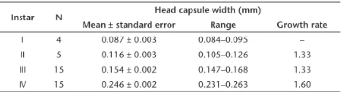
type. There are four instars and two morphotypes; the first form corresponds to the first three instars and the second to the last instar. The first morphotype has a subcylindrical, smooth body, without specialized locomotor structures (Fig. 2). The second morphotype has well-developed calli on thorax and abdomen, and setae of variable sizes distributed throughout the body (Fig. 19). We could not find major morphological differences among instars of the first morphotype. However, they can be identified by their size, since corresponding head capsule widths do not overlap (Table 1). The following exponential growth equation was adjusted for the head capsule width: y = 0.049e0.400x; n = 37; r = 0.99; p < 0.0001.
Penultimate instar. Except for the absence of stemmata, the head of the first morphotype is similar to that of the second one in general color, shape (Figs 3–5), antennae (Fig. 6) and mouth parts (Figs 4, 5, 7, 8), which are described in detail below. The same occurs in relation to thorax and abdomen, including spiracles (Fig. 9). No evident setae were found on the thorax or abdomen of the first morphotype, except for the tenth segment where a pair of conspicuous setae appear dorsolaterally (Figs 10, 11).
Table 1. Variation in size of head capsule width among instars of
Stigmella schinivora reared on Schinus terebinthifolius.
Instar N Head capsule width (mm)
Mean ± standard error Range Growth rate
I 4 0.087 ± 0.003 0.084–0.095 –
II 5 0.116 ± 0.003 0.105–0.126 1.33
III 15 0.154 ± 0.002 0.147–0.168 1.33
Figures 1–11. Egg and third instar of Stigmella schinivora under scanning electron microscopy: (1) egg; (2) general view of larva, lateral; (3–5) head, under dorsal, ventral and anterior views, respectively; (6) antenna, anterior; (7) labrum, anteroventral; (8) labium, showing spinneret in detail, ventral; (9) spiracle of fourth abdominal segment, lateral; (10) last abdominal segment, dorsal; (11) seta of last ab-dominal segment in detail, dorsal. Scale bars: 50, 100, 20, 20, 20, 20, 5, 5, 1, 25, and 10 µm, respectively.
Last instar. Average length ± standard deviation = 1.42 ± 0.21 mm; n = 5. Head light brown, flattened dorsoventrally, partially concealed within the prothorax, with deep epicranial notch. Frontoclypeus rectangular, longer than wide. Labrum
bilobed, with lobes having distal serrated edge, and bearing one pair of short setae mesally (Figs 21, 22); mandibles with well-developed cusps; one long seta on proximal base. Maxilla with well-developed galea and palpi. Labium with tubular
Figures 12–18. Last larval instar and pupal morphology of Stigmella schinivora under light microscopy: (12, 16) larva general, dorsal and ventral views, respectively; (13, 14) detail of tergal and sternal prothoracic plates seen through transparency, dorsal and ventral views, respectively; (15) anal rods of last abdominal segment, dorsal. (17, 18) pupa, dorsal and ventral, respectively. Scale bars: 300, 150, 150, 150, 300, 200, and 200 µm, respectively.
neret (Figs 20, 22), with a pair of setae on proximal base; labial palpi unisegmented, bearing a distal seta. Antenna unisegment-ed, with six apical sensilla; two minute in size, two stout and rounded, and two long and filiform (Fig. 23). A single, circular stemma, posterior to the antenna (Fig. 20). Thorax and abdomen cylindrical, creamy white in preserved material, bearing well-de-veloped filiform setae. Thorax with integument smooth on T1 and sculptured with microtrichia ventrally in T2,3. T1 bearing a light brown shield on the tergum, divided into two elongated, meso-longitudinally arranged plates (Figs 12, 13); a light brown, cup-shaped plate on center of ventral prothoracic sternum (Figs 14, 16). A pair of lateral spiracles without elevated peritreme laterally on prothorax; legs and ambulatory calli absent. T2,3: Dorsal surface smooth; well-developed ambulatory calli ven-trally, with the base wider than the transversally rounded apex, bearing an invagination on middle of the posterior wall (Figs 16, 24, 25). Each callus has the base sculptured with microtrichia and the distal edge smooth (Figs 24, 25). Abdominal segments similar in size from A1 to 8; A9 narrower; A10 smaller and subtriangular in shape. Integument mostly smooth on A1 and A10; partially sculptured with microtrichia dorsally on A3-8, laterally on A8-10 and ventrally on A2-9. Spiracles circular, without elevated peritreme (Fig. 27), laterally, from A1 to 8. Pairs of ambulatory calli present on A2-7, differing from the thoracic ones mainly by not having invagination (Fig. 26). A10 smooth, with a pair
of light brown, longitudinally arranged, distally converging anal rods that are seen by transparency (Fig. 15); two pairs of triangular-shaped projections, one ventrolaterally (Fig. 31), the other on the distal edge of the segment (Figs 29, 31).
Figures 19–31. Last larval instar of Stigmella schinivora under scanning electron microscopy: (19) general view, lateral; (20, 21) head, lat-eral and dorsal views, respectively; (22) mouthparts, anterior; (23) antenna, anterior; (24, 25) mesothoracic ambulatory calli, latlat-eral and ventral views, respectively; (26) ambulatory calli of sixth abdominal segment, ventral; (27) spiracle of eighth abdominal segment, lateral; (28) last abdominal segment, dorsal; (29) detail of tenth abdominal segment showing distal portion of anal rods (indicated by asterisk), posterior projections (indicated by closed arrow) and vestigial setae (indicated by open arrow), dorsal; (30) D2 and L1 setae, indicated by open and closed arrows, respectively, lateral; (31) last abdominal segment showing lateral projection (indicated by closed arrow) and L1 setae (indicated by open arrow). Scale bars: 200, 20, 150, 10, 10, 50, 50, 50, 5, 20, 20, 20, and 20 µm, respectively.
Figures 42–49. Natural history of Stigmella schinivora on Schinus terebinthifolius: (42) host-plant leaf bearing leaf mine on the adaxial sur-face of a foliole (indicated by closed arrow); (43) general view of leaf mine on foliole, showing last instar larva seen through transparence (arrow points to empty egg, and letters indicate position of histological sectioning (treated in Figs 50, 51); (44) empty egg chorium in detail; (45) dissected mine at the final portion, showing last instar larva; (46) exit hole, used by a last instar larva to leave the mine; (47) pupal cocoon adhered to abaxial leaf surface; (48) pupa in detail, after removal of the cocoon, ventral; (49) adult on the leaf, dorsal view. Scale bars: 10, 1, 0.2, 0.5, 0.15, 0.5, 0.5, and 0.5 mm, respectively.
anterior segments, SD and SV group absent. A10 with four pairs of setae. Dorsal group (D) unisetose (represented by D2); two pairs of apparently rudimentary setae (D1 and SD2) (Figs 15, 28, 29). Lateral group (L) unisetose, represented by L1 (Figs 12, 30, 31), longer than D2.
Pupa. Average length ± standard deviation = 1.56 ± 0.03 mm, n = 5. Partially exarate, with distal portion of the wings slightly distant from the abdomen (Fig. 32). Body brownish during early development (darkening later in ontogeny) and flattened dorsoventrally (average maximum width ± standard
elevated peritreme, opened in A1-7 (Fig. 38), partially closed in A8 (Fig. 39). Cremaster absent.
Life history. The egg is usually laid near a lateral vein on the adaxial leaf surface (Fig. 43). After eclosion the first instar larva bores into the leaf and begins to feed on the parenchyma, filling the empty egg with feces (Fig. 44). The serpentine mine is small and narrow, slowly widening throughout larval ontogeny (Figs 42, 43). From the beginning to the end of the mine, the larva feeds only on the palisade parenchyma (Figs 50, 51), which is formed by two layers of overlapping cells in the leaves of S. terebinthifolius (Grisi et al. 2011). The mine is filled fully with feces, which gives it a characteristic blackish appearance (Fig. 43). After completion of development, the last instar larva opens a hole (Figs 45, 46) in distal section of the mine through which it leaves, searching for a pupation site on the abaxial surface either of the same or an adjacent leaf. Cocoon yellowish, cylindrical and silk-made (Figs 40, 41, 47), having flimsy threads, some on the external surface used for attachment to the substrate. It bears a slit on anterior edge through which the pupa projects itself to the outside prior to adult emergence, leaving the exuvium partly protruded from the cocoon (Figs 40, 48, 49).
Densities of S. schinivora are generally low in Laranjeiras do Sul populations of S. terebinthifolius, and in most cases only one mine occurs per leaf and foliole. Mines with mining larvae of S. schinivora were collected mostly during the spring. Appar-ently, more than one generation occurs per year, which should be further explored.
DISCUSSION
Morphology of nepticulid eggs is still controversial, and has not yet been the subject of any detailed study (Davis 1998). According to Johansson et al. (1990), they are covered by a smooth helmet-shaped egg case, supposedly formed by secretion coming from the female colleterial glands. However, Kobayashi (1996) mentioned the existence of a chorion and presence of micropyle canals on the surface of Stigmella castanopsiella (Kuro-ko, 1978). That of S. schinivora is covered by a dome-shaped cap, which can be easily pulled off by physical pressure, thus being detached freely from the remaining egg contents in preserved material. Furthermore, neither micropyles nor aeropyles were found. To better resolve this question, we suggest that oogenesis should be explored in detail for S. schinivora, to test whether or not a true chorion is formed in this species.
The four larval instars found here for S. schinivora fol-low the general pattern recorded for nepticulids in general (Johansson et al. 1990). Although barely mentioned in the recent literature, the existence of two larval morphotypes has been known for a long time in other nepticulids, for example Enteucha acetosae (Stainton, 1854) (Sich 1908, 1909) and Trifur-cula immundella (Zeller, 1939) (Sich 1917). The scarcity of setae on early instars was mentioned by van Nieukerken (2007) for Acalyptris Meyrick, 1921. The absence of true thoracic legs and
Figures 50–51. Transverse histological sections of Schinus terebin-thifolius leaf (indicated by dashed lines “a” and “b” in Fig. 43), showing the organization of Stigmella schinivora mine during larval ontogeny. (50) First instar, initial linear section of mine (position indicated by letter “a” in Fig. 43); (51) last instar, final section of mine (position indicated by letter “b” in Fig. 43). Asterisks indicate leaf mine. (Ab) Abaxial surface of epidermis, (Ad) adaxial surface of epidermis, (Ep) epidermis, (Pp) palisade parenchyma, (Sp) spongy parenchyma. Scale bars: 0.1 and 0.2 mm, respectively.
also of abdominal prolegs bearing chochets, but prominent thoracic and abdominal ambulatory calli instead follows the general pattern found for the last instar of nepticulids (Davis 1987). We associated the existence of these structures with a need for locomotion outside the mine in search for the place to spin the cocoon and pupate.
The prothoracic dorsal shield found in the last instar of S. schinivora is similar to that described for other species of Stigmella by Gustafsson (1985); however, the ventral prothoracic plate shows differences compared to those present in other congeneric species described by him. Thus, this structure may provide diag-nosable taxonomic characters, and should be explored further regarding its use in identification of Stigmella species.
The dorsal sclerotized structures seen by transparency on the last abdominal segment of S. schinivora have received differ-ent names, such “brace rods” (Stehr 1987), “bar-like” (Gustafsson 1981) and “anal rods” (van Nieukerken 2007, van Nieukerken et al. 2011). We opted for anal rods, since given their position it is very likely that these structures are functionally related to the anus. van Nieukerken et al. (2011) reported that the anal rods may be important in the taxonomy of larvae belonging to Acalyptris which should also be further explored in Stigmella.
An interesting characteristic of the first morphotype of S. schinivora is the presence of a single pair of setae in the tenth abdominal segment. van Nieukerken (2007) reported the pres-ence of similar setae, but in A8 (three pairs) in earlier instars in Acalyptris. The large number and size of setae present in the last instar in S. schinivora suggest that these structures may be important from a sensorial perspective when outside the mine prior pupation. They have probably not arisen in this instar in particular, but instead were lost in the previous ones in associ-ation with their endophytic habit.
The comparison of chaetotaxy in Nepticulidae, particularly in Stigmella, showed little variation, suggesting a conserved pat-tern. Compared to the chaetotaxy described by Gustafsson (1981) for Stigmella auromarginella (Richardson, 1890), S. schinivora has in T2-3 absence of L3; in A8 presence of L1 and absence of SV2; and in A9 presence of SV2. In comparison to the chaetotaxy described by Gustafsson (1985) for Stigmella rhomboivora Gustafs-son, 1985, S. schinivora has in T2 absence of L3 and in A9 absence of SV1 and presence of L1. Gustafsson (1981) also states that SV3 is absent in Stigmella plagicolella (Stainton, 1854) and Stigmella paradoxa (Frey, 1858), whereas it is present in S. schinivora.
Two setae have been described in the literature for the last abdominal segment of nepticulid larvae (e.g., Gustafsson 1981, 1985), but they have not been named. The designation of D2 and L1 in this study were inferred by comparing locations of setae in previous abdominal segments. We presume the two pairs of vestigial setae found dorsally in the last abdominal segment of S. schinivora had not been noticed in previous studies due to their reduced size. They are herein tentatively nominated according to Hinton’s system (Stehr 1987), and thus corresponding homologies should be explored further in comparison to other nepticulids.
We are not aware of scanning electron microscopy studies on the pupal morphology of Nepticulidae. The enlarged first antennal segment of S. schinivora stands out, associated with the eye cap in the adult, as well as the absence of any trace of a differentiated process on the head dorsum (= cocoon cutter) and a cremaster on the last abdominal segment. These absences are generally found in the family, as there is no need for the cocoon cutter and cremaster, since, as in S. schinivora, there is usually a slit anteriorly on nepticulid cocoons through which the pupa projects partially to the outside prior to adult emergence (van Nieukerken et al. 2004). Line drawings and description of the pupa of S. plagicolella provided by Patočka and Turčani (2005) are similar to those shown here for S. schinivora. However, these authors do not mention the existence of eight closed abdom-inal spiracles in S. plagicolella, which occurs in S. schinivora. Compared to other nepticulid genera such as Trifurcula Zeller, 1848 (van Nieukerken et al. 2004), Roscidotoga Hoare, 2000 (van Nieukerken et al. 2011) and Acalyptris (van Nieukerken 2007), differences are found in the arrangement of posteriorly directed abdominal spines in A3-7; they form only one row in the ante-rior margin in S. schinivora, contrary to what is found in these genera in which four to five lines of these spines can be found.
The leaf mine of S. schinivora is similar in general shape to congeneric species (e.g. van Nieukerken et al. 2006, Stonis et al. 2013, 2016, Stonis and Remeikis 2017) and to others described for different genera within the Nepticulidae (e.g. van Nieukerken et al. 2011), demonstrating a uniform pattern of the family, even though they may use different host plants. Unfortunately, we did not find other studies addressing histology of mines in Nepticulidae, which precludes comparison with results reported here. Pereira et al. (2017) demonstrated that the damage caused by the gracillariid Leurocephala chilensis Vargas & Moreira, 2016 to the leaves of a plant in the same genus, Schinus molle L., is dif-ferent from that caused by S. schinivora. Most gracillariids show two different kinds of mandibles during development, which may be used initially for slicing and eating only the adaxial epidermis, as is the case of L. chilensis. Chewing mandibles, as in S. schinivora, appear only in latter ontogeny for that species, and are also used to eat the palisade parenchyma until the end of larval development. The different adaptations observed in these two species using closely related hostplants reflect different evolutionary patterns of the families in resource usage (Hering 1951, Menken et al. 2010, Doorenweerd et al. 2016).
Finally, it is important to emphasize that morphology of the immature stages in particular has been increasingly taken into account as an aid in species identification among leaf-miner moths, as for example among gracillariids (Davis and Wagner 2011, Kobayashi et al. 2013, Brito et al. 2017). Information on immature stage morphology is also a precondition for understanding interactions of these stages with host plants, particularly when damage on tissues and histology of the mines are explored in conjunction with ontogeny (e.g., Brito et al. 2012, 2013, Vargas et al. 2015, Pereira et al. 2017). Thus, our
results not only clarify the morphology of S. schinivora imma-ture stages, but also could be used as an integrative framework for characterizing and comparing variation of immature stage morphology and associated host-plant interactions among other nepticulids and beyond.
This is the first report of S. schinivora in Brazil, expanding its geographical distribution that was restricted to the type lo-cality in Argentina. Schinus terebinthifolius is widely distributed in southern South America (see Davis et al. 2011), and thus the range of S. schinivora may be much broader, and should be further explored.
ACKNOWLEDGMENTS
We acknowledge the staff of the Centro de Microscopia Eletrônica/UFRGS for the use of facilities and assistance in scan-ning electron microscopy analyses. We are grateful to Rosângela Brito for assistance in light microscopy photography, and to José Ricardo Assmann Lemes (UFRGS) for helping with fieldwork. We are especially grateful to Erik J. van Nieukerken (Naturalis Biodiversity Center, The Netherlands) for kindly providing lit-erature and for sharing data on morphology of the egg and first instar of nepticulids. We thank Lafayette Eaton for editing the text. We are also grateful to the reviewers Jonas Rimantas Stonis (Lithuanian University of Educational Sciences, Lithuania) and Germán San Blas (Universidad Nacional de La Pampa, Argentina) for their comments and suggestions. Financial support for this study came in part from project UTA-MAYOR 9718-17 granted to H.A. Vargas by Universidad de Tarapacá, Chile. C.M. Pereira and G.R.P.Moreira were supported, by a doctoral scholarship and research grant from CNPq, Brazil (Process numbers 140496/2015-7 and 16/2551-0000485-4 PRONEX, respectively).
LITERATURE CITED
Brito R, Gonçalves GL, Vargas HA, Moreira GRP (2012) A new species of Phyllocnistis Zeller (Lepidoptera: Gracillariidae) from southern Brazil, with life-history description and genetic com-parison to congeneric species. Zootaxa 3582: 1–16.
Brito R, Gonçalves GL, Vargas HA, Moreira GRP (2013) A new Bra-zilian Passiflora leafminer: Spinivalva gaucha gen. n., sp. nov. (Lepidoptera, Gracillariidae, Gracillariinae), the first gracillariid without a sap-feeding instar. ZooKeys 291: 1–26. https://doi. org/10.3897/zookeys.291.4910
Brito R, Mielke OHH, Gonçalves GL, Moreira GRP (2017) Descrip-tion of three new species of Phyllocnistis Zeller, 1848 (Lepidop-tera: Gracillariidae) from the Atlantic Forest, South Brazil, with notes on natural history and phylogeny. Austral Entomology. https://doi.org/10.1111/aen.12298
Braun AF (1917) Nepticulidae of North America. Transactions of the American Entomological Society 43: 155–209.
Davis DR (1987) Nepticulidae. In: Stehr FW (Ed.) Immature Insects. Kendall/Hunt Publishing Company, Dubuque, vol. 1, 350–351.
Davis DR (1998) The Monotrysian Heteroneura. In: Kristensen NP (Ed.) Handbook of zoology, Lepidoptera, moths and butterflies. Walter de Gruyter, Berlin, vol. 1, 65–90.
Davis DR, Mc Kay F, Oleiro M, Vitorino MD, Wheeler GS (2011) Bi-ology and systematics of the leaf mining Gracillariidae of Brazil-ian Pepper Tree, Schinus terebinthifolius Raddi, with descriptions of a new genus and four new species. Journal of Lepidopterists’ Society 65: 61–93. https://doi.org/10.18473/lepi.v65i2.a1 Davis DR, Wagner DL (2011) Biology and systematics of the New
World Phyllocnistis Zeller leafminers of the avocado genus Per-sea (Lepidoptera, Gracillariidae). ZooKeys 97: 39–73. https:// doi.org/10.3897/zookeys.97.753
Doorenweerd C, Nieukerken EJ van, Hoare RJB (2016) Phyloge-ny, classification and divergence times of pygmy leaf-mining moths (Lepidoptera: Nepticulidae): the earliest lepidopteran radiation on Angiosperms? Systematic Entomology 42: 267– 287. https://doi.org/10.1111/syen.12212
Grisi FA, Angelo AC, Boeger MRT, Leitão CAE, Galvão SF, Wendling I (2011) Morfoanatomia foliar em mudas de Schinus terebin-thifolius sob diferentes níveis de saturação hídrica. Floresta 41: 881–894. https://doi.org/10.5380/rf.v41i4.25351
Gustafsson B (1981) Characters of systematic impor-tance in European Nepticulidae larvae (Lepidoptera). In-sect Systematics & Evolution 12: 109–117. https://doi. org/10.1163/187631281X00436
Gustafsson B (1985) New species of Stigmella from the Gambia (Lepidoptera, Nepticulidae). Tijdschrift voor Entomologie 127: 165–177.
Hering EM (1951) Biology of the leaf miners. Junk, The Hague, 420 pp. https://doi.org/10.1007/978-94-015-7196-8
Johansson R, Nielsen ES, Nieukerken EJ van, Gustafsson B (1990) The Nepticulidae and Opostegidae (Lepidoptera) of north west Europe. Fauna Entomologica Scandinavica 23: 1–739.
Kobayashi S, Huang GH, Nakamura A, Hirowatari T (2013) Four new species of Gracillariidae (Lepidoptera) from China and Ja-pan, and description of the pupal morphology of the genera Co-rythoxestis, Eumetriochroa, Guttigera, and Metriochroa. Zootaxa 3619: 101–129. https://doi.org/10.11646/zootaxa.3619.2.1 Kobayashi Y (1996) Gross embryology of a monotrisian
hetero-neuran moth, Stigmella castopsiella Kuroko (Nepticulidae, Lep-idoptera) and its phylogenetic significance. The Lepidoptero-logical Society of Japan 47: 194–200.
Menken SBJ, Boomsma JJ, Nieukerken EJ van (2010) Large-scale evolutionary patterns of host plant associations in the Lepi-doptera. Evolution 64: 1098–1119. https://doi.org/10.1111/ j.1558-5646.2009.00889.x
Myers N, Mittermeier RA, Mittermeier CG, Fonseca GAB, Kent J (2000). Biodiversity hotspots for conservation priorities. Nature 403: 853–858. https://doi.org/10.1038/35002501
Patočka J, Turčani M (2005) Lepidoptera Pupae: Central European Species. Apollo Books, Stenstrup, vol. 1, 321 pp.
Gracillariidae) from the Azapa Valley, northern Chilean Ataca-ma Desert, with notes on life-history. Revista Brasileira de En-tomologia 6: 6–15. https://doi.org/10.1016/j.rbe.2016.11.003 Puplesis R, Robinson GS (2000) A review of the Central and South
American Nepticulidae (Lepidoptera) with special reference to Belize. Bulletin of the Natural History Museum 69: 3–114. Sich A (1908) Notes on the life-history of Nepticula acetosae Stt.
Entomologist’s Record and Journal of Variation 20: 248–252. Sich A (1909) Notes on the life-history of Nepticula acetosae Stt.
Entomologist’s Record and Journal of Variation 21: 103–106. Sich A (1917) Notes on the ovum and larva of Trifurcula
immun-della, Zeller. Entomologist’s Record and Journal of Variation 29: 117–121.
Šimkevičiūtė A, Stonis JR, Diškus A (2009) Taxonomic checklist of Nepticulidae of Mexico, with the description of three new spe-cies from the Pacific Coast (Insecta, Lepidoptera). Acta Zoolog-ica LituanZoolog-ica 19: 268–277. https://doi.org/10.2478/v10043-009-0037-0
Stehr FW (1987) Order Lepidoptera. In: Stehr FW (Ed.) Immature Insects. Kendall/Hunt Publishing Company, Dubuque, vol. 1, 288–305.
Stonis JR, Remeikis A (2016) Southern Andean Stigmella sinuosa
complex (Lepidoptera, Nepticulidae): unraveling problematic taxonomy with a pictorial key of adults? Zootaxa 4136: 309-322. https://doi.org/10.11646/zootaxa.4136.2.3
Stonis JR, Remeikis A (2017) Anacardiaceae-feeding Nepticulidae in the Neotropics: description of Stigmella lilliputica sp. nov. from Argentina, a pest on Chilean peppertree Schinus polyg-ama. Biologija 63: 230–237. https://doi.org/10.6001/biologi-ja.v63i3.3577
Stonis JR, Remeikis A, Diškus A, Noreika R (2013) New Nepticulidae species (Insecta: Lepidoptera) from the Yucatán Peninsula (SE Mexico). Zootaxa 3609: 223–230. https://doi.org/10.11646/ zootaxa.3609.2.8
Stonis JR, Remeikis A, Davis DR (2014) Ten new species from the Patagonian Andes (Argentina and Chile), mostly belonging to a newly designated Stigmella purpurimaculae group (Lepidop-tera: Nepticulidae). Zootaxa 3887: 321–353.
van Nieukerken EJ (2007) Acalyptris Meyrick: revision of the pla-tani and staticis groups in Europe and the Mediterranean (Lepidoptera: Nepticulidae). Zootaxa 1436: 1–48. https://doi. org/10.11646/zootaxa.1436.1.1
van Nieukerken EJ (2018) Nepticulidae and Opostegidae of the world, version 2.0. Scratchpads, biodiversity online. http:// nepticuloidea.info [Accessed 09/01/18]
van Nieukerken EJ, Berg C van den, Hoare RJB (2011) A new spe-cies of the endemic Australian genus Roscidotoga Hoare from rainforests in southern Queensland (Lepidoptera: Nepticuli-dae). Tijdschrift voor Entomologie 154: 193–201. https://doi. org/10.1163/004074912X13397496981508
van Nieukerken EJ, Doorenweerd C, Hoare RJB, Davis DR (2016a). Revised classification and catalogue of global Nepticulidae and Opostegidae (Lepidoptera, Nepticuloidea). ZooKeys 628: 65– 246. https://doi.org/10.3897/zookeys.628.9799
van Nieukerken EJ, Doorenweerd C, Nishida K, Snyers C (2016b) New taxa, including three new genera show uniqueness of Neotropical Nepticulidae (Lepidoptera). ZooKeys 628: 1–63. https://doi.org/10.3897/zookeys.628.9805
van Nieukerken EJ, Mazurkiewicz A, Pałka K (2004) Trifurcula pal-lidella (Duponchel, 1843) (Nepticulidae): distribution, biology and immature stages, particularly in Poland. Nota Lepidopter-ologica 27: 159–178.
van Nieukerken EJ, Schreurs AEP, Stiphout ML van, Ellis WN (2006)
Stigmella aceris (Lepidoptera: Nepticulidae), een nieuwe mi-neermot van esdoorns in Nederland en België. Entomologische Berichten 66: 174–180.
Vargas HA, Brito R, Basilio DS, Moreira GRP (2015) A morpho-logical reappraisal of the immature stages and life history of Elachista synethes Meyrick (Lepidoptera, Elachistidae), an Australian leaf miner alien to Chile. Revista Brasileira de Entomologia 59: 265–273. https://doi.org/10.1016/j. rbe.2015.07.002
Submitted: February 15, 2018 Accepted: April 4, 2018
Available online: October 16, 2018
Editorial responsibility: Gabriel L.F. Mejdalani
Author Contributions: GRPM and CMP conceptualized the study. CMP and DSS made the illustrations. CMP, HAV, and GRPM performed the descriptions. CMP, GRPM, HAV, and DSS wrote the first draft. GRPM reviewed and edited the first version of the manuscript.
Competing Interests: The authors have declared that no competing interests exist.
© 2018 Sociedade Brasileira de Zoologia. Published by Pensoft Publishers at https://zoologia.pensoft.net