UNIVERSIDADE DA BEIRA INTERIOR
Ciências da Saúde
Evaluation of dopaminergic degeneration
influence on endothelial activity in experimental
models of Parkinson's Disease
Patrícia Vanessa Almeida Lindeza
Dissertação para obtenção do Grau de Mestre em
Ciências Biomédicas
(2º ciclo de estudos)
Orientadora: Doutora Ana Clara Braz Cristóvão
Coorientadora: Profª. Doutora Liliana Bernardino
Coorientador: Prof. Doutor Yoon-Seong Kim
iii
“Para ser grande, sê inteiro; nada teu exagera ou exclui; sê todo em cada coisa; põe quanto és no mínimo que fazes; assim em cada lago, a lua toda brilha porque alta vive.”
v
Acknowledgments
There are lots of people I would like to thank for a huge variety of reasons. This dissertation involved about 9 months, which seemed much more to me, given the way I was welcomed by all members of Brain Repair Group.
It is a pleasure to express my truthful gratitude to all who made this thesis possible with words of encouragement. It will be easy to name all the people that helped to get this done, but it will be tough to thank them enough. I will nonetheless try…
Firstly, I would like to thank my Supervisor, Doctor Ana Clara Cristóvão, for accepted me to integrate the best investigation group ever, for her friendship, guidance, help and support during the course of all this work. Her wise advices help me to grow as an investigator. I also acknowledge her for having believed in my capabilities all the time.
I would like to extend my gratitude to Doctor Liliana Bernardino, my Co-supervisor, for doing so much more than would be possible to do for me. Her friendship, prompt way of being and her work capacity are remarkable characteristics to pursue. Her optimism is an example of life. I would also like to thank to Doctor Yoon-Seong Kim for his help and support. I could not have imagined having a better Supervisor and Co-supervisors. It has been a privilege to be able to learn from their example.
I would like to thank my entire colleagues group for their support, knowledge sharing, force and courage in less good moments, but also for all the moments of fun and happiness that we had together, without a doubt, I have been blessed with a friendly and cheerful group. I would like to special thank Márcia Fonseca, my snowflake, who as a very good friend, was always willing to help and give her best suggestions. It would have been a lonely lab without her. I thank you for have been present from the beginning and show me that even with a broken foot, we can achieve anything we want. Besides, that's why we're here!
To the exceptional Raquel Ferreira and the “impecável” Tiago Santos, a very special thank, they were part of this. It was because of their prompt availability and help that part of this project went forward. The lab was always more fun with you both! Is impossible to forget such good friends! I’ve learned so much! Thank you!
To Cláudia Saraiva and Catarina Rebelo, my favorite phD students, to all friendship, encouragement, advices for the future and help for the help in the laboratory work. For you, I wish all the best!
vi
To my family, thanks for your endless love, support and encouragement. Thanks for never give up on believing me and for all your kindness even when you can’t understand what I am doing so many hours in the lab. Mom, Dad, Sister and Samuel, I love you all.
To Luís Martins, my deep love, for all the care and support throughout my life. Thank you for giving me strength to reach for the stars and chase my dreams. You were part of this. I love you.
vii
Resumo
A doença de Parkinson é uma doença neurodegenerativa cujas causas não se encontram totalmente compreendidas. Os modelos animais de Parkinson são ferramentas fundamentais em investigação permitindo o entendimento dos mecanismos envolvidos na patogénese da doença. O modelo animal ideal deve reproduzir muitas, se não todas, as características da doença de Parkinson. Os modelos atuais de Parkinson para além de não mimetizarem os modelos de exposição a toxinas que se pensa ocorrer nos humanos, não apresentam todas as características moleculares e bioquímicas fundamentais da doença de Parkinson, como por exemplo a acumulação de alfa-sinucleína, tornando-se assim restritivos no que se refere à sua aplicação. Deste modo, é necessário o permanente desenvolvimento de novos modelos que mimetizem a doença de Parkinson nos humanos de uma forma mais consistente. Neste projeto, desenvolvemos um novo modelo através da administração crónica de paraquato, via difusão lenta e prolongada de pequenas doses de paraquato assegurada por cápsulas de difusão osmótica. Este modelo desenvolvido por nós reproduz características importantes da doença humana, como é o caso da acumulação de alfa-sínucleina que não se observa nos anteriores modelos animais da doença de Parkinson induzidos por esta toxina. Em paralelo, estudos recentes têm-se focado no estudo da barreira hemato-encefálica que é uma estrutura pouco explorada nesta doença neurodegenerativa. Evidências clinicas demonstram que as disfunções nesta barreira estão associadas a um número elevado de doenças do sistema nervoso central. O HMGB1 tem sido demonstrado como um sinalizador de inflamação, sendo libertado por células necróticas durante estes processos, e tem como recetor, entre outros, o RAGE. Este recetor entre outras células está presente nas células endoteliais, que são o principal componente da barreira hemato-encefálica, e está envolvido em processos inflamatórios através da promoção da proliferação e migração celular. Apesar da importância deste recetor em doenças como AVC, tumores cerebrais e Alzheimer, não existe informação consistente acerca do seu envolvimento na doença de Parkinson. Em estudos anteriores, outro ligando do recetor RAGE (S100B) apresentou-se aumentando em doentes de Parkinson, e existem evidências de que a neuroinflamação observada em Parkinson compromete o funcionamento normal da barreira hemato-encefálica. Deste modo, existem boas indicações para analisar os níveis de expressão de HMGB1 e do recetor RAGE em modelos experimentais de Parkinson. Os resultados obtidos revelam um aumento da expressão de HMGB1 e RAGE com mais significado nos modelos in vivo de PQ e 6-OHDA. Os resultados obtidos sugerem que o papel do ligando HMGB1 tal como do seu recetor na doença de Parkinson, deve ser explorado como forma de perceber se serão marcadores consistentes da neuroinflamação observada nesta doença e se poderão constituir importantes alvos terapêuticos.
viii
Palavras-Chave
Doença de Parkinson, Modelos animais, Paraquato, Neurónios dopaminérgicos, Barreira hemato-encefálica, RAGE, HMGB1
ix
Resumo Alargado
No ano de 1817, James Parkinson descreveu pela primeira vez a doença de Parkinson. Esta patologia caracteriza-se por uma degeneração progressiva da via nigrostriatal dopaminérgica. Os mecanismos moleculares e celulares responsáveis pelo desenvolvimento da doença de Parkinson não se encontram totalmente compreendidos devido, em parte, ao facto de não existir um modelo que mimetize o que se passa nos humanos. Vários estudos apontam para que esta doença tenha uma causa multifatorial, incluindo disfunção mitocondrial, aumentos de stress oxidativo, neuroinflamação, disfunção do proteossoma e agregação de proteínas, como responsáveis pela degeneração dos neurónios dopaminérgicos na substantia nigra pars
compacta. Esta doença foi a primeira doença neurológica a ter um modelo animal de estudo e
a ser tratada através de terapia de substituição de neurotransmissores. Diversos estudos têm demonstrado uma relação entre a exposição a neurotoxinas ambientais e o desenvolvimento da doença de Parkinson. O Paraquato é um herbicida capaz de induzir toxicidade seletiva nos neurónios dopaminérgicos, levando à sua degeneração podendo assim ser utilizado para mimetizar o ambiente patológico observado na doença. Ainda outros modelos de Parkinson incluem o tratamento com 1-metil-4-fenil-1,2,3,tetraidropiridina (MPTP) e 6-hidroxidopamina. Contudo, até à data, não existe um modelo capaz de reproduzir completamente as características observadas na doença humana, que apresenta um caracter crónico. Nenhum dos modelos até hoje desenvolvidos apresenta consistentemente a característica fulcral da doença, a alfa-sínicleinopatia, limitando o estudo dos seus mecanismos patogénicos. O modelo ideal deve incluir, se não todas, as principais caraterísticas da patologia em estudo. Este é um desafio difícil, uma vez que surgem continuamente novos dados acerca da patologia. O desenvolvimento de um modelo que se aproximasse da doença crónica observada em humanos representaria um avanço para o estudo dos mecanismos envolvidos na doença de Parkinson. Neste projeto, foi desenvolvido um novo modelo animal da doença de Parkinson através da administração lenta e continua de paraquato ao longo de 4 semanas. Para este efeito recorreu-se a um método que por difusão osmótica garantiu a libertação gradual de pequenas doses do pesticida. Este novo modelo de Parkinson reflete caraterísticas fulcrais da patologia incluindo perda neuronal dopaminérgica e acumulação e agregação de alfa-sinucleína. O modelo animal desenvolvido representa uma ferramenta atrativa para estudar não só os diversos mecanismos envolvidos na doença, mas também, a aplicação de diversas terapias para a doença de Parkinson.
O ambiente inflamatório crónico observado em doentes de Parkinson leva à libertação de citocinas pro-inflamatórias que resultam em claros sinais de inflamação central e periférica do sistema nervoso. De acordo com a literatura, existem evidências de que a inflamação observada em Parkinson pode comprometer o normal funcionamento da barreira hemato-encefálica. Nos últimos anos, o envolvimento desta barreira tem sido estudado em diversas
x
patologias como o AVC, tumores cerebrais ou a doença de Alzheimer. Em ambientes de inflamação a libertação de citocinas tem-se revelado um forte alvo terapêutico. A molécula HMGB1 trata-se de um mediador de inflamação presente na maioria das células humanas e tem-se revelado fortemente envolvida em processos inflamatórios de diferentes naturezas. O seu recetor principal, RAGE, é um multi-ligando da superfamília das imunoglobulinas descrito pela primeira vez em 1992. Este é amplamente expresso em vários tipos de células, incluindo as células endoteliais conhecidas pelo seu papel fundamental na barreira hemato-encefálica. Os seus diversos ligandos são denominados de moléculas associadas ao dano e são libertadas por células necróticas. Após a sua ativação, esta molécula induz proliferação e migração celular. Estudos recentes têm desvendado o papel deste recetor em doenças como Alzheimer contudo, apesar da importância deste recetor em diversas doenças neurodegenerativas não existe nenhuma informação acerca do seu envolvimento na doença de Parkinson. A literatura indica ainda que as espécies reativas de oxigénio geradas na doença de Parkinson conduzem ao aumento de HMGB1, por outro lado, um estudo envolvendo outro ligando do recetor RAGE (S100B) revelou um aumento da sua expressão em doentes de Parkinson. Deste modo e de acordo com a literatura, existem fortes evidências que reforçam a necessidade de aprofundar a análise da expressão de HMGB1 e RAGE em modelos experimentais de Parkinson. Neste projeto avaliou-se a expressão de HMGB1 e RAGE em modelos in vivo e in vitro de Parkinson. Os níveis de expressão foram obtidos por Western Blot de tecidos das regiões substantia nigra e estriado, recolhidos dos cérebros de animais expostos a diferentes neurotoxinas (paraquato, MPTP e 6-hidroxidopamina), bem como de células neuronais dopaminérgicas (células da linha celular N27) expostas a paraquato, MPP+ e 6-hidroxidopamina e células endoteliais (células
endoteliais da veia umbilical humana) expostas a 6-hidroxidopamina. Os resultados obtidos, revelam um aumento mais expressivo de HMGB1 e RAGE nos modelos in vivo de paraquato e 6-hidroxidopamina.
Deste modo, os resultados obtidos indicam que um estudo mais detalhado do papel tanto do ligando HMGB1 como do seu recetor RAGE na doença de Parkinson, bem como dos seus mecanismos celulares e moleculares associados, seria uma mais-valia para melhor perceber se estes têm potencial para servirem como moléculas para monitorizar a inflamação neuronal, bem como para testar potenciais abordagens terapêuticas para o tratamento desta doença.
xi
Abstract
Parkinson’s disease (PD) is a common neurodegenerative disorder with no known cure. The animal models, in particular the PD models, are important tools in experimental medical science to better understand the mechanisms involved in disease pathogenesis. The ideal animal model for PD should recapitulate most, if not all, features of the human disease. Actual PD animal models available do not show prominent alpha-synucleinopathy, an hallmark of the disease, leading to their restrictive application to undisclosed important molecular mechanisms responsible for PD development. In this way, the implementation of a new model that mimic PD in a more consistent way will be an important step to improve future investigations. In this work, we developed a novel paraquat (PQ)-based chronic PD model by using osmotic minipumps to assure the continuous administration of low doses of PQ for a longer period. Besides the fact that the exposure paradigm mimics in a closer way what happen in humans, this model also reproduces several key characteristics of the human PD, including the important alpha-synucleinopathy. Recent studies have implicated the Blood-Brain Barrier (BBB) as one of the underexplored brain structures in PD. Clinical evidences indicate that BBB dysfunctions are associated with a number of serious CNS diseases. HMGB1 has been shown to be a long-searched-for nuclear danger signal passively released by necrotic cells inducing inflammation. Its receptor, RAGE, is expressed in various cells, including endothelial cells that are largely present in BBB being involved in chronic inflammation and cell proliferation and migration. Despite the importance of this receptor in several diseases such as multiple sclerosis, stroke, brain tumors, AD and cancer, there is no consistent information about this receptor in Parkinson's disease neuroinflammation. In previous studies, another RAGE ligand (S100B) have been reported to be overexpressed in PD patients, despite there are evidences that neuroinflammation associated with PD can compromise the BBB. In this way, it will be of great importance to analyse the expression levels of HMGB1 and RAGE within PD experimental models. Our results revealed an increase of HMGB1 and RAGE expression levels with more significance in vivo models, specifically in PQ and 6-hydroxidopamine models. In this way, the obtained results indicates that a deeper study of the role of both HMGB1 and RAGE in PD will be of great interest to understand they role in PD neuroinflammation and to know if they targeting may serve as a neuroprotective approaches against the development and/or progression of PD.
xii
Keywords
Parkinson’s Disease, Animal models, Paraquat, Dopaminergic neurons, Blood-brain barrier, RAGE, HMBG1.
xiii
Index
Chapter 1-Introduction... 1
1.1. Parkinson´s Disease (PD) ... 1
1.1.1. Definition and Pathophysiology ... 1
1.1.2. Mechanism of Neurodegeneration ... 2 1.1.3. Inflammatory Response ... 3 1.1.4. Animal models ... 4 PQ Model ... 5 MPTP Model ... 6 6-OHDA Model ... 6 1.2. HMBG1-RAGE ... 7 1.3. Blood-Brain Barrier (BBB) ... 9 1.3.1. Blood-brain barrier in PD ... 10 1.4. Main Goals ... 12
Chapter 2-Material and Methods ... 13
2.1. Animal Models and Treatment Paradigm ... 13
2.1.1. PD rat model induced by chronic exposure to low doses of PQ ... 13
2.1.2. MPTP Model ... 14
2.1.3. 6-Hydroxydopamine Model ... 14
2.2. Immunohistochemistry ... 15
2.3. Protein Extraction and Western Blot Analysis... 16
2.4. Cell Cultures and Treatments ... 16
2.4.1. Human Umbilical Vein Endothelial Cells (HUVECs) ... 16
2.4.2. Immortalized rat mesencephalic dopaminergic cell culture (N27 Dopaminergic Cells) .. ... 17
2.4.3. Co-Culture ... 17
2.4.4. N27 Cell Toxins Treatments ... 18
2.5. MTT Reduction Assay ... 19
xiv
Chapter 3- Results ... 21
3.1. Chronic exposure to PQ induced the key features of PD in a novel animal model .... ... 21
3.1.1. Chronic exposure to PQ by osmotic minipumps induced dopaminergic neurotoxicity ... 21
3.1.2. Chronic exposure to paraquat by osmotic minipumps induced alpha-synuclein phosphorylation ... 23
3.2. Expression of HMGB1 and RAGE in PD Animal Models ... 24
3.2.1. In vivo chronic exposure to PQ induced significant changes in the expression levels of HMGB1 in ST and RAGE in both SN and ST... 24
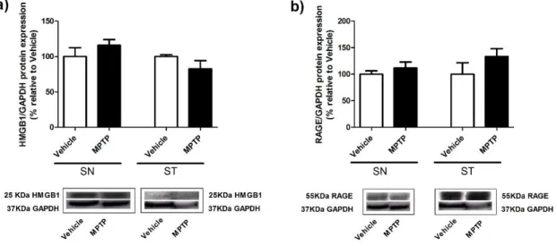
3.2.2. The in vivo acute MPTP exposure does not induces changes in the HMGB1 and RAGE protein expression profile in both SN and ST ... 25
3.2.3. Dopaminergic damage induced by the intrastriatal injection of 6-OHDA increased both HMGB1 and RAGE expression in ST ... 26
3.3. Dopaminergic damage induced by 6-OHDA in vitro show a tendency to increase the expression levels of RAGE in Co-Culture ... 28
3.4.
Toxins-mediated N27 cell death ... 29
3.5. Dopaminergic degeneration induced by toxins increased the expression of HMGB1-RAGE in N27 cells ... 30
Chapter 4- Discussion ... 33
Chapter 5- Conclusions ... 39
xv
Index of figures
Chapter I – Introduction
Figure 1- Schematic representation of PD hallmarks.. ... 1
Figure 2 - Key molecular mechanisms that result in neurodegenerative processes in PD.. ... 3
Figure 3- PQ, rotenone, 6-OHDA and MPP+ toxicity mechanism . ... 5
Figure 4- RAGE signalling resulte in sustained inflammation ... 8
Figure 5- The BBB composition. ... 9
Figure 6- The BBB in AD. ... 11
Chapter II- Materials and Methods
Figure 7- Timeline experiments for PQ chronic administration carried out in vivo ... 14Figure 8- Timeline experiments for MPTP performed in vivo. ... 14
Figure 9- Timeline experiments for 6-hydroxydopamine administration carried out in vivo. .. 15
Figure 10 – Schematic representation of co-culture with N27 cells and HUVEC treatment. ... 18
Figure 11- Toxin treatments applied to N27 Cells. ... 18
Chapter III- Results
Figure 12- Dopaminergic neuronal degeneration induced by chronic exposure to PQ.. ... 22Figure 13- Levels of alpha-synuclein phosphorylated at serine 129 in the SN of rats exposed to chronic dose of PQ.. ... 23
Figure 14- Expression of HMGB1 and RAGE in tissues from SN and ST of rat brain treated with chronic administration of PQ ... 25
Figure 15- Expression of HMGB1 and RAGE in tissues from SN and ST of mice brain treated with acute administration of MPTP. ... 26
Figure 16 - Expression of HMGB1 and RAGE in tissues from SN and ST of mice brain treated with acute administration of 6-OHDA. ... 27
Figure 17- Expression of HMGB1 and RAGE in tissues from SN and ST of mice brain treated with chronic administration of 6-OHDA. ... 28
Figure 18- Expression of RAGE on HUVEC and N27 cells. ... 29
Figure 19- Toxins-mediated N27 cells death. ... 30
xvi
xvii
List of acronyms
6-0HDA- 6-Hydroxidopamine AD- Alzheimer’s Disease
AGE- Advanced glycation end products ANOVA- Analysis of variance
Aβ- β- Amyloid peptide BBB- Blood-brain barrier CNS- Central nervous system DA- Dopamine
DAT - Dopamine active transporter HMGB1 - High-mobility group protein 1 LRP-1- Lipoprotein receptor-related protein 1 MPP+- 1-methyl-4-phenylpyridinium
MPTP- 1-methyl-4-phenyl-1, 2,3,6-tetrahydropyridine
MTT- (3-(4,5-Dimethylthiazol-2-yl)-2,5-Diphenyltetrazolium Bromide) NF-kB- Nuclear factor kappa B
PD- Parkinson’s Disease PQ -Paraquat
RAGE- Receptor for advanced glycation end products ROS- Reactive oxigen species
SN- Substantia nigra ST- Striatum
SNpc- Substantia nigra pars compacta TH- Tyrosine hydroxylase
Evaluation of dopaminergic degeneration influence on endothelial activity in experimental models of Parkinson’s Disease
1
Chapter 1- Introduction
1.1. Parkinson´s Disease (PD)
1.1.1. Definition and Pathophysiology
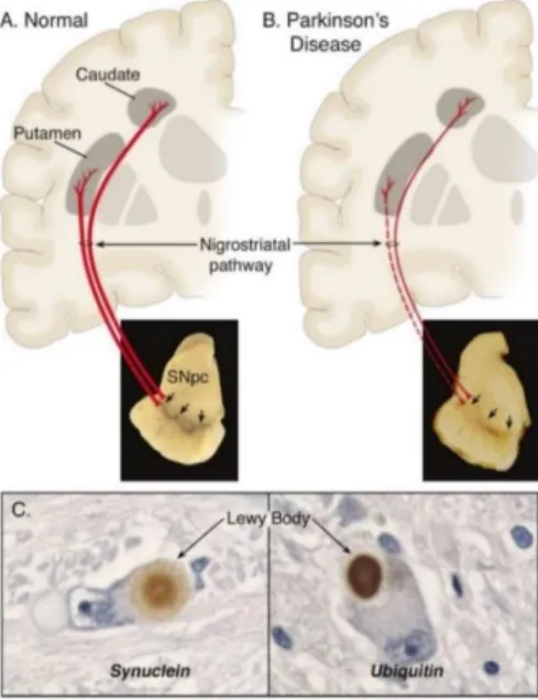
Parkinson’s disease (PD), described in 1817 by James Parkinson [1-3]. This neurodegenerative disease affect an important fraction of world population. It is estimated that 1-2% of the population over 55 years of age is affected by PD and its prevalence dramatically increases after this age, illustrating the effect of aging in this disease [4-6]. In the US it is prevised that in 2040, the population aged 65 years and older will be as high as 80 million [4, 7, 8]. There is no curative treatment [4, 9] and the current management is limited to supportive care and treatment that partially alleviates disease symptoms but does not slow the disease progression [10]. PD is the second most common neurodegenerative disease and is characterized by a severe loss (~50-70%) of dopaminergic neurons in the substantia nigra (SN) [1, 11] and of its fiber projections in putamen and caudate nucleus with the consequent loss of dopamine (DA) levels in the striatum (ST), resulting in motor control impairment [2] (Figure 1A and B). Another pathological hallmark of PD is the observation of intracytoplasmic inclusions called Lewy bodies, containing alpha-synuclein and ubiquitin in dopaminergic neuron, [1, 2, 4, 11] (Figure 1C).
Figure 1- Schematic representation of PD hallmarks. (A) In a normal nigrostriatal pathway the pigmentation of the SNpc produced by neuromelanin within the DA neurons is projected to the ST. (B) In PD the degeneration of the nigrostriatal pathway result in loss of dark-brown pigment neuromelanin due to marked loss of DA neurons and decrease of fibers projecting to ST. (C) Intraneuronal inclusions (Lewy bodies) showing alpha-synuclein and ubiquitin. Figure from Dauer and Przedborski (2003) [12].
Evaluation of dopaminergic degeneration influence on endothelial activity in experimental models of Parkinson’s Disease
2
The PD pathogenesis is characterized by motor problems of patients and several non-motor features [3]. Impaired motor function is typically used to establish the clinical diagnosis of PD and result from nigral neuron degeneration and consequent decrease in dopaminergic striatal innervation, (Figure 1) [6, 11]. The main symptoms are bradykinesia, rigidity, falls, tremor, speech and swallowing difficulties and postural instability with an asymmetric onset spreading to become bilateral with time [3, 13]. The non-motor symptoms such as depression [14], anxiety, apathy [15, 16], psychosis [17], and sleep disturbance, have greater significance when assessed by quality-of-life measures or health economics [3, 13, 18].
The precise ethiology of PD has been under investigation for almost two centuries [19]. Approximately 95% of PD cases are sporadic with no apparent genetic linkage to the pathology resulting in idiopathic PD. There are some risk factors for PD, such as, ageing or environmental exposure to toxins like the herbicides in example (i.e) paraquat (PQ) and the synthetic heroin analogue 1-methyl-4-phenyl-1, 2,3,6-tetrahydropyridine (MPTP) resulting in acquired PD [1, 10, 19] . On the other hand, genetic risk factors include mutations in an ever increasing list of genes which affect either protein metabolism or mitochondrial function, such as Pink1 (PARK6) [20], Parkin (PARK 2) [21], DJ-1 (PARK7) [22] and alpha-synuclein (PARK 1) [23], thus highlighting that the dysfunction in either is sufficient to cause PD in 5% of the cases [12, 19, 24].
1.1.2. Mechanism of Neurodegeneration
Decades of investigation of toxin-based models and genetic models of PD, as well as of sporadic PD patients, have unveiled a number of potential molecular biomarkers of pathology that include protein aggregation [25], proteasomal stress, oxidative stress [26], mitochondrial dysfunction, lysosomal dysfunction and aberrant autophagy proving that PD is a multisystem disorder [6, 27].
Oxidative stress remains a keystone of the concepts underlying the loss of dopaminergic neurons in PD [6, 28, 29]. The mitochondria produces an important amount of reactive oxygen species (ROS). In addition, SN neurons are subjected to oxidative stress derived from their own endogenously occurring dopamine metabolism [30]. Also, it is known that Nox1-generated superoxide is implicated in the oxidative stress elicited by PQ in DA cells [29]. Oxidative stress may induce PD due to altered accumulation of iron in SNpc, changes in calcium channel activity, altered proteolysis (proteasomal and lysosomal), changes in alpha-synuclein aggregation, and the presence of mutant proteins (i.e. DJ-1) [6].
Evaluation of dopaminergic degeneration influence on endothelial activity in experimental models of Parkinson’s Disease
3 The effect of mitochondrial dysfunction has been discussed for over 3 decades and is considered an important contributor to the pathogenesis of PD [6]. The discovery that MPTP impair the mitochondrial electron transport chain by inhibiting complex I lead to subsequent studies that allowed the identification of abnormalities in the activity of this complex resulting in cellular oxidative stress [2].
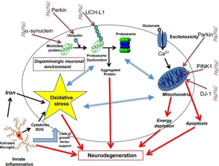
There are several molecular mechanism contributing to the neurodegenerative process of dopaminergic neurons in the SN in PD. Some of these mechanism are represented in (Figure 2). It is important to consider not only the oxidative stress and mitochondrial dysfunction, but also the changes occurring in the inflammatory environment as important contributors to PD pathogenesis [30].
Figure 2 - Key molecular mechanisms that result in neurodegenerative processes in PD. The innate inflammation plays an important role trough activated microglia that in association with proteasome and mitochondrial disjunction lead to oxidative stress. In the cause of these dysfunctions are genetic mutations or neurotoxins exposure. Figure from D.T Dexter and P. Jenner (2013) [6].
1.1.3. Inflammatory Response
The term “inflammation” refer the complex biological response of the immune system to cell injury and tissue damage. Also, this response occurs after exposure to toxic proteins, infection, or abnormal molecular signals [31]. Chronic inflammation is an important feature of this neurodegenerative disease [32]. In PD patients, pro-inflammatory cytokines are increased in the brain and cerebrospinal fluid. Also in pre-clinical animal models of PD there are obvious signs of central and peripheral inflammation [33].
Microglia is thought to play a several role in the Central Nervous System (CNS) innate immune response [31]. They are the resident innate immune cells in the brain being only 5-15% of the
Evaluation of dopaminergic degeneration influence on endothelial activity in experimental models of Parkinson’s Disease
4
whole population of cells [32]. In healthy conditions, microglia generally exhibit a surveying phenotype and perform a scavenging role by removing debris and waste material from the parenchyma. These actions are enhanced following infection, tissue damage, accumulation of toxic protein, or other triggering signal [31, 34].
Activated microglia produce ROS during neuroinflammatory process through intracellular peroxidases, cell surface NADPH oxidase activity and oxidative processes in mitochondria [26, 34]. High levels ROS can damage or inactivate proteins leading to aberrant intracellular signalling, cellular degeneration and death [31]. Pro-inflammatory molecules released by activated microglia such as HMGB1 [35], interleukin-1 (IL)-1, tumour necrosis factor-alpha (TNF-α), and nitric oxide (NO) can be neurotoxic. For instance, the NO reacts with superoxide (O2-), produced by activated microglia, producing highly reactive peroxynitrite anions (ONOO-)
leading to DNA base modifications. These events lead to a disruption of enzymatic function by altered transcription due to DNA damage resulting in loss of structural protein integrity, which can generate cellular apoptosis or necrosis [26].
1.1.4. Animal models
Animal models of PD have been widely used to explore the pathogenesis and pathophysiology of this neurodegenerative disorder [7, 36]. Various pesticides, herbicides and drugs have been used in animals and in vitro models of PD [37]. Animal models are essential tools in experimental biomedical science to better understand pathogenesis of human diseases [28, 38], providing the opportunity to test different therapeutic approaches [28, 38]. The classification of these models depends on systemic or local (intracerebral) administration of neurotoxins that are capable to reproduce most of the pathological and phenotypic features of PD in mammals [36]. Actually, the major animal models used for PD are the ones induced by the toxins rotenone, PQ, MPTP and 6-Hydroxidopamine (6-OHDA), (Figure 3).
The ideal model should reproduce the clinical and pathological features of PD such as progressive loss of dopaminergic neurons and deposition of LB-like inclusions in brain, however no animal model reproduce all the features of the human disease [18]. Naturally, actual animal models of PD have their own specificities and limitations, which must be carefully taken into consideration when choosing the one to be used [12, 36].
Evaluation of dopaminergic degeneration influence on endothelial activity in experimental models of Parkinson’s Disease
5 Figure 3- Molecules like PQ, rotenone, 6-OHDA and MPP+ are currently used to produce experimental
models of PD. These neurotoxins easily cross cell membrane through the dopamine transporter (DAT) thus inducing the formation of alpha-synuclein aggregates and mitochondrial impairment. These events result in ROS production and eventually in cell death. Figure adapted from Cabezas, R et al. (2013) [37].
PQ Model
Exposure to the herbicide 1,1’-dimethyl-4,4’-bipyridinium or PQ, used in agriculture, is considered a putative risk factor for PD [39]. The toxicity of PQ appears to be mediated by the formation of superoxide radicals [12, 40]. The effects of PQ through oxidative stress mediated by redox cycling, generates ROS. The superoxide radical, hydrogen peroxide, and hydroxyl radicals can lead to the damage of lipids, proteins, DNA and RNA [7, 41]. In this way, PQ can induce PD-like lesions in certain mouse strains and rats [24].
Recent studies have shown fundamental features of PD induced by PQ exposure, such as, the selective degeneration of dopaminergic neurons [42], dopamine depletion in the ST, alpha-synuclein up-regulation [43], as well as lipid peroxidation [44]. In result, the PQ-based model represent great importance to PD research due to its ability to induce increases in alpha-synuclein and Lewy body-like inclusions in dopaminergic neurons in the SN [12, 43].
PQ is structurally similar to 1-methyl-4-phenylpyridinium (MPP+), the active metabolite of
MPTP [7, 24, 28, 41] however their biochemical lesions are substantially different. The primary mechanism of MPP+ toxicity is the impairment of mitochondria. This impairment
Evaluation of dopaminergic degeneration influence on endothelial activity in experimental models of Parkinson’s Disease
6
induced by PQ is mainly by cellular redox cycling [24, 28, 41]. There are reports revealing that PQ also target complex I and III of mitochondria [24].
MPTP Model
The MPTP administration is one of the most common animal models used to study PD and has shown to produce permanent parkinsonism in humans, non-human primates and rodents, by exerting an effect primarily of mitochondrial complex I function [24, 28, 45]. The mechanism of MPTP toxicity has been extensively studied and characterized, [12, 46]. MPTP can rapidly cross the blood-brain barrier due to his lipophilic nature [12] being posteriorly metabolized in astrocytes by monoamine oxidase-B, and subsequently converted to the active toxic cation MPP+ that is released from the nigral and striatal astrocytes through the organic cation
transporter 3 into the extracellular space [12, 46]. MPP+ is a polar molecule that is not able to
enter dopaminergic cells freely, thus, its uptake depends on active plasma membrane carrier systems, being taken up by neighbouring dopaminergic neurons and terminals through the DAT [12, 46, 47]. Within the mitochondria, MPP+ lead to production of ROS and decreases the
synthesis of adenosine triphosphate by blocking the complex I that interrupt the transfer of electrons from complex I to ubiquinone [12].
The MPTP model of PD have some limitations, most of protocols of MPTP administration apply acute drug treatments and do not mimic the progressive nature of PD [28, 36]. The chronic MPTP model may overcome this limitation however long-term administration of MPTP in smaller doses may result in the recovery of motor behaviour deficits. In addition, the MPTP model does not directly mimic the systemic mitochondrial impairment found in PD [28, 36].
6-OHDA Model
The classic model based on local (i.e. intracerebral) injection of a neurotoxin is the 6-OHDA model, which was also the first PD animal model ever generated [36, 38]. This model was used to cause lesion of the nigrostriatal dopaminergic pathway in the rat, being used today for both in vitro and in vivo investigations [7, 48].
The neurotoxin, 6-OHDA, is structurally similar to dopamine and norepinephrine presenting high affinity for the plasma membrane transporters of these catecholamines [45]. 6-OHDA cannot cross the blood-brain barrier [12, 38], being most commonly injected unilaterally to the SN, medial forebrain bundle, or ST [46]. When delivered to the ST, 6-OHDA induces progressive and partial damage to the nigrostriatal pathway [46].
The 6-OHDA, once inside of neurons is readily oxidized and produces hydrogen peroxide and paraquinone, both of which are highly toxic and 24 hours after 6-OHDA injections
Evaluation of dopaminergic degeneration influence on endothelial activity in experimental models of Parkinson’s Disease
7 dopaminergic neurons start degenerating and die [12, 45]. The degree of loss is dependent on the injection location and dose of the toxin, as well as the survival time following the lesion. However, like many other PD models, this model lacks the progressive, age-dependent effects of PD and does not produce extra-nigral pathology or Lewy body-like inclusions [45, 46]. PD is a chronic disease developed gradually with the typical symptoms expressed over a long period of time [28]. Neurotoxins such as PQ, 6-OHDA and MPTP used to induce dopaminergic neurodegeneration, mainly by ROS generation, have received the most attention [12]. Few models so far reproduce the progression of extranigral and alpha-synuclein pathology pathology that characterizes PD [28, 45]. Eventually, the ideal model would exhibit all the clinical and pathologic features of PD, but this may be a difficult challenge.
1.2.
HMBG1-RAGE
The HMGB1 previously known as amphoterin, is a DNA binding protein and an important mediator of inflammation via receptors of the innate immune system that are present in the cell nucleus of most mammalian cells [49-51].
There are a few number of identified receptors for HMGB1, among them the principals are the toll-like receptor (TLR) 4 and receptor for advanced glycation end products (RAGE) [52, 53]. These receptors are increased in neurons and glia cells resulting in acute and chronic CNS injuries [52, 54].
The functional role of HMBG1 depends upon its location. Inside the nucleus, HMGB1 acts as an architectural protein that binds DNA, where it exerts different roles and functions, once outside the cell it acts as a pro-inflammatory cytokine [55]. As an extracellular protein, HMGB1 exerts autocrine and paracrine effects. It is responsible for activation of nuclear factor kappa B (NF-κB), diffuse endothelial activation, systemic activation of inflammatory cells, stimulation of innate immune cell migration and activation [55]. To act as inflammatory mediator, HMGB1 must be released by active secretion from living inflammatory cells or from necrotic cells. HMGB1 is the only nuclear protein that have the capacity to induce cytokines and activate inflammatory cells when it is applied extracellularly [56]. After its release from stressed and necrotic cells, HMGB1 triggers inflammation, induces cell proliferation, migration and survival, mainly through interactions with RAGE [57].
RAGE was first described in 1992 [58] as a multi-ligand receptor of the immunoglobulin superfamily of cell surface molecules that can interact with several ligands named
damage-Evaluation of dopaminergic degeneration influence on endothelial activity in experimental models of Parkinson’s Disease
8
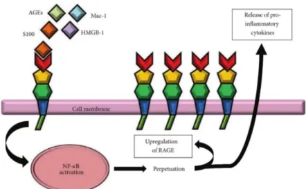
associated molecular pattern molecules, released by dying or necrotic cells during tissue damage [57, 59-62]. The β-amyloid peptide (Aβ), S100B, HMGB1 and advanced glycation end products (AGE) are examples of these molecules and are represented in (Figure 4) [59-61]. AGEs are a product of a series of reactions, the initial one being nonenzymatic glycoxidation that is followed by mechanisms involving ROS [61]. This receptor is expressed in cerebral endothelial cells, neurons, macrophages, monocytes and microglia [63-65]. In humans and mice, the gene encoding RAGE is located on chromosome 6 resulting in a protein with a molecular weight of about 55 kDa [66].
RAGE acts like a pattern recognition receptor being involved in inflammation resolution responsible for tissue repair or alternatively, through its perpetuation, may results in chronic inflammation [62]. Recently, a study developed by Li and colleagues [64], suggest that intracerebral Aβ interaction with RAGE at the blood-brain barrier (BBB) up-regulates endothelial cognate ligand chemokine ligand 5 expression and causes circulating T cell infiltration in Alzheimer Disease (AD) brain [64]. The RAGE-ligand interaction lead to up-regulation of RAGE (Figure 4) via positive feedback loop. The RAGE activation increases pro-inflammatory cytokines secretion promoting pro-inflammatory cell recruitment. RAGE also activates pathways responsible for acute and chronic inflammation [66] that have been associated with various diseases such as vascular disease, diabetes, cancer, and neurodegenerative disorders like AD [67, 68] suggesting that RAGE might be an effective target to treat many different diseases [62, 66].
Figure 4- RAGE signalling resulting in sustained inflammation by activation of the transcription factor NF-kB. Figure from Chuah Y. et. al. (2013) [66].
Evaluation of dopaminergic degeneration influence on endothelial activity in experimental models of Parkinson’s Disease
9
1.3. Blood-Brain Barrier (BBB)
Recently, the role of the pathophysiological importance of the BBB in neurological disorders and the influence of its physiological changes in boosting the neurodegenerative process has been intensively investigated [69].
The BBB is localized at the interface between the blood and the cerebral tissue [70, 71], and it is formed by endothelial cells of cerebral blood vessels (Figure 5) which present intercellular tight junctions and the polarized expression of many transport systems. The transport systems at BBB are the carrier-mediated transport (glucose, amino acids, water-soluble vitamins), the active-efflux transport (low-molecular-mass metabolic products) and receptor-mediated transcriptosis (peptide-specific receptors) [70, 72-74]. The surface area of these microvessels is the largest interface for blood–brain exchange [72]. The BBB endothelial cells, together with pericytes, astrocytes and microglia, separates the components of the circulating blood from neurons and forms the functional neurovascular unit [75]. The crossing of components through the BBB in an uncontrolled way generates neurotoxic products compromising synaptic and neuronal dysfunction. This can happen due to an ischemic injury, intracerebral hemorrhage, neurodegenerative process, inflammation, or vascular disorder, this typically [75]. This barrier have several other functions in the brain. Some of that functions are the supply of the brain with essential nutrients and the efflux of waste products. Also, the BBB protects the brain from fluctuations in ionic composition that can occur after meals or exercise, which may disturb the synaptic and axonal signalling. Globally, the BBB plays an important role to maintain a tightly regulated microenvironment for reliable neuronal signalling [69, 72, 76].
Figure 5- The BBB is composed by endothelial cells, basement membrane, astrocytes, microglia, neurons and pericytes. Brain microvascular endothelial cells acts as mediators between blood and brain interacting with the basement membrane and cells of the neurovascular unit such as neurons, astrocytes and microglia. Figure from Cardoso, F. et al. (2010) [73].
Evaluation of dopaminergic degeneration influence on endothelial activity in experimental models of Parkinson’s Disease
10
1.3.1. Blood-brain barrier in PD
BBB is one of the underexplored brain structures in ageing and PD and its dysfunction is associated to a number of CNS diseases such as multiple sclerosis, stroke, brain tumors, epilepsy or AD [70].
Recently, in vivo studies have shown that BBB dysfunction is related to the course of PD [77]. A study developed by Gray, M. et al. [77], used histologic markers of serum protein, iron, and erythrocyte extravasation to demonstrate significant increased permeability of the BBB in the postcommissural putamen of PD patients. Another investigation in PD patients, reported an elevated uptake of the P-glycoprotein (Pgp) substrate [11C] verapamil in the midbrain, which
is consistent with disturbed Pgp function described by other authors such as Bartels, A. L., et
al. [76, 78]. Altogether, these events could facilitate the accumulation of toxic compounds in
the brain.
Taking as an example animal models of AD, Aβ accumulation is first seen in the neighbourhood of blood vessels. The toxicity including the endothelium and astrocytes is observed before significant neuronal loss and disturbances of CNS homeostasis. These events happens as a result of barrier deficiencies contributing to exacerbate the later neuropathology [76]. Moreover, observations of post-mortem brain tissue from AD patients revealed a number of brain endothelium alterations, such as decreased number of mitochondria, increased number of pinocytosis vesicles, collagen accumulation in basal lamina and necrosis [70].
More recently, studies reached the identification and functional characterization of peptides and proteins transport through the BBB. The transport of Aβ through cerebral endothelium is now well known, as shown in (Figure 6), Aβ peptide influx into the brain is dependent on Aβ chaperones and mediated by RAGE [79]. Due to a lower expression of lipoprotein receptor-related protein 1 (LRP-1) and increased expression of RAGE [79-81], the influx of Aβ increases into the brain. Moreover, soluble forms of LRP-1 are detected in lower amounts conditioning the normal sequester of Aβ peptide that increase in the brain [81]. In summary, these observations strongly suggest that AD progression may involve the age-dependent alteration of Aβ transport across the BBB via RAGE and LRP-1 pathways [70].
Evaluation of dopaminergic degeneration influence on endothelial activity in experimental models of Parkinson’s Disease
11 Figure 6- The BBB in AD. The Aβ peptide is transported to brain by RAGE and cleared from the brain to the blood by LRPs, in healthy conditions (right). In AD (left), RAGE is overexpressed and the expression of LRPs is decreased, leading to the accumulation of Aβ in the brain. Figure from Weiss, N. et al. (2009) [70].
In PD the involvement of ROS and inflammatory processes in neurodegeneration is evident [61]. Recent studies suggest that AGE-RAGE–induced cytosolic ROS production facilitates mitochondrial superoxide production. This fact show the evident role of the advanced glycation pathway in the development of disorders such as diabetic or nephropathy [82]. One of the principal factors linked to the induction of inflammation in PD pathogenesis is the NF-kB [83-85]. According to previous studies, RAGE ligation leads to a sustained activation of NF-kB pathway [86]. Due to an enhanced level of RAGE ligands in chronic disorders, this receptor is hypothesized to have a causative effect in a range of inflammatory diseases [57]. Moreover, ROS involved in PD pathogenesis such as increased levels of hydrogen peroxide, induces the secretion and release of HMGB1 by macrophages, monocytes [56, 87]. The use of antioxidants such as ethyl pyruvate [88] or green tea [89] have shown a protective effect in inflammatory response, by decreasing systemic HMGB1 accumulation. Although the interaction of HMGB1 with RAGE was shown to play a major role in oxidative stress-associated diseases its role in PD pathogenesis remains unclear [90].
Evaluation of dopaminergic degeneration influence on endothelial activity in experimental models of Parkinson’s Disease
12
1.4. Main Goals
There are several models for PD, however few in vivo models clearly exhibit the most important hallmarks of the disease which is alpha-synuclein pathology. With this in mind, we proposed to develop a new PQ-based rat PD model that closely recapitulates cardinal features of PD including dopaminergic neuronal loss and alpha-synuclein pathology markers providing an attractive tool to evaluate the pathologic mechanism as well as various therapeutics approaches for PD. In order to better understand PD pathogenesis, we also propose to evaluate the interplay between the BBB and the dopaminergic neurodegenerative process, induced by the activation of RAGE by its ligand HMGB1, which is so far underexplored in PD. The main goals of this study were:
Development a new animal model for PD by chronic administration of PQ using osmotic minipumps;
Characterize the protein expression profile of HMGB1 and RAGE in experimental models of PD.
Evaluation of the dopaminergic degeneration influence on endothelial activity, through HMGB1-RAGE pathway.
Evaluation of dopaminergic degeneration influence on endothelial activity in experimental models of Parkinson’s Disease
13
Chapter 2- Material and Methods
2.1. Animal Models and Treatment Paradigm
The experiments were carried out on rats and mice, in accordance with protocols approved by the national ethical requirements for animal research, and in accordance with the Directive 2010/63/EU of the European Parliament and the Council on the protection of animals used for scientific purposes. The experiments in vivo involved 8-10 weeks old male wistar rats for the PQ model and 8-12 weeks old C57BL6 mice for MPTP and 6-OHDA model. All animals were kept in appropriate cages, under temperature/humidity-controlled environment on a 12-hr light/dark cycle with free access to food and water. All efforts were made to reduce the number of animals to be used for the study and to minimize their suffering.
2.1.1. PD rat model induced by chronic exposure to low doses of
PQ
The chronic administration of PQ was carried out using osmotic minipumps (Alzet Durect, Cupertino, CA) at a dose of 2.5 mg/kg/day with a fluid delivery rate of 0.25 µL/h for a period of four weeks (Alzet model 2004, large pumps). The pumps were implanted subcutaneously on the back, slightly posterior to the scapulae (shoulder blades). All the rats were weighed at day 1 and every other day for 5 weeks. One week after the end of infusion (5 weeks after implantation), all animals were anesthetized with 5 µl/g of ketamine and xylazine (900 and 500 µg in 4,9 ml 0,9% NaCL total volume, respectively) euthanized and the brains were then recovered (Figure 7). For Western blot analysis, eleven (5 saline and 6 treated) brains were collected and total protein lysates from SN and ST were prepared. For immunohistochemistry studies, animals were anesthetized, euthanized by transcardial perfusion with 0.9% NaCl followed by perfusion with 4% paraformaldehyde (PFA). Following perfusion with saline and 4% PFA, brains were removed, and immersion-fixed in 4% PFA overnight and cryoprotected in 30% sucrose. Serial coronal sections (40 m) were cut on a cryostat, collected in cryopreservative solution, and stored at -20ºC until processed for immunohistostainings.
Evaluation of dopaminergic degeneration influence on endothelial activity in experimental models of Parkinson’s Disease
14
Figure 7- Timeline experiments for PQ chronic administration carried out in vivo.
2.1.2. MPTP Model
MPTP (Sigma-Aldrich, St. Louis, Missouri, USA) was dissolved in sterile 0.9% NaCl and injected intraperitoneally (i.p) four times in the same day, each injection separated by 2 h intervals. The experiments were carried out on 8-12 weeks old male C57BL6 mice and the dose used was 15 mg/kg body weight, been the total dose after the 4 injections of 60 mg/kg [91]. Saline group (four mice) were exposed to the same procedure, receiving an equivalent volume of sterile 0.9% NaCl. Seven days after the MPTP exposure (Figure 8), all animals were anesthetized with 5 µl/g of ketamine and xylazine (900 and 500 µg in 4,9 ml 0,9% NaCL total volume, respectively) and euthanized by transcardial perfusion with 0.9% NaCl and the brains were then recovered. Afterward, for Western-blot analysis, brains were collected and total protein lysates from SN and ST were prepared.
Figure 8- Timeline experiments for MPTP performed in vivo.
2.1.3. 6-Hydroxydopamine Model
6-OHDA (Sigma-Aldrich) was dissolved in sterile 0.02 % ascorbic acid. Mice were deeply anesthetized with 5 µl/g of ketamine and xylazine (900 and 500 µg in 4,9 ml 0,9% NaCL total volume, respectively) and placed in a mice stereotaxic apparatus, and a site in the right ST (coordinate: anteroposterior (AP), –0.6 mm; dorsolateral (DL), -2mm; dorsoventral (DV),–3 mm relative to bregma, in accordance to [92]), was selected to inject 10 g of 6-OHDA at a rate was 0.2 μl/min [93]. Afterward, the syringe was kept in place for additional 5 min before
Evaluation of dopaminergic degeneration influence on endothelial activity in experimental models of Parkinson’s Disease
15 being slowly retracted. Mice were sacrificed after 3 and 7 days (3 and 2 mice respectively). All animals were anesthetized and euthanized by transcardial perfusion with 0.9% NaCl and the brains were then recovered (Figure 9). For Western blot analysis, brains were collected and total protein lysates from SN and ST Ipsi and Contralateral hemispheres were prepared. The ipsilateral side was considered the brain hemisphere injected with 6-OHDA and the contralateral side the non-injected one.
Figure 9- Timeline experiments for 6-hydroxydopamine administration carried out in vivo.
2.2. Immunohistochemistry
The TH immunohistostaining was carried out to determine the number of dopaminergic neurons in the SNpc of the PD rat model induced by PQ developed in the present project, by stereological count. We have also evaluated by immunohistochemistry the reactivity of p129 alpha-synuclein antibody in the SN of treated and untreated rats. In this way, sections were incubated at room temperature with blocking solution for 1 hour (5% FBS and 0.3 % Triton X-100 in PBS, pH 7.5). The endogenous peroxidase were inactivated by incubating the section in 3% H2O2 for 30 min and afterward with primary antibodies overnight. Finally, sections were
incubated with secondary antibodies in blocking solution at room temperature for 1 hour. The primary antibodies used were mouse anti-TH (1:10,000) and rabbit anti-p129alpha-synuclein (1:250). The secondary antibodies used were, respectively, mouse and rabbit biotinylated secondary antibodies (1:200). The staining procedure was performed by the manufacturer’s protocol (Vectastain ABC kit, Vectorlab, Burlingame, CA) and the reaction was visualized using 3,3’-diaminobenzidine (DAB) reagent in Tris buffer saline containing 0.02% H2O2.
Unbiased counting of TH-positive dopaminergic neurons was performed in the SNpc using the optical fractionator method. For each rat brain, the TH+ neurons were count in the SNpc of eight coronal sections, serially selected with 200 m apart representing the whole SNpc.
Evaluation of dopaminergic degeneration influence on endothelial activity in experimental models of Parkinson’s Disease
16
2.3. Protein Extraction and Western Blot Analysis
For the western blot protein analysis, the dissected SN and ST brain tissues (in vivo setting), and the N27 cells lines (in vitro setting), were lysed on ice in RIPA buffer (0.15 M NaCl, 0.05 M Tris-Base, 5mM E ethylene glycol tetraacetic acid, 1% Triton X-100 (Fisher Chemicals, Hampton, New Hampshire), 0.5% deoxycholic acid 0.1% SDS, 10mM dichlorodiphenyltrichloroethane containing a cocktail of proteinase inhibitors). The total protein concentration from lysates was determined using the Thermo Scientific Pierce BCA Protein Assay Kit (Massachusetts, USA) following the manufacturing instructions. To perform the western blot, samples (100 µg of protein of cell lysates or 40 µg of brain tissue lysates) were loaded to each lane of a 12% bis-acrylamide gel (Applichem, Darmstadt, Germany). The proteins were separated by a sodium dodecyl sulfate-polyacrylamigel gel. The running buffer used was Tris-glycine SDS: 25mM Tris, 192 mM glycine, 0.1% SDS, pH 8.3). After electrophoresis at 120 V proteins were transferred at constant 300 mA onto a polyvinylidene difluoride membrane (GE, Heathcare, Little Chanfont, UK), using transfer buffer (10 mM Tris-glycine and 20% methanol (Fisher Chemicals). Afterwards, the membranes were blocked for 1 hour or 15 min in 5% low fat milk or 0.1% gelatin (Fluka, St.Louis, Missouri, USA), respectively, in tris-buffer saline containing tween-20 (0.1%) (Fisher Scientific, Massachusetts, USA). The membranes were incubated overnight at 4ºC with the following primary antibodies: mouse anti-HMGB1 (1:500) from (HMGBiotech srl, Milano, Italy); goat anti-RAGE (1:300) and rabbit anti-p129alpha-synuclein (1:500) both from Santa Cruz Biotechnologies; mouse anti--actin (1:5000) and mouse anti-GAPDH (1:1000) from Millipore. Specific protein bands were detected using the appropriate secondary antibodies (goat anti-mouse (1:5000), donkey anti-rabbit (1:5000) or chicken anti-goat (1:5000) all from Santa Cruz Biotechnologies) conjugated to horseradish peroxidase and detected by Enhanced Chemiluminescence detection (Millipore). Densitometric analyses of the protein bands were performed using the ImageLab software (Bio-Rad, Hercules, CA, USA).
2.4. Cell Cultures and Treatments
2.4.1. Human Umbilical Vein Endothelial Cells (HUVECs)
The HUVECs were grown in EGM Plus Growth Medium (Lonza, USA) containing 2% FBS and BBE, 100 units penicillin, and 50 μg/ml streptomycin, growth factors, cytokines and supplements. The cells were maintained in a humidified atmosphere of 5 % CO2 at 37°C. HUVEC cultures
Evaluation of dopaminergic degeneration influence on endothelial activity in experimental models of Parkinson’s Disease
17 were prepared for experiments by counting the number of viable cells by trypan blue-excluding cells and plating the cells on polystyrene tissue culture dishes at a density of 2x10⁵
cells/well in 6 well culture plates for 24 hours.
2.4.2. Immortalized rat mesencephalic dopaminergic cell culture
(N27 Dopaminergic Cells)
The immortalized rat mesencephalic dopaminergic cell (N27 dopaminergic cells) were grown in RPMI 1640 Medium (Sigma-Aldrich) containing 10% foetal bovine serum, 100 units penicillin and 50 μg/ml streptomycin (Invitrogen, Barcelona, Spain), in a humidified atmosphere of 5 % CO2 at 37°C. N27 cultures were prepared for experiments by counting the number of viable
cells by trypan blue-excluding cells and plating the cells on polystyrene tissue culture dishes at a density of 0.5x10⁴ cells/well in 96 well culture plates and 3x10⁵ cells/well in 6 well
culture plates for 24 hours.
2.4.3. Co-Culture
A co-culture can be defined as the growth of more than one distinct cell type in a combined culture. Such in vitro models can provide a more physiologically relevant way of demonstrating in vivo-like tissue morphology and function. Co-cultures can be employed to monitor intercellular communication between distinct brain cell types. In order to understand the intracellular communication between injured dopaminergic cells (N27 cells) and endothelial cells (HUVECs) both cells were growth in a co-culture system. The HUVECs and N27 cells were plated separately, both in 6 well plate, at 2x10⁵ per well. N27 cells were then
growth in glass coverslip supported in solid paraffin droplets, and 24 hours after transferred to the 6 well plate containing HUVECs, resulting in co-culture. The co-cultures were then treated with 100 μM of 6-OHDA and kept for additional 24 hours (Figure 10). The cells were maintained in a humidified atmosphere of 5 % CO2 at 37°C.
Evaluation of dopaminergic degeneration influence on endothelial activity in experimental models of Parkinson’s Disease
18
Figure 10 – Schematic representation of co-culture with N27 cells and HUVECs treatment.
2.4.4. N27 Cell Toxins Treatments
To estimate the response of the N27 cells to MPP+, PQ and 6-OHDA (Sigma-Aldrich), these
cells were grown at a density of 3x10⁵ cells/well in 6 well culture plates and then treated for
3 hours with 30 μM of MPP+, 500 μM of PQ and 10, 25 and 50 μM of 6-OHDA. The treatments
were prepared in RPMI 1640. Three hours after starting the treatment, all media containing toxins was removed, cells were quickly washed with sterile phosphate buffered saline (PBS) and fresh medium was added to the wells. Afterward, cells were incubated for further 21 hours (Figure 11). After this time, the cells were used to perform the cytotoxic studies using the MTT reduction assay or were collected to prepare cell lysates for western blot.
Evaluation of dopaminergic degeneration influence on endothelial activity in experimental models of Parkinson’s Disease
19
2.5. MTT Reduction Assay
To assess N27 cells viability after exposure to toxins, the levels of MTT reduction were measured. N27 cells were plated at a density of 0.5x10⁴ cells/well in 96 well culture plates.
After exposure to MPP+ 30 µM, PQ 500 µM or 6-OHDA 10, 25 and 50 µM for 3 hours the medium
was replaced by new cell culture medium. The cells was incubated until preamble 24 hours. For the assay using MTT reduction cells were incubated with 0.5 mg/ml of MTT for 4 hours at 37ºC. MTT is converted by viable cells to a water-insoluble precipitate (formazan, presumably directly proportional to the number of viable cells) that is dissolved in 10% sodium dodecyl sulfate-hydrochloric acid (Across Organics) and colorimetrically quantified (O.D. 570-690 nm) using a microplate spectrophotometer (Xmark microplate spectrophotometer, Bio-rad) after overnight incubation at 37ºC with 5 % CO2.. When cells die, they lose the ability to convert
MTT into formazan, thus color formation serves as a useful marker for viable cells only.
2.6. Statistical Analysis
Statistical analysis was carried out with GraphPad Prism v.5 (GraphPad Software Inc., San Diego, CA). Data are expressed as mean ± standard error of mean (SEM) of at least three animals (in vivo studies) or at least three experiments in independent cell cultures. Statistical significance was determined by using one-way ANOVA or Student’s t-test followed by Bonferroni’s post hoc test for comparison with control. P<0.05 were considered to represent statistical significance.
Evaluation of dopaminergic degeneration influence on endothelial activity in experimental models of Parkinson’s Disease
Evaluation of dopaminergic degeneration influence on endothelial activity in experimental models of Parkinson’s Disease
21
Chapter 3- Results
In vivo assays
3.1. Chronic exposure to PQ induced the key features of PD in
a novel animal model
Extensive efforts have been made to establish experimental animal models that recapitulate key pathologic hallmarks of PD, in order to obtain greater insight into the pathogenesis of disease as well as to test new therapeutic strategies. Here, we characterized important features of a novel animal model of PD. Specifically, we evaluated dopaminergic cell death by counting TH+ neurons in SN and the distribution of these TH+ neurons in sequential regions of
SNpc.
The deposition of alpha-synuclein into Lewy bodies (LBs) is a major pathological feature of PD [94] and only a few rodent PD models recapitulate this pathological characteristic [43, 46, 95]. In order to evaluate if our new model developed LB-like alpha-synuclein aggregation, we also investigated the phosphorylated form of alpha-synuclein (pS129) expression and aggregation in the SN that was never achieved in any model before.
3.1.1. Chronic exposure to PQ by osmotic minipumps induced
dopaminergic neurotoxicity
To investigate the extent of dopaminergic neuronal loss in our new PD rat model induced by slow infusion of a low dose of PQ, the number of tyrosine hydroxylase (TH)-positive dopaminergic neurons in the SNpc were counted using unbiased stereological method.
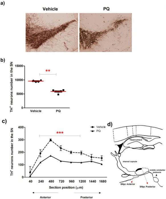
As shown in (Figure 12 a and b), a significant decrease of 39 ±3.3% in dopaminergic neurons was observed in rats exposed to PQ, when compared with the ones exposed to vehicle. To evaluate if a specific region of the SNpc was preferentially affected by chronic exposure to PQ, we sequentially counted dopaminergic neurons in the representative sections of the entire SN from anterior to posterior (Figure 12 d). Even though significant a DA neuronal loss was observed over the entire SNpc, the most prominent loss (~2 fold reduction) was found in the middle sections of the SNpc (Figure 12 c) when compared with the same sections in vehicle animals.
Evaluation of dopaminergic degeneration influence on endothelial activity in experimental models of Parkinson’s Disease
22
Figure 12- Dopaminergic neuronal degeneration induced by chronic exposure to PQ. (a) Representative photomicrographs of TH-immunostaining and quantitative analysis (b) of the number of TH-positive dopaminergic neurons in the SN of rats after 5 weeks exposure to PQ. TH-positive neurons were stereologically counted. (c) Quantitative analysis of TH-positive neurons, along the SN in section from anterior to posterior, collected with 240 µm apart. Scale bars = 500 m. Data are shown as the mean ± SEM. (d) Sagittal representation of the rat brain depicting, among others, the SNpc. Adapted from [96]. Statistical analysis was performed using the Student t test. *P<0.05; **P<0.01 and ***P<0.001.
Evaluation of dopaminergic degeneration influence on endothelial activity in experimental models of Parkinson’s Disease
23
3.1.2. Chronic exposure to paraquat by osmotic minipumps
induced alpha-synuclein phosphorylation
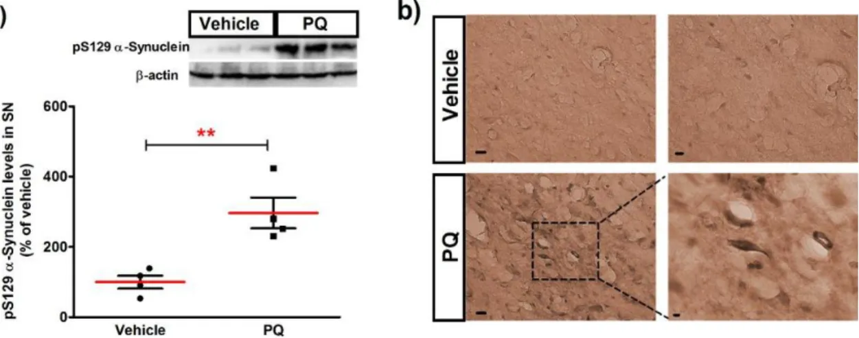
To investigate the expression of the phosphorylated form of alpha-synucleinin our new PD rat model, we then quantified the expression levels of pS129 alpha-synuclein by western blot. In parallel we obtained representative photomicrographs of pS129 alpha-synuclein immunoreactivity in the SN.
W
estern blot analysis of the SN tissues obtained by macroscopic dissection showed that PQ significantly increased the level of pS129 alpha-synuclein protein (296.7 ± 43.62%; n=4;P<0.01), when compared with vehicle (100.0 ± 18.49%) as shown in (Figure 13 a). The increase
in pS129 alpha-synuclein protein was further confirmed in the SNpc region of the coronal tissue sections by immunohistochemistry (Figure 13 b). Taken together, these results show that a low-dose PQ induced dopaminergic neuronal loss in the SNpc and cytoplasmic inclusions of pS129 alpha-synuclein aggregates.
Figure 13- Levels of alpha-synuclein phosphorylated at serine 129 in the SN of rats exposed to chronic dose of PQ are increased. (a) Representative immunoblot and quantitative analysis of pS129 alpha-synuclein protein levels. pS129 alpha-alpha-synuclein protein was determined in total lysates of the rats SN tissues by immunoblot analysis. (b) Representative photomicrographs of pS129 alpha-synuclein immunoreactivity showing increased levels in the SN. Scale bars are: in the left column 20 µm, in the upper panel of the right column 10 mm and in the lower panel 5 m. The results are expressed as percentage of vehicle (n=4). Data are shown as the mean ± SEM. Statistical analysis was performed using the Student t test. **P<0.01.
Evaluation of dopaminergic degeneration influence on endothelial activity in experimental models of Parkinson’s Disease
24
3.2. Expression of HMGB1 and RAGE in PD Animal Models
The dopaminergic cell death observed in PD lead the activation and development of neuroinflammatory reactions. In the inflammatory environment, there is an increased release of pro-inflammatory cytokines that activate immune cell proliferation and migration to the brain. This is a cycling mechanism that easily culminates in the activation of more inflammatory cascades increasing the susceptibility of neuronal cells to further degeneration. In spite of clear evidences regarding the importance of HMGB1 expression in inflammatory processes and the role of RAGE in neurodegenerative diseases, there is no consistent information concerning the expression of this receptor and its ligands in PD. Here we present the results of the protein expression levels of HMGB1 and RAGE in three different PD models: the novel model of PQ develop in this project, the classic MPTP model and the 6-OHDA model. To our knowledge, this is the first time that animal models of PD are evaluated to the expression of this ligand and receptor. This characterization will be helpful to understand the role of HMGB1 and RAGE in the neuroinflammation observed in PD.
3.2.1. In vivo chronic exposure to PQ induced significant changes
in the expression levels of HMGB1 in ST and RAGE in both SN
and ST
In order to characterize the behaviour of HMGB1 and RAGE protein expression levels in our new PD rat model induced by PQ, we tested the effect of a low dose of PQ infused at concentration of 2.5 mg/kg/day during 4 weeks, on the expression of these molecules in the SN and ST.
In the presence of PQ, the expression of HMBG1 does not show differences statistical significant, being slightly decreased (88.94 ± 10.02%; n=5) when compared to vehicle (100.0 ± 16.69%; n=4) in SN. In the ST, the expression of this ligand is significantly reduced (68.68 ± 6.732%; n=5; P<0.01) comparative to vehicle (100.0 ± 2.577%; n=4) as presented in (Figure 14 a). However, a significant increase of the expression of RAGE in SN was observed (142.0 ± 14.88%; n=5; P<0.05) relatively to the vehicle (100% ± 12.05%; n=4). The expression levels of RAGE in ST (179.0 ± 20.00%; n=5; P<0.01) was also significantly increased when compared to vehicle (100.0 ± 9.836%; n=4) as showed in (Figure 14 b).
These results suggest that in our new PD rat model the expression of HMGB1 decreases in both in SN and ST. The same does not happened with RAGE expression that may be influenced by the dopaminergic degeneration induced by chronic exposure to PQ.