UNIVERSIDADE DA BEIRA INTERIOR
Ciências da Saúde
Citotoxicidade in vitro da ticlopidina
em linhas celulares Hep G2 e Caco 2
Experiência profissionalizante nas áreas de Investigação,
Farmácia Hospitalar e Farmácia Comunitária
Catarina Isabel da Silva Martins
Dissertação para obtenção do Grau de Mestre em
Ciências Farmacêuticas
(ciclo de estudos integrado)
Orientador: Doutora Ana Isabel de Jesus Martinho Co-orientador: Professora Doutora Maria Eugenia Gallardo Alba
UNIVERSITY OF BEIRA INTERIOR
Health Sciences
In vitro cytotoxicity of ticlopidine
in Hep G2 and Caco 2 cell lines
Professionalizing experience in the areas of Research,
Hospital Pharmacy and Community Pharmacy
Catarina Isabel da Silva Martins
Dissertation to obtain the Master Degree in
Pharmaceutical Sciences
(integrated cycle of studies)
Supervisor: Ana Isabel de Jesus Martinho, PhD Co-supervisor: Professor Maria Eugenia Gallardo Alba, PhD
“Se não sais de ti, não chegas a saber quem és.” José Saramago
This work was carried out under the “Programa Operacional Regional do Centro 2007-2013 QREN (Programa Mais Centro)” through the project CENTRO-07-ST24-FEDER-002012 entitled “Therapeutic drug monitoring on age related diseases”, COMPETE program and Portuguese Foundation for Science and Technology through the project PEst-OE/SAU/UI0709/2014.
Acknowledgements
At the end of this hard journey, some special people who positively contributed for its success deserve a special word and a thank you.
Ana, the best supervisor I could have had, even when you had to scold. For the knowledge transmitted but also for the company and the conversations in between the work.
Prof. Eugenia, my co-supervisor, for suggesting Ana as a supervisor and for being one of the greatest professors I had, you inspired me.
Sara Silva and Joana Norte, the lab mates, for the help and also the talks.
Prof. Luiza Granadeiro (from CICS-UBI), who kindly provided the Hep G2 cells used in the study.
Prof. Samuel and Dr. Musicco, for allowing me an international experience, which I loved and where I learned a lot. “La bella Italia” is indeed beautiful.
In Italy, Dr. Angelo Tarantino, Dr. Lidia di Cerbo, Dr. Elisa Marchesini, Dr. Elisabetta Umana, Dr. Antonia La Malfa and especially Dr. Nicoletta Jannitti, who inspired me most of all, I want to be a pharmacist like you one day. Also, thanks everyone for the hospitality and for being so nice and friendly.
From the Nunes Feijão Pharmacy, Dr. Isabel Feijão, Andreia Rodrigues and Dr. Cátia Silva. And the pool parties friends in Barreiro.
Mom and Dad, for allowing me this 6 year adventure. Thanks for letting me pursue the dream and for the support in (most of) my projects.
Sara, my little sister, for everything and especially for your contribution in this work. My Grandparents, your pride in me makes me so proud.
EncantaTuna, my second family, college time was better with all you sisters.
Joana Mendonça, the best person I found in pharmaceutical sciences, for being the best mate ever. For the study times and for the drinks and nights out, together is better.
Fábio, always pushing me to aim for more: to know better, to do better, to be better. Always there, even when far, my best friend.
Abstract
The present dissertation is divided into 3 parts.
In the Part I is described the research work conducted at the Health Sciences Research Center of the University of Beira Interior (CICS–UBI, Covilhã) with the aim of evaluating the cytotoxicity of ticlopidine and Hypericum perforatum extractin hepatic and intestinal epithelium cell lines.
Ticlopidine is a prodrug mainly used in the prophylaxis of thromboembolic complications in patients with thromboembolic disease, especially if they are aspirin-intolerant. It is associated with multiple drug interactions, mostly via cytochrome P450 (CYP)-inhibition.
H. perforatum has been widely used as an antidepressant, anti-inflammatory and
antimicrobial agent, being considered, however, responsible for numerous herb-drug interactions due to the induction of CYP enzymes and P-glycoprotein expression.
Given the predisposition of both these compounds to induce interactions when co-administered with other substances, it was considered relevant to study their cytotoxicity, alone and in combination, in hepatic – Hep G2 – and intestinal epithelium – Caco 2 – cell lines. The cells were incubated with various concentrations of ticlopidine and/or H. perforatum extract at various periods of time and the putative cytotoxicity induced by each incubation condition was assessed through cellular viability (MTT) assays.
Overall, the results showed that ticlopidine may be cytotoxic in both Hep G2 and Caco 2 cells, depending on its concentration and period of incubation, and that the simultaneous incubation of cells with both the compounds promotes a similar pattern to that observed when cells are incubated with the extract alone and it is dose-, time- and cell line-dependent.
The Part II refers to the hospital pharmacy internship performed at the Istituti
Fisioterapici Ospedalieri, in Rome, between February 2nd and April 29th 2015.
Finally, the Part III refers to the community pharmacy internship carried out in the Nunes Feijão Pharmacy, near Barreiro, between May 11th and August 10th 2015.
Keywords
Resumo alargado
A presente dissertação encontra-se dividida em 3 partes.
Na Parte I encontra-se descrito o trabalho de investigação realizado no Centro de Investigação em Ciências da Saúde da Universidade da Beira Interior (CICS-UBI, Covilhã) com o objetivo de avaliar a citotoxicidade da ticlopidina e de um extrato de hipericão em células hepáticas e do epitélio intestinal.
A ticlopidina é um pró-fármaco oral apresentado apenas na forma de comprimidos e utilizado essencialmente na profilaxia de eventos cardiovasculares major em pessoas com doença tromboembólica, especialmente se forem intolerantes ao ácido acetilsalicílico. Requer bioativação in vivo pelas enzimas do citocromo P450 (CYPs) para formar o seu metabolito ativo, que se liga irreversivelmente ao recetor P2Y12 nas plaquetas, inibindo a sua ativação e agregação induzidas pela adenosina difosfato (ADP) de uma forma tempo- e dose-dependente. Tratando-se de um potente inibidor mecanismo-dependente de algumas CYPs, inibindo até o seu próprio metabolismo, a ticlopidina está associada a múltiplas interações farmacológicas e não farmacológicas. Além disso, apresenta diversas reações adversas hematológicas, pelo que o seu uso deve ser monitorizado nos primeiros três meses de tratamento. Os efeitos adversos são reversíveis após a retirada do fármaco.
O hipericão, uma das mais antigas e bem estudadas plantas medicinais, tem sido amplamente utilizado como agente antidepressivo, anti-inflamatório e antimicrobiano. Apresenta-se sob diversas formas farmacêuticas, sendo os extratos considerados como a mais importante. Barato e fácil de obter, torna-se uma alternativa às terapias farmacológicas comuns, sem apresentar efeitos adversos major.
Encontram-se identificados mais de 150 constituintes: a hipericina é considerada o composto farmacologicamente mais importante, enquanto à hiperforina é atribuída a responsabilidade pela maioria das interações medicamentosas. De facto, o hipericão é um potente indutor das CYPs e da expressão da glicoproteína-P (P-gp). De acordo com o efeito farmacológico demonstra diversos mecanismos de ação; enquanto antidepressivo, atua de uma forma semelhante aos inibidores seletivos da recaptação de serotonina, podendo originar um síndrome serotoninérgico ou interações farmacodinâmicas se coadministrado com fármacos que aumentam a sinalização deste neurotransmissor.
Dada a predisposição de ambos os referidos compostos para induzir interações quando coadministrados com outras substâncias considerou-se relevante estudar a sua citotoxicidade, sozinhos e em associação, em células hepáticas – Hep G2 – e do epitélio intestinal – Caco 2. As células foram incubadas com várias concentrações de ticlopidina e/ou de extrato de hipericão durante vários períodos de tempo e a citotoxicidade putativa induzida por cada condição de incubação foi avaliada através de ensaios de viabilidade celular (MTT).
In vitro cytotoxicity of ticlopidine in Hep G2 and Caco 2 cell lines
No geral, a ticlopidina mostrou-se significativamente citotóxica em ambas as linhas celulares apenas na concentração de 200 μM; todavia, as células revelaram uma recuperação da viabilidade celular após 24 h de incubação.
Quanto ao hipericão, apresentou maior toxicidade na linha celular hepática, comparativamente aos resultados obtidos para a linha celular intestinal. Mais uma vez, as células recuperaram e voltaram a proliferar após 72 h de incubação.
Da incubação com ambos os compostos obtiveram-se resultados semelhantes aos obtidos na incubação apenas com hipericão, principalmente nas células Caco 2. Nas células Hep G2, a incubação com ambos os compostos mostrou-se menos citotóxica que a incubação com hipericão sozinho.
A recuperação da viabilidade celular apresentada por ambas as linhas celulares na incubação com os compostos sozinhos ou em combinação sugere, nas células Hep G2, a formação de conjugados de glutationa não tóxicos e, nas células Caco 2, um mecanismo de efluxo pela P-gp.
Em suma, os resultados obtidos mostraram que a ticlopidina pode ser citotóxica em ambas as linhas celulares Hep G2 e Caco 2, dependendo da sua concentração e do período de incubação, e que a incubação simultânea das células com ambos os compostos promove um padrão similar ao observado quando as células foram incubadas com o extrato sozinho, sendo a toxicidade dependente da dose, do tempo e da linha celular.
A Parte II refere-se ao estágio em farmácia hospitalar realizado nos Istituti Fisioterapici
Ospedalieri, em Roma, entre 2 de fevereiro e 29 de abril de 2015.
Finalmente, a Parte III refere-se ao estágio em farmácia comunitária feito na Farmácia Nunes Feijão, perto do Barreiro, entre 11 de maio e 10 de agosto de 2015.
Palavras-chave
Index
Acknowledgements ... vii
Abstract... ix
Resumo ... xi
Index ... xiii
Index of Figures ... xvii
Index of Tables ... xix
List of Acronyms ... xxi
Part I – In vitro cytotoxicity of ticlopidine in Hep G2 and Caco 2 cell lines ... 1
1. Introduction ... 1
1.1. Ticlopidine ... 1
1.1.1. Historical context and general characterization ... 1
1.1.2. Physical and chemical characteristics ... 1
1.1.3. Pharmacokinetics ... 2
1.1.4. Pharmacodynamics ... 4
1.1.5. Therapeutic applications ... 6
1.1.6. Adverse and toxic effects ... 6
1.1.7. Pharmacological and non-pharmacological interactions ... 7
1.2. Hypericum perforatum ... 8
1.2.1. General characterization ... 8
1.2.2. Bioactive compounds ... 9
1.2.3. Pharmacodynamics ... 9
1.2.4. Therapeutic applications ... 10
1.2.5. Adverse and toxic effects ... 11
1.2.6. Pharmacological interactions ... 11
2. Aims ... 15
3. Material and methods ... 17
3.1. Material ... 17
3.1.1. Compounds, solutions and culture media ... 17
3.1.2. Equipment ... 17 3.2. Cell lines ... 18 3.2.1. Hep G2 cells ... 18 3.2.2. Caco 2 cells ... 18 3.3. Methods ... 19 3.3.1. Cell cultures ... 19
3.3.1.1. Characterization and preparation of culture media ... 20
3.3.1.2. Thawing and cell counting ... 20
In vitro cytotoxicity of ticlopidine in Hep G2 and Caco 2 cell lines
3.3.1.4. Freezing ... 22
3.3.2. Incubation of the cells with ticlopidine and/or Hypericum perforatum ... 22
3.3.2.1. Preparation of the solutions ... 22
3.3.2.2. Incubation procedure ... 22
3.3.3. MTT assays ... 22
3.3.3.1. Background ... 22
3.3.3.2. Experimental procedure ... 23
3.4. Statistical data analysis ... 23
4. Results and discussion ... 25
4.1. Hep G2 cells ... 26
4.2. Caco 2 cells ... 29
5. Conclusions and perspectives ... 33
Part II – Internship report: hospital pharmacy ... 35
1. Introduction ... 35
2. Background ... 35
3. General characterization of the Istituti Fisioterapici Ospitalieri ... 36
3.1. IFO’s pharmaceutical services: internal hospital pharmacy and external hospital pharmacy ... 36
3.1.1. Location, facilities and working hours ... 36
3.1.2. Human resources ... 37
4. Organization and management ... 37
4.1. Provision ... 37
4.1.1. Hospital Therapeutic Handbook ... 37
4.1.2. Selection system and acquisition ... 38
4.1.2.1. Drugs not included in the Corporate Therapeutic Handbook ... 38
4.1.2.2. Narcotic and psychotropic drugs ... 38
4.1.2.3. Drugs not registered in Italy ... 39
4.1.3. Reception and verification ... 40
4.1.3.1. Reception and verification of narcotic and psychotropic drugs ... 40
4.2. Storage ... 40
7. Clinical trials ... 48
7.1. Experimental medicines circuit ... 48
8. Pharmacovigilance ... 50
9. Technical committees ... 51
10. Conclusions ... 52
Part III – Internship report: community pharmacy ... 53
1. Introduction ... 53
2. General characterization of the pharmacy ... 53
2.1. Location, working hours and external appearance ... 53
2.2. Facilities ... 54
2.3. Human resources ... 55
2.4. Scientific documents and information support ... 55
3. Organization and management ... 56
3.1. Provision ... 56
3.1.1. Orders management ... 56
3.1.1.1. Order submitting ... 56
3.1.1.2. Reception and verification of the order ... 57
3.2. Price marking ... 57
3.3. Expiring dates control ... 58
3.4. Storage ... 58
3.5. Returns ... 59
4. Medicines and other healthcare products ... 60
4.1. Medicines subjected to medical prescription ... 61
4.1.1. Generic medicines ... 63
4.1.2. Reimbursement ... 64
4.1.3. Special medicines ... 66
4.2. Medicines not subjected to medical prescription ... 66
4.3. Suspended and credit sales ... 67
4.4. Advising and dispensing other healthcare products ... 67
4.4.1. Medicines and other products for veterinary use ... 67
4.4.2. Dietary products ... 68
4.4.3. Phytotherapy products and food supplements ... 69
4.4.4. Cosmetic, dermocosmetic and hygiene products ... 69
4.4.5. Medical devices ... 70
4.5. Galenic preparations ... 70
4.5.1. Raw materials ... 71
4.5.2. Material and equipment ... 72
4.5.3. Protocols and techniques ... 72
4.5.4. Quality control ... 73
In vitro cytotoxicity of ticlopidine in Hep G2 and Caco 2 cell lines
4.5.6. Extemporaneous preparations ... 74
5. Pharmacist-patient-medication relationship ... 74
5.1. Ethical issues ... 74
5.2. Pharmacovigilance ... 75
5.3. Recycling expired medicines ... 75
6. Self-medication ... 76
7. Other services ... 77
7.1. Anthropometry and body mass index ... 77
7.2. Blood pressure ... 78
7.3. Capillary blood glucose and total cholesterol ... 78
8. Accounting and management ... 79
9. Conclusions ... 80
References ... 81
Index of Figures
Figure 1. Chemical structure of ticlopidine. 2
Figure 2. Metabolic pathways of ticlopidine. 3
Figure 3. ADP platelet receptor signaling pathways and site of action of ticlopidine. 5
Figure 4. Possible mechanism for irreversible inhibition of CYP2B6 by ticlopidine. 7
Figure 5. Chemical structures of hypericin, pseudohypericin, and hyperforin. 9
Figure 6. Hep G2 cells morphology under optical microscope. 18
Figure 7. Caco 2 cells morphology under optical microscope. 19
Figure 8. Grid layout of the Neubauer improved hemocytometer under optical
microscope.
21
Figure 9. Graphical representation of the relative percentage of the viable Hep G2
cells after incubation with various concentrations of ticlopidine (1, 10, 100 and 200 μM) for different periods of incubation (12, 24, 48 and 72 h). (* p < 0,05)
26
Figure 10. Graphical representation of the relative percentage of the viable Hep G2
cells after incubation with various concentrations of H. perforatum extract (1 and 10 μM) for different periods of incubation (12, 24, 48 and 72 h). (* p < 0,05)
28
Figure 11. Graphical representation of the relative percentage of the viable Hep G2
cells after incubation with various concentrations of the combination of ticlopidine and H. perforatum extract (1 + 1 and 10 + 10 μM) for different periods of incubation (12, 24, 48 and 72 h). (* p < 0,05)
29
Figure 12. Graphical representation of the relative percentage of the viable Caco 2
cells after incubation with various concentrations of ticlopidine (1, 10, 100 and 200 μM) for different periods of incubation (12, 24, 48 and 72 h). (* p < 0,05)
30
Figure 13. Graphical representation of the relative percentage of the viable Caco 2
cells after incubation with various concentrations of H. perforatum extract (1 and 10 μM) for different periods of incubation (12, 24, 48 and 72 h). (* p < 0,05)
31
Figure 14. Graphical representation of the relative percentage of the viable Hep G2
cells after incubation with various concentrations of the combination of ticlopidine and H. perforatum extract (1 + 1 and 10 + 10 μM) for different periods of incubation (12, 24, 48 and 72 h). (* p < 0,05)
Index of Tables
Table 1. Summary of pharmacokinetic characteristics of ticlopidine. 4
Table 2. Summary of the drug-drug interactions associated with ticlopidine and its
downregulation of the expression of various CYPs.
7
Table 3. Important biological compounds found in H. perforatum. 11
Table 4. Pharmacological interactions with H. perforatum. 12
Table 5. Summary of the literature research to choose the ticlopidine concentrations
to include in the study.
25
Table 6. Relative percentage of the viable Hep G2 cells plus various concentrations
of ticlopidine (1, 10, 100 and 200 μM) for different periods of incubation (12, 24, 48 and 72 h).
26
Table 7. Relative percentage of the viable Hep G2 cells plus various concentrations
of H. perforatum extract (1 and 10 μM) for different periods of incubation (12, 24, 48 and 72 h).
27
Table 8. Relative percentage of the viable Hep G2 cells plus various concentrations
of the combination of ticlopidine and H. perforatum extract (1 + 1 and 10 + 10 μM) for different periods of incubation (12, 24, 48 and 72 h).
28
Table 9. Relative percentage of the viable Caco 2 cells plus various concentrations of
ticlopidine (1, 10, 100 and 200 μM) for different periods of incubation (12, 24, 48 and 72 h).
29
Table 10. Relative percentage of the viable Caco 2 cells plus various concentrations
of H. perforatum extract (1 and 10 μM) for different periods of incubation (12, 24, 48 and 72 h).
31
Table 11. Relative percentage of the viable Caco 2 cells plus various concentrations
of the combination of ticlopidine and H. perforatum extract (1 + 1 and 10 + 10 μM) for different periods of incubation (12, 24, 48 and 72 h).
32
Table 12. The 5 levels of therapeutic classification. 61
Table 13. Pathologies and medicines covered and values of reimbursement for the
special regimen “O”.
List of Acronyms
5-HT Serotonin
AD Anno Domini
ADP Adenosine diphosphate ADR Adverse drug reaction AIFA Agenzia Italiana del Farmaco
API Active pharmaceutical ingredient ATC Anatomical Therapeutic Chemical AUC Area under curve
BMI Body mass index BSA Body surface area CCK Cholecystokinin
Cmax Peak plasma concentrations
CNS Central nervous system CO2 Carbon dioxide
COMT Cathecol-O-methyltransferase CYPs Cytochrome P450 enzymes DH Day Hospital
DMEM-C Dulbecco’s Modified Eagle’s Medium – high glucose complete DMEM-I Dulbecco’s Modified Eagle’s Medium – high glucose incomplete DMSO Dimethyl sulfoxide
EDTA Ethylenediamine tetraacetic acid FBS Fetal bovine serum
FDA Food and Drug Administration GABA Gamma-aminobutyric acid GSH Glutathione
HbA1C Glycated hemoglobin A1c HCl Hydrochloric acid
HMG-CoA 3-hydroxy-3-methyl-glutaryl-CoA
H. perforatum Hypericum perforatum
IFO Istituti Fisioterapici Ospitalieri
INFARMED National Authority of Medicines and Health Products IRCCS Istituti di Ricovero e Cura a Carattere Scientifico
IRE Istituto Nazionale Tumori Regina Elena
ISG Istituto Dermatologico San Gallicano
LDL Low-density lipoprotein MAO Monoamine oxidase
In vitro cytotoxicity of ticlopidine in Hep G2 and Caco 2 cell lines
MPO Myeloperoxidases
MTT 3-(4,5-dimethylthiazol-2-yl)-2,5-diphenyltetrazolium bromide NFP Nunes Feijão Pharmacy
NHS National Health System
OATP-B Organic anion transporting polypeptide B OTC Over the counter
P-gp P-glycoprotein
PAF Platelet activating factor PBS Phosphate-saline buffer
RNF Rete Nazionale di Farmacovigilanza
RPMI-C Roswell Park Memorial Institute 1640 Medium complete RPMI-I Roswell Park Memorial Institute 1640 Medium incomplete SSRI Selective serotonin reuptake inhibitor
t1/2 Elimination half-life
UMaCA Unità di Manipolazione di Chemioterapici Antiblastici
UV Ultraviolet
TRPA1 Transient receptor potential ankyrin1 VAT Value-added tax
VEGF Vascular endothelial growth factor WHO World Health Organization
Part I – In vitro cytotoxicity of ticlopidine in Hep G2 and
Caco 2 cell lines
1. Introduction
1.1. Ticlopidine
1.1.1. Historical context and general characterization
Ticlopidine is a first generation thienopyridine [1], synthesized for the first time and patented in Germany in 1974 [2, 3]. Thienopyridines are heterocyclic compounds containing a thiophene ring fused to a pyridine ring [4].
It was first used in 1978 [5] and introduced in the German market in 1979 [6]. In the United States of America, ticlopidine was patented in 1977 [3] and received its market approval by the Food and Drug Administration (FDA) in October 1991 [7, 8], being available for general clinical use in December 1991 [9, 10]. Nevertheless, in Portugal, it was approved only since May 15th 1981 [11].
It was studied as an anti-inflammatory agent, but its potent antiplatelet effects were more remarkable [1], being therapeutically used in the prophylaxis of thromboembolic complications in patients with thromboembolic disease, especially if they are intolerant to aspirin [12].
The recommended dosage is 250 mg orally, twice a day with meals [6, 13]. Commercially, the unique available form is a 250 mg tablet of a hydrochloride salt [13, 14]. In Portugal, it is presented as coated tablets and film-coated tablets [14] and the commercial name is Tiklyd®
[11].
Due to the reported idiosyncratic adverse reactions, especially blood disorders and drug induced liver injuries, attributed to ticlopidine, it was given a FDA black box warning for life threatening hematological adverse reactions [8, 15]. It is not genotoxic or cardiogenic [16].
It was referred by the Portuguese National Pharmacovigilance newsletter in 1997 due to an adverse reaction identified, a bone marrow depression [17], and in 1999 in the form of a reminder that its use must be monitored in the first 3 months of treatment (when adverse reactions are most likely to occur) [18].
1.1.2. Physical and chemical characteristics
Chemically, ticlopidine is the 5-(o-chlorobenzyl)-4,5,6,7-tetrahydrothieno[3,2-c]pyridine [3].
In vitro cytotoxicity of ticlopidine in Hep G2 and Caco 2 cell lines
Figure 1. Chemical structure of ticlopidine (adapted from [19]).
Ticlopidine is a highly hydrophobic molecule [20], positively charged at physiological pH [21]. It exhibits low aqueous solubility and it improves the rheological properties of blood [22, 23]. Its commercially available formula, ticlopidine hydrochloride, is freely soluble in water and in methanol, moderately soluble in methylene chloride and ethanol and slightly soluble in acetone [13].
1.1.3. Pharmacokinetics
The thienopyridines, and thus ticlopidine, are oral prodrugs that require in vivo bioactivation, consisting in the conversion into a pharmacologically active metabolite containing a thiol moiety. This thiol metabolite binds specifically and irreversibly to the P2Y12 receptor, inhibiting platelet activation and aggregation in a time- and dose-dependent manner [6, 24].
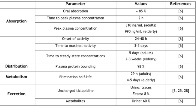
Ticlopidine exhibits a non-linear pharmacokinetics [6]. When administered orally, approximately 85 % is absorbed, with a peak plasma concentration (Cmax) after 2 h [6].
Bioavailability is decreased in about 18 % by co-administration with antacids and increased approximately 20 % if the drug is co-administrated with food [13, 24].
The median Cmax after the first dose is 310 ng/mL, increasing to 990 ng/mL after 21 days
of treatment [6]. The onset of activity of the recommended daily dose of ticlopidine (250 mg twice a day) is observed after 24 to 48 h, reaching its maximal activity within 3 to 5 days [6]. Previous authors verified that the delayed onset of action is related with its need for in vivo bioactivation [24].
The steady-state concentrations in the plasma are reached in 5 days in adults and in 2 to 3 weeks in elderly people [6]. The vast majority of ticlopidine (98 %) is bound to plasma proteins [6], mainly to serum albumin and lipoproteins [13]. The median elimination half-life (t1/2) after
multiple dosing is 29 h in adults, while in older patients it is of 4 to 5 days [6]. These data suggest that ticlopidine inhibits its own metabolism, probably by inhibiting the cytochrome
intermediate remain unknown, as well as the ones which contribute to the N-dealkylation of ticlopidine to 2-chlorohippuric acid and tetrahydrothienopyridine [6].
Figure 2. Metabolic pathways of ticlopidine (ROT:
[1-(2-chloro-benzyl)-4-thioxo-piperidin-(3Z)-ylidene]-acetic acid, TSOD: ticlopidine S-oxide dimer, metabolites found in vitro only; adapted from [28]).
The reactive intermediates of ticlopidine formed by thiophene-epoxidation, S-oxidation and epoxidation of the chlorophenyl ring can be trapped by glutathione (GSH), leading to protein covalent bindings [25, 28]. Additionally, one of the S-oxide GSH-conjugates is transformed in the correspondent mercapturic acid [28].
In vitro cytotoxicity of ticlopidine in Hep G2 and Caco 2 cell lines
In activated neutrophils, myeloperoxidases (MPO) also contribute to the metabolism of ticlopidine; the major metabolite attained by this pathway is a dehydro-ticlopidine [30]. Through this mechanism, the parent drug is oxidized by MPO to a reactive intermediate, thiophene-S-chloride, that can be rearranged to form 2-chloroticlopidine or be trapped by GSH to form a conjugate [30]. Although this pathway does not significantly contribute to the clearance of ticlopidine, the reactive intermediate is likely to be responsible for the ticlopidine-induced bone marrow toxicity [30].
The clearance of ticlopidine diminishes significantly with repeated dosage [6]. N-dealkylated and N-oxide metabolites appear rapidly in the urine [6, 28]. About 60 % of the dose is excreted in the urine and unchanged ticlopidine is present only in trace amounts [6, 25, 28]. Ticlopidine is a substrate of the P-glycoprotein (P-gp) and it is subject to intestinal absorption [4]. Also, almost 8 % of the unchanged drug is eliminated in the feces, through excretion in the bile and/or due to lack of absorption [6].
Table 1. Summary of pharmacokinetic characteristics of ticlopidine.
Parameter Values References
Absorption
Oral absorption ≈ 85 % [6]
Time to peak plasma concentration 2 h [6]
Peak plasma concentration 310 ng/mL (adults)
990 ng/mL (elderly) [6]
Onset of activity 24-48 h [6]
Time to maximal activity 3-5 days [6]
Time to steady-state concentrations 5 days (adults)
2-3 weeks (elderly) [6]
Distribution Plasma protein bounding 98 % [6]
Metabolism Elimination half-life 29 h (adults)
4-5 days (elderly) [6]
Excretion Unchanged ticlopidine
Urine: traces
Feces: 8 % [6, 25, 28]
Metabolites Urine: 60 % [6]
Thienopyridines inhibit shear- and platelet activating factor (PAF)-induced platelet aggregation and enhance the susceptibility of platelet aggregates to disaggregate [24, 32, 33]. They also decrease circulating levels of fibrinogen; inhibit erythrocyte aggregation, the expression of tissue factor on endothelial cells and fibronectin synthesis and stimulate nitric oxide production [24].
Additionally, due to the irreversible nature of the interaction with ticlopidine [26], platelets are inhibited even when no active metabolite is detected in plasma [19] and the platelet function is recovered only 11 to 13 days after discontinuation of the treatment [26].
The collagen- and the thrombin-induced platelet aggregation are also inhibited, but only to some extent, probably due to blockade of amplification [8, 34]. Ticlopidine also exhibits a vasomodulatory actions that apparently contribute to its therapeutic effects in ischemic syndromes and coronary events [35].
Figure 3. ADP platelet receptor signaling pathways and site of action of ticlopidine (adapted from [36]).
Recently, it has been found that ticlopidine activates the Ca2+-permeable ion channel
transient receptor potential ankyrin1 (TRPA1), which can thereafter trigger the release of serotonin (5-HT) and/or cholecystokinin (CCK) [37]. A case report from Matsui et al. (2004) also stated that ticlopidine decreased vascular endothelial growth factor (VEGF) levels in POEMS syndrome through inhibition of ADP-induced platelet aggregation, resulting in an α-granule release and a consequent reduction in VEGF releasing from platelets [38].
In vitro cytotoxicity of ticlopidine in Hep G2 and Caco 2 cell lines
1.1.5. Therapeutic applications
Ticlopidine is indicated and widely used in the secondary prevention of major vascular events, namely stroke, in patients with a history of cerebrovascular, coronary or peripheral artery disease or at high risk for thrombotic stroke, especially if they are intolerant or allergic to aspirin [8, 24, 39].
It is also effective in the long-term management of ischemic stroke and claudication [40], as well as in the treatment of unstable angina, coronary artery bypass graft and acute occlusion after angioplasty [41].
Ticlopidine is contraindicated in patients with active bleeding, preexisting neutropenia or thrombocytopenia, history of thrombotic thrombocytopenic purpura or aplastic anemia, severe hepatic dysfunction or hypersensitivity to the drug [39].
1.1.6. Adverse and toxic effects
In general, drugs used at higher daily doses (more than 50 mg) exhibit an increased incidence of idiosyncratic drug reactions [27]. Also, drugs with considerable protein binding should be taken in lower doses [27]. The dose of ticlopidine is 250 mg twice a day and it circulates almost completely bond to plasma proteins (98 %), so it applies to both these cases. The peak incidence of adverse reactions to ticlopidine is at 3 to 6 weeks of treatment and they become rare after 3 months [8, 42]. During the first 3 months, hematological monitoring should be performed every 2 weeks [8, 42].
The most commonly reported adverse effects are nausea, vomiting and diarrhea, frequently accompanied by abdominal cramp [9, 24], and a recent study showed that these adverse effects may be due to the activation of TRPA1, with the subsequent release of 5-HT and possibly CCK [37].
More serious and rarer effects include bleeding (minor or major), bone marrow suppression – neutropenia and agranulocytosis, thrombocytopenia (including thrombotic thrombocytopenic purpura), severe aplastic anemia, cholestatic alterations, colitis, hepatotoxicity, rash and arthritis [9, 11, 24]. Regarding the hepatotoxicity of ticlopidine, a previous study showed that it is probably due to a GSH saturation and depletion, leading to an increased concentration of reactive metabolites [43].
1.1.7. Pharmacological and non-pharmacological interactions
Ticlopidine proved to be a potent mechanism-based inhibitor of the CYP2B6 and the CYP2C19, in vitro [6, 44, 45]. These authors also observed that the 2-oxo-ticlopidine (thiolactone metabolite) inhibits the CYP2B6 isoform similarly to ticlopidine [6, 45]. The parent drug also inhibits the CYP1A2 and the CYP2D6 [6]. A mechanism-based inhibition involves the metabolic activation of ticlopidine by the CYP2C19 and the CYP2B6 to a reactive intermediate, 2-oxo-ticlopidine, which can irreversibly modify the CYPs by forming a disulfide bond with an available cysteine, either after hydrolysis or after another cycle of CYP-dependent oxidation [45, 46]. Frequently, this results in drug-drug interactions, as the inactivated CYPs have to be replaced by newly synthesized ones [45].
Figure 4. Possible mechanism for the irreversible inhibition of the CYP2B6 and the CYP2C19 by
ticlopidine (adapted from [46]).
Table 2. Summary of the drug-drug interactions associated with ticlopidine and its downregulation of
the expression of various CYPs.
Drug Inhibited CYP Effect of the interaction with ticlopidine References
Theophylline CYP1A2 Decreases the clearance and increases the elimination t1/2 [6]
Phenytoin CYP2C19 Inhibits the clearance and elevates the plasma concentrations,
with consequent symptomatic toxicity [6, 21, 47]
Omeprazole CYP2C19 Significant decrease in the apparent clearance [6]
Bupropion CYP2B6 Inhibits its metabolism (possible occurrence of seizures) [6, 48, 49]
Tramadol CYP2B6 and/or
CYP2D6
Decreases the renal clearance and inhibits the metabolism,
with increased exposure to tramadol [50]
Carbamazepine CYP3A4 Increases the levels of carbamazepine [42, 51]
In vitro cytotoxicity of ticlopidine in Hep G2 and Caco 2 cell lines
Various interactions have been described in literature. In fact, ticlopidine interacts with digoxin, cimetidine, phenobarbital and propranolol [8, 11, 13]. Despite their competition for the CYP2C19 isoform, proton-pump inhibitors are commonly administered with ticlopidine for a prophylactic purpose, to help reduce the risk of gastrointestinal bleeding [54, 55].
Nonetheless, not all the pharmacological interactions with ticlopidine are adverse; some have synergistic effects. That is the case with aspirin, a nonsteroidal anti-inflammatory drug (NSAID), commonly used in dual antiplatelet treatment with ticlopidine [33, 56]. They may potentiate each other by the combination of different mechanisms of action [57]: while ticlopidine inhibits platelet aggregation induced by ADP and PAF, aspirin inhibits platelet aggregation induced by arachidonic acid – all the three mechanisms involved in platelet activation [32, 33]. However, this therapy is frequently associated with hemorrhagic adverse events, namely lower gastrointestinal tract bleeding [33, 58] and, in some cases, a triple therapy (ticlopidine, aspirin and warfarin) may be preferential, although the increased risk of hemorrhagic complications [56].
The concomitant administration of ergoloid mesylates with ticlopidine decreases plasma ticlopidine concentrations by inhibiting its absorption and, thus, its bioavailability [59]. Also, caffeine, the most widely used psychoactive substance and with extensively described neuroprotective properties, has its CYP1A2-catalysed metabolism inhibited by ticlopidine [60]. Regarding the herb-drug interactions, the concomitant use of Ginkgo biloba with ticlopidine increases the antithrombotic and antiplatelet effects of the drug, with reduced tendency for adverse events and no significant alterations in the pharmacokinetic parameters of ticlopidine [59, 61]. In fact, Ginkgo biloba has neuroprotective and antioxidant properties and it is a direct inhibitor of platelet aggregation [61].
1.2. Hypericum perforatum
1.2.1. General characterization
Hypericum perforatum (H. perforatum), also known as St. John’s wort, is one of the
oldest and best studied medicinal plants [62, 63]. It has been used since the Greek and the Roman civilizations [64] and it is commercially available in several forms: capsules, liquid extracts, oils, ointments and dried extracts, which are considered the most important
1.2.2. Bioactive compounds
Until now, more than 150 constituents have been identified in H. perforatum.
As shown in Figure 5, its main constituents are: naphthodianthrones (hypericin and pseudohypericin), xanthones, flavonoids and phloroglucinols (hyperforin and adhyperforin) [62]. Amongst them, hypericin is considered the most pharmacologically important compound and hyperforin is assumed as the responsible for most of the H. perforatum interactions [62, 63].
Figure 5. Chemical structures of hypericin, pseudohypericin, and hyperforin (adapted from [62]).
The concentrations of the bioactive compounds differ between the different parts of the plant. Most clinical studies have used H. perforatum extracts standardized to 0,2 % to 0,3 % hypericin. The required daily dose of dry extract to achieve antidepressant effects is 900 mg, ranging from 600 mg to 1200 mg, three times a day [62].
1.2.3. Pharmacodynamics
Despite the structural similarity between hypericin, pseudohypericin and hyperforin, these components are pharmacodinamically different [62].
H. perforatum extract and hyperforin alone inhibit the reuptake of several
neurotransmitters, including 5-HT, noradrenaline, dopamine, glutamate and gamma-aminobutyric acid (GABA). These are involved in the regulation of mood, motivation and reward [62]. As an antidepressant, the proposed mechanisms of action also include inhibition of monoamine oxidase (MAO) by hypericin and by the flavonoids fraction of the extract, inhibition of cathecol-O-methyltransferase (COMT) and suppression of the release of interleukin-6 (which modulates the release of cortisol) [62, 63]. The subchronic treatment with H. perforatum triggers a downregulation of β-adrenergic receptors [62, 63]. In the central nervous system (CNS), the extract may also interfere with the processes of polymerization of β-amyloid, a peptide responsible for the onset and progression of Alzheimer’s disease, thus preventing it [62].
In vitro cytotoxicity of ticlopidine in Hep G2 and Caco 2 cell lines
The antiviral effects are associated with photoactivated hypericin. It requires an intact membrane or cell surface and it acts at the level of the anchor assembly or virus, binding to the lipid membrane by inhibition of viral fusion and syncytium formation [62, 65]. Other possible mechanisms are a direct action against the capsid of the virus by inhibiting its mobility, inhibition of protein kinase C phosphorylation of the receptor involved in CD4 and inhibition of tyrosine kinase [62]. Substances with antimicrobial, antibacterial and antifungal activity (essential oils, phloroglucinols, flavonoids and tannins) are also present in the extract, giving it antibacterial activity, even in topical use [62].
Previous studies also reported anticancer properties in H. perforatum extracts, due to its capacity to inhibit the nuclear factor κB and to block the endothelium migration in response to inflammatory cytokines [62].
1.2.4. Therapeutic applications
H. perforatum is used in the treatment of mild to moderate depression, demonstrating
a similar activity comparing to other conventional antidepressants, without presenting its major side effects [62, 63]. It can also be effective in the treatment of generalized anxiety, somatoform, sleep, obsessive–compulsive and seasonal affective disorders [62, 63].
Additionally, various studies support its burn- and wound-healing, antimicrobial and antiviral properties and potential in the treatment of cancer and inflammatory disorders and its use as an antioxidant and neuroprotective agent [62]. In gastritis and gastric ulcer, H.
Table 3. Important biological compounds found in H. perforatum (adapted from [63]).
Active compounds Examples Action
Naphthodianthrones
(lipophilic)
hypericin
pseudohypericin antidepressant, antiviral, photosensitizer
Phloroglucinols
(lipophilic)
hyperforin
adhyperforin antidepressant, antibiotic
Flavonoids (lipophilic/hydrophilic) quercetin hyperoside quercitrin isoquercitrin rutin antidepressant, anti-inflammatory Biflavonoids
(lipophilic) biapigenin sedative, anti-inflammatory
Procyanidins (hydrophilic) procyanidin catechin epicatechin anti-inflammatory, antioxidant Essential oils (lipophilic) terpenes alcohols n.d. Amino acids
(hydrophilic) GABA antidepressant
Phenylpropanes
(hydrophilic)
caffeic acid chlorogenic acid
Xanthons
(lipophilic) norathyriol antidepressant
(n.d. – not determined)
1.2.5. Adverse and toxic effects
H. perforatum appears to be well tolerated [62, 63]. Nonetheless, previous studies
reported some adverse effects, including: gastrointestinal complaints, nausea, rash, asthenia, dizziness, allergic reactions and, rarely, photosensitivity reactions [62, 63]; the latter are attributed to the presence of hypericin and pseudohypericin (naphthodianthrones) [62, 63].
Furthermore, confusion, restlessness, lethargy, dry mouth and psychotic events were also side effects reported to H. perforatum [62]. The serotonin syndrome was also already described as an adverse effect of this plant as it acts as a selective serotonin reuptake inhibitor (SSRI), increasing the serotonergic stimulation [62, 63].
1.2.6. Pharmacological interactions
According to literature, H. perforatum strongly increases the CYP3A4, CYP2E1, CYP2C19 and the P-gp expression. Moreover, several studies reported pharmacokinetic interactions when it is used in combination with drugs that are metabolized by these enzymes or transported by the P-gp [62, 66]. Additionally, pharmacodynamic interactions may arise when the extract is concomitantly used with drugs that increase serotonin signaling in the brain [66].
In vitro cytotoxicity of ticlopidine in Hep G2 and Caco 2 cell lines
As a non-prescription medicine, it is frequently consumed/administrated in combination with other drugs, increasing the potential for the occurrence of several herb-drug interactions [62], some of them already identified as summarized and presented on Table 4.
Table 4. Pharmacological interactions with H. perforatum (adapted from [62]).
Prescribed drug Possible mechanism
Effects of the interaction with H.
perforatum
CNS
SSRIs, buspirone and bupropione
Additive effects on 5-HT reuptake
inhibition
Possible serotonin syndrome
Benzodiazepines (alprazolam and midazolam)
Induction of CYP3A4 and CYP2C19
Reduction of the AUC and t1/2, significant
increase of oral clearance
Amitriptyline and nortriptyline
Induction of CYP3A4
and CYP2C19 Reduced AUC
Phenytoin Induction of
CYP2C19
Increased urinary excretion of phenytoin metabolites
Zolpidem Induction of CYP3A4 Decreased plasma concentration
Clozapine Induction of CYP1A2
and CYP3A4 Decreased AUC
General anesthetics
Fentanyl, propofol
and sevoflurane Delayed onset of action
Opioids
Methadone and
pethidine Induction of CYP2D2
Reduction of plasmatic concentration and abstinence syndrome
Dextromethorphan
and oxycodone Induction of CYP3A4 Reduction of plasmatic concentration NSAIDs Ibuprofen Increased expression
of glycoprotein G Reduction of plasmatic concentration
Corticosteroids
Dexamethasone, prednisone and budesonide
Induction of CYP3A4 Reduction of plasmatic concentration
Antihistamines Fexofenadine Induction of P-gp Increased maximum plasma concentration and decreased oral clearance
Table 4 (continued). Pharmacological interactions with H. perforatum (adapted from [62]).
Prescribed drug Possible mechanism
Effects of the interaction with H.
perforatum Cardiovascular Warfarin Particle formation with H. perforatum in aqueous solution, induction of CYP3A4
Loss of the anticoagulant effect, significant reduce in the pharmacologic effect of racemic
warfarin
Phenprocoumon Induction of CYP3A4 Decreased plasma levels of phenprocoumon
Nifedipine Induction of CYP3A4
and CYP2C19
Induced metabolism with increased plasma concentrations of dehydronifedipine
Verapamil
Induction of first-pass CYP3A4
metabolism
Reduced bioavailability of verapamil
Digoxin Induction of P-gp Decreased intestinal absorption and reduction of plasma AUC and Cmax
Hypolipidemic
Atorvastatin Increases CYP3A4
and P-gp activity Increased LDL, increased total cholesterol
Simvastatin Decreases plasma
concentrations Increased LDL
Hypoglicemic Gliclazide and tolbutamide
Induction of CYP and P-gp
Decreased plasma concentrations of gliclazide and tolbutamide Gastrointestinal Omeprazole, esomeprazole and pantoprazole Induction of
CYP2C19 Decreased plasma concentration
Gastrointestinal Loperamide
Induces a MAO inhibitor-drug
reaction
Brief episode of delirium
Oral
contraceptives
Etinilestradiol and desogestrel
Induction of CYP3A4
Reduction of plasmatic concentration, bleeding and pregnancies
Etinilestradiol and noretindrone
Increased clearance of noretindrone and decreased t1/2 of etinilestradiol; increased
metabolism of both
Immuno-suppressants
Cyclosporine and tacrolimus
Induction of CYP and P-gp
Decreased plasma concentration and organ rejection
Antineoplastic Imatinib, irinotecan and docetaxel
Induction of CYP3A4 and P-gp
Decreased plasma concentration, altered hepatic metabolism, decreased clinical efficacy
Antimicrobial
Voriconazole Induction of CYP3A4,
CYP2C19 and CYP2C9 Decreased AUC
Erythromycin Induction of CYP3A4 Increased metabolism (decreased AUC)
2. Aims
Even though ticlopidine was synthesized for the first time more than 40 years ago, it is not an extensively studied drug. Due to its bone marrow toxicity, it was almost completely substituted by clopidogrel, a second generation thienopyridine, and most recently by new generation thienopyridines, as prasugrel, cangrelor and ticagrelor. H. perforatum, on the other hand, is a widely used herbal extract with plenty of pharmacokinetic and pharmacodynamic interactions documented, some of which with drugs also used in the cardiovascular system.
The main aim of this study was to evaluate and to determine the cytotoxicity of ticlopidine and H. perforatum hydroalcoholic extract in hepatic – Hep G2 – and intestinal – Caco 2 – cell lines incubated with various concentrations and for different periods of time. Moreover, the co-incubation of cells with both compounds also allowed the identification of a putative joint action promoted by these compounds in these cell lines.
For that purpose, MTT assays were performed, aiming to determine the cellular viability/cytotoxicity of the cells following their incubation with the mentioned compounds alone and combined.
3. Material and methods
3.1. Material
3.1.1. Compounds, solutions and culture media
3-(4,5-dimethylthiazol-2-yl)-2,5-diphenyltetrazolium bromide (MTT, Sigma-Aldrich Co. LLC)
70 % ethanol
100 % ethanol
Dimethyl sulfoxide (DMSO, Sigma Aldrich Co. LLC)
Dulbecco’s Modified Eagle’s Medium – high glucose complete (DMEM-C, Sigma-Aldrich Co. LLC)
Dulbecco’s Modified Eagle’s Medium – high glucose incomplete (DMEM-I, Sigma-Aldrich Co. LLC)
HCl 40 mM in isopropanol
Hydroalcoholic extract of H. perforatum (0,3 % hypericin, EPO S.r.l.)
Mixture of antibiotics and antimycotic (penicillin, streptomycin and amphotericin B, Sigma-Aldrich Co. LLC)
Phosphate-buffered saline 1X (PBS 1X)
Roswell Park Memorial Institute 1640 Medium complete (RPMI-C, Sigma-Aldrich Co. LLC)
Roswell Park Memorial Institute 1640 Medium incomplete (RPMI-I, Sigma-Aldrich Co. LLC)
Ticlopidine (Sigma-Aldrich Co. LLC)
Trypan blue dye 0,4 %
Trypsin-EDTA 0,25 % (Sigma-Aldrich Co. LLC)
3.1.2. Equipment
Centrifuge Mikro 20 Hettich
Centrifuge Sigma 3K18C Bioblock Scientific
Greenhouse NuAire DHD Autoflow CO2 Air-Jacketed Incubator
Hemocytometer or Neubauer chamber
Laminar flow cabinet NuAire Class II
Optical microscope Olympus CK40
In vitro cytotoxicity of ticlopidine in Hep G2 and Caco 2 cell lines
3.2. Cell lines
3.2.1. Hep G2 cells
The human hepatoma cell line, Hep G2, was isolated in 1979 by Aden et al. from a primary hepatoma of an 11-year old Argentine male [67] and it has been widely used for in
vitro toxicity assays. These cells have an epithelial-like morphology, resembling the liver
parenchymal cells, and synthesize and secrete many of the characteristic plasma proteins of normal human liver cells [67]. However, it has been stated that the overall drug metabolizing enzymatic activity is lower than in the normal liver cells [67]. Even though, it can predict non-metabolism mediated hepatotoxicity with an 80 % sensitivity and a 90 % specificity [68]. Additionally, it presents the advantages that these cells are easily maintained in culture (in Dulbecco’s Modified Eagle’s Medium [67]), stable, human derived, organ specific [68] and quite versatile: they are used in a variety of mutagenicity test systems and studies related with some mechanisms of viral infections and gene expression, transcription and cytotoxicity [67].
Figure 6. Hep G2 cells morphology under an optical microscope (scale bar = 100 μm, adapted from
[69]).
absorption through the intestinal membrane. These cells spontaneously differentiate in normal culture conditions, with a progressive development of brush border, tight junctions and efflux and uptake transporters, ultimately resembling the human small intestinal mucosa cells. They express some CYPs, phase II conjugating enzymes and membrane efflux proteins, including the P-gp, enabling in vitro prediction of the likely gastrointestinal permeability of drugs. This cell line is also used to predict the solubility and bioavailability of drugs and the possibility of drug-drug or herb-drug-drug interactions in the gut lumen [70].
Figure 7. Caco 2 cells morphology under an optical microscope (scale bar = 100 μm, adapted from [71]).
Caco 2 cells were maintained in RPMI-C medium (RPMI medium supplemented with 10 % of FBS and 100 μg/mL of mixture of antibiotics and antimycotic). To perform the MTT assays, the cells were incubated in RPMI-I medium (which differs from RPMI-C due to the absence of FBS).
3.3. Methods
3.3.1. Cell cultures
Before starting to use the laminar flow cabinet, the UV light must be turned on for 15 min to sterilize the inside of the cabinet. About 5 min before starting to work, one should turn on the vertical flow and the light inside the cabinet, raise the front cover up until the indicated threshold, sprinkle the cabinet with 70 % ethanol and clean everything with paper laboratory towels.
All culture media and solutions must be pre-heated in a bath at 37 ºC. One should always wear gloves in the culture room, sprinkled with 70 % ethanol every time before putting the hands inside the cabinet. Also, all the material to be used inside the laminar flow cabinet must be sprinkled with 70 % ethanol before placing it inside the cabinet.
In vitro cytotoxicity of ticlopidine in Hep G2 and Caco 2 cell lines
After the end of the work, one must pass bleach through the vacuum system, empty the residues reservoir, clean the cabinet with 70 % ethanol and paper laboratory towels, close it and turn on the UV light on for an additional 15 min.
3.3.1.1. Characterization and preparation of culture media
The basic components of culture media are water, inorganic salts, carbohydrates, vitamins, essential amino acids, lipids and various supplements (hormones, serum, proteins/peptides and/or antibiotics). If needed, tampon systems or pH indicators may be added.
In the maintaining of the cell cultures, the culture media is supplemented with FBS, because it contains vital elements for cell growth: growth factors, albumin, cell adhesion factors, Fe3+ ion carriers, antiproteases, etc.
The composition of each culture medium must be adequate to the cell line with which one is working, taking into account the nutritional needs of the cells.
When performing the assays (incubation with the compounds and MTT assays), FBS is withdrawn because it contains some not quantified components that may interfere with the studies.
The culture media used in this study, above referred, have a distinct composition and 2 of them are supplemented with serum:
DMEM-C culture medium;
DMEM-I culture medium;
RPMI-C culture medium;
RPMI-I culture medium.
3.3.1.2. Thawing and cell counting
The cells were removed from liquid nitrogen and thawed in a thermostatic bath at 37 ºC. Then, they were transferred to a 15 mL tube containing 4 to 5 mL of the appropriate culture medium and centrifuged at 230 rpm for 5 min. The supernatant was discarded, the pellet suspended and the volume equivalent to 1x10⁶ cells was added to a 25 cm2 culture flask, making
Figure 8. Grid layout of the Neubauer chamber (hemocytometer) under an optical microscope [72].
3.3.1.3. Trypsinization
When Hep G2 and Caco 2 cell lines reached 90-95 % and 85-90 % of confluence, respectively, a trypsinization procedure was performed. For that, the culture medium of the cells-containing flasks was discarded and the cells were washed with PBS 1X by adding this solution to the flask and gently stirring it over them. PBS 1X was discarded and trypsin-EDTA 0,25 % was added (2 mL in a 25 cm2 culture flask, 4 mL in a 75 cm2 culture flask and 8 mL in a
175 cm2 culture flask), letting it to act for approximately 5 min inside an incubator at 37 ºC, 5
% CO2 and 95 % humidity. The process was sped up with gently sharp raps to help the cells to
detach from the bottom of the flask.
When the cells were no longer adhered to the culture flask, the trypsin-EDTA reaction was stopped by adding twice its volume of the appropriate complete culture medium. The cell suspension was recovered to a 15 mL tube and centrifuged at 230 rpm for 5 min. Then, the supernatant was discarded and the pellet was suspended in 1 mL of the appropriate complete culture medium. The suspension of cells was then transferred to one or more culture flasks, previously identified with the cell line designation, the passage number, the user acronym and the date. The number of cells per flask was 3x10⁵ in 25 cm2 flasks (with 5 mL of culture
medium), 2x10⁶ in 75 cm2 flasks (with 15 mL of culture medium) and 1x10⁷ in 175 cm2 flasks
(with 30 mL of culture medium).
When the aim was to trypsinize the cells to a 96-well plate, the process was similar to the described above with the exception that, in the end, the cells were transferred directly to each well (2,5x10⁴ and 2x10⁴ cells per well, for the Hep G2 and the Caco 2 cells, respectively).
In vitro cytotoxicity of ticlopidine in Hep G2 and Caco 2 cell lines
3.3.1.4. Freezing
To freeze the cells, the process was similar to the described above for trypsinization. The difference was that, after the cell counting, 1x10⁶ cells were frozen in a cryopreservation
tube, in adequate culture medium containing 10 % of DMSO.
3.3.2. Incubation of the cells with ticlopidine and/or Hypericum perforatum
3.3.2.1. Preparation of the solutions
To perform this study, the stock solutions of ticlopidine and H. perforatum extract were previously prepared, from which the intermediate and final solutions of both the compounds were prepared.
The stock solution of ticlopidine 10 mM was prepared in DMSO; successive dilutions, in the incomplete culture medium, were performed to obtain the working concentrations: 1, 10, 100 and 200 μM.
The stock solution of H. perforatum extract was prepared through the dilution of a hydroalcoholic extract in a 5 % DMSO and 0,1 % HCl 1 M solution to a final concentration of 10 mM. Similarly, successive dilutions in the appropriate incomplete culture medium were prepared: 1 and 10 μM.
3.3.2.2. Incubation procedure
Cells were incubated with various concentrations of ticlopidine and/or H. perforatum extract and for various periods of time. As mentioned, the concentrations tested for ticlopidine were: 1, 10, 100 and 200 μM. For the H. perforatum extract, the concentrations used were: 1 and 10 μM. Finally, for the combinations of both compounds, the concentrations used were: 1 + 1 and 10 + 10 μM. The periods of incubation studied were of 12, 24, 48 and 72 h.
Approximately 12 h before incubating the cells with the compounds, the culture medium was replaced for the same but without FBS – incomplete culture medium. After 12 h, the incomplete medium was discarded and the cells were incubated with the drug and/or the extract according to the defined concentrations and periods of incubation. At the end of the incubation with the compounds, the medium was discarded and MTT assays were performed.
metabolic activity. The metabolic reduction of MTT by the mitochondrial dehydrogenases associated to NADPH and NADH results in the production of water-insoluble formazan crystals (purple-blue/violet colored) inside the cells, which are quantified after its solubilization. This reduction occurs only when the reductases of the cells are active – and thus, the cells are viable – and so it is commonly used as a measure of cell proliferation/viability.
When determined compounds contact with the cells, the MTT assay allows the evaluation of their ability to inhibit cell proliferation/viability.
3.3.3.2. Experimental procedure
After the incubation of the cells with ticlopidine and/or H. perforatum extract, the culture medium was discarded and 120 μL of MTT 0,5 mg/mL were added to each cells-containing well (all this procedure was carried out protecting the MTT from the light). Then, the multiwell plate was wrapped in aluminum paper and incubated in an incubator at 37 ºC, 5 % CO2 and 95 % humidity for approximately 2 h.
Next, the MTT solution in each well was discarded (to a specific container), each well was carefully washed twice with PBS 1X and 120 μL of HCl 40 mM in isopropanol were added to solubilize the formazan crystals. The content of each well was then transferred to a clean multiwell plate to minimize the interference of the color of the extract. Finally, the absorbance was read in a spectrophotometer (microplate reader) at 570 nm.
3.4. Statistical data analysis
Data were analyzed using the Microsoft Office Excel 2013®.
The percentage of viable cells after the incubation with the compounds in study was calculated through the absorbance’s values obtained at the end of the MTT assay, according to the following formula:
cell viability (%) =average of the absorvances after icubation with drug/extract average of the control absorvances × 100 Student’s t-test was performed in order to compare the cellular viability of the control (cells not treated with any of the compounds) and the treated cells and to achieve putative significant differences between the groups. Statistical significance was considered when p < 0.05 (*).
4. Results and discussion
It is well known that most of the pharmacokinetic and pharmacodynamic interactions are due to the drugs or other substances which act as a substrate for the metabolizing enzymes, particularly CYPs. Additionally, the metabolism of a compound may be altered by the co-administration of another substance, which may be clinically significant [73]. Both ticlopidine and H. perforatum are metabolized by CYPs [6, 62, 66], suggesting a possible herb-drug interaction between them when co-administrated.
The transport across the gastrointestinal membrane also plays a key role in the biotransformation of compounds, namely through the P-gp, which is actively involved in the transport of molecules back into the gastrointestinal lumen. Several interactions occur due to the upregulation or inhibition of the transporter efflux pumps [73]. In fact, H. perforatum strongly increases the P-gp expression, which is the cause for many of the herb-drug interactions described in literature [66].
Hep G2 and Caco 2 cells were incubated with different concentrations of ticlopidine and
H. perforatum hydroalcoholic extract, alone or in combination, and the cellular
viability/cytotoxicity was evaluated.
In a first approach, it was required to choose the concentrations and incubation times to include in the subsequent assays. Based on literature (Table 5), the initial concentrations considered were: 1, 10, 50 and 100 μM for ticlopidine; 1 and 10 μM for H. perforatum and, for their combinations, 1 + 1 and 10 + 10 μM. However, ticlopidine concentrations were changed to 1, 10, 100 and 200 μM after the first assays because the analysis of the data revealed that the highest concentration used exhibited significant cytotoxicity only for the longer periods of incubation.
Table 5. Summary of the literature research to choose the ticlopidine concentrations to include in the
study.
Concentration Justification Reference
1-3 μM Therapeutic steady-state plasma concentrations [47]
3 μM Peak plasma concentrations of the active metabolites after a dose
of 250 mg twice daily [37]
4 μM Main serum concentrations for the recommended maintenance dose
(250 mg every 12 h) at steady-state [74]
5 μM Relevant concentrations of ticlopidine following a multiple dose
administration [75]
2,5-10 μM Therapeutically relevant concentrations [76]
> 10 μM Dose-dependent cytotoxicity, namely concentration-dependent
granulocytes toxicity [29, 74]
50 μM Identify all possible metabolites produced in vitro [75]
60 μM Concentration of the prodrug in the blood after oral or IV
administration [77]
In vitro cytotoxicity of ticlopidine in Hep G2 and Caco 2 cell lines
Regarding the incubation times, they were chosen based on previous studies conducted by the research group. Therefore, for each compound and each concentration tested, the MTT assays were carried out after an incubation of 12, 24, 48 or 72 h with the compounds of interest.
The results are showed and discussed in the following sections.
4.1. Hep G2 cells
As mentioned, the Hep G2 cell line, although metabolically poor, allows the prediction of non-metabolism mediated hepatotoxicity [67, 68].
The results of the incubation of ticlopidine alone on Hep G2 cells are shown in Table 6 and in Figure 9. After 12 and 24 h of incubation, only 200 μM of ticlopidine decreased the cell viability. Furthermore, after 48 and 72 h of incubation, there was an increase in the relative percentage of cell viability for all concentrations tested, suggesting that ticlopidine is only slightly cytotoxic for these cells and, for incubations longer than 24 h (48 and 72 h) cells seemed to have the capacity to recover from the initial marginal cytotoxicity promoted by this drug. Table 6. Relative percentage of the viable Hep G2 cells plus various concentrations of ticlopidine (1, 10,
100 and 200 μM) for different periods of incubation (12, 24, 48 and 72 h).
Incubation (h) Concentration (µM) 12 24 48 72 0 100 100 100 100 1 101,09 ± 9,06 93,59 ± 11,00 140,15 ± 7,52 160,85 ± 4,04 10 85,54 ± 9,98 95,17 ± 7,47 130,21 ± 7,57 138,85 ± 2,50 100 82,65 ± 7,75 100,52 ± 10,61 151,50 ± 8,01 233,53 ± 4,35 200 69,14 ± 6,42 75,69 ± 8,13 138,98 ± 7,19 267,45 ± 4,46
![Figure 2. Metabolic pathways of ticlopidine (ROT: [1-(2-chloro-benzyl)-4-thioxo-piperidin-(3Z)-ylidene]- [1-(2-chloro-benzyl)-4-thioxo-piperidin-(3Z)-ylidene]-acetic acid, TSOD: ticlopidine S-oxide dimer, metabolites found in vitro only; adapted from [28])](https://thumb-eu.123doks.com/thumbv2/123dok_br/18675403.914143/27.892.156.781.180.962/figure-metabolic-pathways-ticlopidine-piperidin-piperidin-ticlopidine-metabolites.webp)
![Figure 3. ADP platelet receptor signaling pathways and site of action of ticlopidine (adapted from [36])](https://thumb-eu.123doks.com/thumbv2/123dok_br/18675403.914143/29.892.159.784.442.889/figure-platelet-receptor-signaling-pathways-action-ticlopidine-adapted.webp)
![Figure 4. Possible mechanism for the irreversible inhibition of the CYP2B6 and the CYP2C19 by ticlopidine (adapted from [46])](https://thumb-eu.123doks.com/thumbv2/123dok_br/18675403.914143/31.892.264.668.402.721/figure-possible-mechanism-irreversible-inhibition-cyp-ticlopidine-adapted.webp)
![Figure 5. Chemical structures of hypericin, pseudohypericin, and hyperforin (adapted from [62])](https://thumb-eu.123doks.com/thumbv2/123dok_br/18675403.914143/33.892.183.754.336.562/figure-chemical-structures-hypericin-pseudohypericin-hyperforin-adapted.webp)
![Table 3. Important biological compounds found in H. perforatum (adapted from [63]).](https://thumb-eu.123doks.com/thumbv2/123dok_br/18675403.914143/35.892.169.760.126.657/table-important-biological-compounds-h-perforatum-adapted.webp)
![Table 4. Pharmacological interactions with H. perforatum (adapted from [62]).](https://thumb-eu.123doks.com/thumbv2/123dok_br/18675403.914143/36.892.107.745.222.925/table-pharmacological-interactions-h-perforatum-adapted.webp)
![Figure 6. Hep G2 cells morphology under an optical microscope (scale bar = 100 μm, adapted from [69])](https://thumb-eu.123doks.com/thumbv2/123dok_br/18675403.914143/42.892.159.693.513.817/figure-hep-cells-morphology-optical-microscope-scale-adapted.webp)
![Figure 7. Caco 2 cells morphology under an optical microscope (scale bar = 100 μm, adapted from [71])](https://thumb-eu.123doks.com/thumbv2/123dok_br/18675403.914143/43.892.203.733.307.635/figure-caco-cells-morphology-optical-microscope-scale-adapted.webp)
![Figure 8. Grid layout of the Neubauer chamber (hemocytometer) under an optical microscope [72]](https://thumb-eu.123doks.com/thumbv2/123dok_br/18675403.914143/45.892.297.637.105.454/figure-grid-layout-neubauer-chamber-hemocytometer-optical-microscope.webp)