UNIVERSIDADE DE LISBOA
FACULDADE DE MEDICINA DENTÁRIA
COMPARATIVE ANALYSIS OF ROOT
CANAL ANATOMY AFTER
MECHANICAL PREPARATION
WITH HYFLEX CM
™AND HYFLEX EDM
™Luana Silva de Amorim
DISSERTAÇÃO
MESTRADO INTEGRADO EM MEDICINA DENTÁRIA
UNIVERSIDADE DE LISBOA
FACULDADE DE MEDICINA DENTÁRIA
COMPARATIVE ANALYSIS OF ROOT
CANAL ANATOMY AFTER
MECHANICAL PREPARATION
WITH HYFLEX CM
™AND HYFLEX EDM
™Luana Silva de Amorim
Dissertação orientada pelo Prof. Doutor António Ginjeira
MESTRADO INTEGRADO EM MEDICINA DENTÁRIA
“Não é o trabalho, mas o saber trabalhar, que é o segredo do êxito no trabalho. Saber trabalhar quer dizer: não fazer um esforço inútil, persistir no esforço até ao fim, e saber reconstruir uma orientação quando se verificou que ela era, ou se tornou, errada.”
AGRADECIMENTOS
A todos aqueles que de alguma forma deram a sua contribuição para o meu percurso académico e também para esta etapa final, a realização da dissertação de Mestrado, deixo aqui o meu agradecimento sincero.
Ao Professor António Ginjeira, pela orientação, pela simpatia e disponibilidade durante todo este trabalho. Por aquilo que com ele aprendi.
Ao Diogo, um agradecimento especial pelo carinho e apoio. Pelos conselhos e pela transmissão de confiança e de força, em todos os momentos.
Às minhas grandes amigas Patrícia e Fátima, presentes nestes 5 anos tão importantes, pelos bons momentos e apoio. E principalmente pela disponibilidade e ajuda na realização deste projeto.
Aos colegas e amigos da faculdade, em particular ao João, ao Pedro, ao Joel, à Carolina, presentes em todos estes anos, pelos momentos de amizade partilhados, pelo apoio e conselhos.
À Filipa, pelo apoio e ajuda neste projeto.
À minha família, em especial aos meus pais e ao meu irmão, pela vida que me proporcionaram e por aquilo que sou hoje. Pelo apoio e incentivo.
i
RESUMO
INTRODUÇÃO: A limpeza e a instrumentação dos canais radiculares são passos muitos importantes no tratamento endodôntico. Um dos principais objetivos da instrumentação é seguir a anatomia do canal, mantendo a curvatura do canal e a posição do foramen apical. A introdução de instrumentos em Níquel-Titânio (Ni-Ti) vem tornar a instrumentação mais eficaz e segura. As ligas de NiTi usadas nos tratamentos endodônticos contêm aproximadamente 56% de níquel e 44% de titânio, conferindo a estes instrumentos maior flexibilidade e maior resistência à fratura por torção comparados com os instrumentos de aço. Avanços tecnológicos permitiram adaptar estes instrume ntos a novos designs e técnicas mais fáceis e rápidas, que preservam a forma original do canal, reduzindo os erros iatrogénicos.
As limas HyFlex® são um novo tipo de limas mecanizadas de NiTi comercializadas desde 2011. Uma das grandes diferenças para os outros instrumentos de NiTi é a quantidade de níquel (52% comparado com 54,5-57%). HyFlex CM™ é um sistema composto por várias limas, submetidas a um tratamento de superfície de controle de memória (CM) que aumenta a sua resistência à fadiga, comparando com limas Ni-Ti submetidas ao tratamento convencional ou ao tratamento M-Wire®. Devido à baixa
quantidade de níquel e a processos específicos de fabricação, as limas HyFlex CM™ não voltam à sua forma original durante a instrumentação, ao contrário dos outros instrumentos de NiTi. Esta capacidade combinada com a sua maior flexibilidade reduz o risco de degraus, transporte apical ou perfurações, sendo ideais para a preparação de canais curvos. Segundo o fabricante, as limas recuperam a sua forma original após a esterilização em autoclave, a aproximadamente 134ºC, e as que não recuperarem devem ser descartadas. O sistema HyFlex EDM™ é uma nova ferramenta para a preparação de canais, usando a técnica de lima única em rotação contínua. As limas HyFlex EDM™ OneFile são submetidas a um processo de fabrico inovador que usa máquinas de eletroerosão, o que resulta numa maior dureza e resistência à fratura. Segundo o fabricante, as limas HyFlex EDM™ são 700% mais resistentes à fadiga cíclica do que as limas NiTi tradicionais. Tais como as limas HyFlex CM™, estas limas possuem o efeito CM e propriedades regenerativas, seguindo e mantendo a anatomia original do canal. Também estas após a esterilização em autoclave voltam à sua forma original.
ii
OBJETIVOS: Comparar a manutenção da anatomia original do canal após diferentes preparações mecânicas: HyFlex CM™ e HyFlex EDM™. Verificar a influência da esterilização na eficiência de corte de cada um dos sistemas.
MATERIAIS E MÉTODOS: A amostra era constituída por 30 canais com curvatura em forma de S. Foram divididos em 2 grupos de 15 blocos, cada um preparado com um sistema de limas mecanizadas: Grupo A – HyFlex EDM™; Grupo B – HyFlex CM™. No Grupo A, a lima Glidepath foi usada com uma rotação de 300 rpm e um binário de 1.8Ncm e a OneFile a uma rotação de 500rpm e um binário de 2.5Ncm (configurações sugeridas pelo fabricante). No Grupo B, as limas foram usadas com uma rotação de 500 rpm e 1.5Ncm de binário (configurações sugeridas pelo fabricante), sendo a última lima usada a lima de diâmetro 25 com 4% de conicidade. O comprimento de trabalho em todos os canais simulados foi de 16mm. Após a instrumentação de cada 3 canais, as limas foram esterilizadas em autoclave, a aproximadamente 134ºC, sendo que os últimos 3 blocos de cada grupo foram instrumentados com limas que passaram por 4 ciclos de esterilização. Para se proceder a uma análise quantitativa, fotografou-se cada amostra antes e após a preparação mecânica, imagens que foram depois sobrepostas e tratadas no programa Rhinoceros Software. Neste programa de tratamento de imagem, foi determinado o eixo médio do canal e identificados os pontos de medição correspondentes às curvaturas coronal e apical, através da interceção de duas retas tangentes a cada curva. O alargamento do canal provocado pela instrumentação foi medido através da distância entre a margem do canal pré-instrumentado e a margem do canal pós-instrumentado através de uma aplicação de dimensões existente no programa. Estes valores, obtidos à escala real, permitem fazer a avaliação quantitativa da modificação da anatomia inic ia l do canal.
A análise estatística foi feita com auxílio do programa SPSS IBM®, com recurso ao teste Shapiro-Wilk, para identificação da distribuição normal dos resultados, e o teste Levene, para análise da homogeneidade da distribuição normal. Foi ainda usado o teste não paramétrico Mann-Whitney U, para a análise dos resultados com distribuição não normal ou com distribuição normal não homogénea, e o teste paramétrico t-student, para análise dos valores com distribuição normal homogénea, considerando os valores estatisticamente significativos com p<0,05. A análise descritiva dos resultados inclui médias e desvio padrão.
iii
RESULTADOS: As diferenças na preparação do canal que os dois sistemas de limas causam na porção convexa e na porção côncava, de ambas as curvaturas (coronal e apical) são estatisticamente significativas, pelo que o sistema HyFlex EDM™ provoca um maior alargamento do canal do que o sistema HyFlex CM™.
No Grupo A, o sistema HyFlex EDM™ provocou um maior alargamento na curvatura coronal do que na curvatura apical, sendo este alargamento estatisticame nte significativo. No entanto, as diferenças entre as duas porções da curvatura coronal não são estatisticamente significativas. Na curvatura apical, este sistema provocou um maior alargamento estatisticamente significativo na porção côncava da curvatura.
No Grupo B, o sistema HyFlex CM™ provocou um maior alargamento na porção convexa da curvatura coronal, sendo apenas estatisticamente significativa quando comparado com a porção convexa da curvatura apical. Na curvatura apical, este sistema provocou um maior alargamento na porção côncava da curvatura comparativamente à porção convexa, sendo que esta diferença foi estatisticamente relevante.
Em relação à influência da esterilização, no Grupo A foram registadas diferenças estatisticamente significativas nas porções côncavas das curvaturas coronal e apical, enquanto no Grupo B apenas se registaram diferenças estatisticamente significativas na porção côncava da curvatura coronal.
DISCUSSÃO: A lima OneFile do sistema HyFlex EDM™ tem 0.25mm de diâmetro na ponta, e para uma justa comparação, a última lima usada do sistema HyFlex CM™ na preparação dos canais foi a lima com 0.25mm de diâmetro de ponta e 4% de conicidade. Os resultados deste estudo mostraram que o sistema HyFlex EDM™ provoca um maior alargamento significativo do canal do que o sistema HyFlex CM™ (p<0,05), sendo maior alteração da anatomia original do canal nas porções côncavas e convexas de ambas as curvaturas. Isto pode ser justificado pelas diferenças nas conicidades das últimas limas usadas na preparação do canal (HyFlex EDM™ OneFile tem conicidade variável enquando a HyFlex CM™ tem 4%) e pelas diferenças do tratamento de superfície (HyFlex EDM™ é submetida a um tratamento de eletroerosão), que pode afetar a flexibilidade das limas. Estes sistemas de limas provocaram um maior alargamento estatisticame nte significativo na curvatura coronal do que na apical, contrariando os resultados obtidos por Neto em 2015. Isto pode estar relacionado com a diferença de flexibilidade das limas usadas no estudo referido (ProTaper Next™, ProTaper Gold™ e ProTaper Universal™).
iv
A esterilização das limas após a instrumentação de cada três canais simulados tem o objetivo de representar um molar com três canais radiculares, simulando um ato clínico. Os resultados obtidos neste estudo sobre a influência da esterilização na eficiência de corte das limas foram inconclusivos, provavelmente devido à pequena amostra usada e à incapacidade de controlar a padronização da instrumentação. Estes resultados não estão de acordo com a literatura, onde estudos sugerem que os ciclos de esterilização não influenciam a capacidade de corte das limas HyFlex CM™.
Neste estudo foram usados canais simulados em blocos de resina para analisar modificações da anatomia original do canal após instrumentação com diferentes sistemas rotativos de limas. Os blocos de resina com canais em forma de S têm sido descritos como uma boa técnica para avaliar as diferenças no desempenho dos instrumentos, provavelmente por provocarem maior dificuldade na instrumentação. Contudo, estes blocos nem sempre refletem a verdadeira ação dos instrumentos em canais radiculares de dentes reais.
Na análise destes resultados é necessário ter em conta a possível introdução de vieses devido: ao grau de incerteza do Software Rhinoceros, considerando 0,006, e aos dados dependentes da precisão do operador durante o procedimento experimenta l (instrumentação padronizada, manutenção do comprimento de trabalho exata e estabilização do bloco de resina durante a preparação mecânica).
CONCLUSÃO: De acordo com as limitações deste estudo, o sistema HyFlex CM™ foi o sistema rotativo que melhor manteve a original anatomia do canal simulado, revelando maior flexibilidade que o sistema HyFlex EDM™.
Os resultados sobre a influência da esterilização na eficiência de corte das limas dos sistemas HyFlex EDM™ e HyFlex CM™ foram inconclusivos, sendo necessário estudos com amostras maiores.
Durante a prática clínica, os médicos devem escolher os instrumentos apropriados, adaptando um sistema rotativo a cada caso específico. É importante respeitar a anatomia original do canal, reduzindo assim o risco de provocar degraus, perfurações e transporte apical, e contribuindo para um melhor prognóstico do tratamento endodôntico.
PALAVRAS-PASSE: HyFlex CM; HyFlex EDM; instrumentos de níquel-titâ nio; instrumentação canal radicular; endodontia.
v
ABSTRACT
INTRODUCTION: One of the primary goals of root canal preparation is following the original anatomy of the canal, maintaining the root canal curvature and the spatial relationship of the apical foramen to periapical tissues and root surface. The aims of this study are to evaluate maintenance of the original canal anatomy when comparing two different rotary systems, HyFlex CM™ and HyFlex EDM™, and to verify the influence of sterilization on the cutting efficiency of the same systems.
MATERIAL AND METHODS: A quantitative analysis was made by measuring canals of 30 samples, distributed by two groups of 15 samples each (Group A - HyFlex EDM™, Group B – HyFlex CM™), by superimposed images of pre and post instrumentation using Rhinoceros Software. The statistical analysis was obtained using Shapiro-Wilk test, Levene’s test, Mann-Whitney U test and student’s t-test, with a significance of p<0,05.
RESULTS: The HyFlex EDM™ system caused significantly greater widening of canals than HyFlex CM™, especially at coronal curvature. Regarding sterilizations’ influe nce, on HyFlex EDM™ system the differences statically significant are on inner of both curvature, while on HyFlex CM™ system the only difference statically significant is on inner of coronal curvature.
DISCUSSION AND CONCLUSIONS: Within the limitations of this study, the results suggest that HyFlex CM™ was the system that more respect original canal anatomy. Higher flexibility might be the predominant propriety responsible by these results. The results of influence of sterilization on cutting efficiency of HyFlex CM™ and HyFlex EDM™ were inconclusive, being necessary studies with a larger sample.
KEYWORDS: HyFlex CM; HyFlex EDM; nickel-titanium instruments; root canal shaping; endodontics.
INDEX
RESUMO ... I ABSTRACT...V FIGURES INDEX... VIII TABLES INDEX... IX
1 INTRODUCTION ... 1
1.1 AIMS OF ENDODONTICS TREATMENT ... 1
1.2 NITI INSTRUMENTS ... 1
1.2.1 HyFlex® ... 2
1.2.1.1 HyFlex® CM™ ... 2
1.2.1.2 HyFlex® EDM (Electrical Discharge Machining)™ ... 3
2 AIMS... 5
3 MATERIALS AND METHODS ... 7
3.1 CANAL INSTRUMENTATION ... 7 3.2 IMAGE ANALYSIS... 10 3.3 STATISTICAL ANALYSIS... 12 4 RESULTS ... 13 5 DISCUSSION ... 17 6 CONCLUSION ... 19 REFERENCES ...X APENDIX A ... XIII ... XIII APPENDIX B ... XV ABBREVIATIONS... XV SYMBOLS ... XV UNITS... XV
viii
FIGURES INDEX
Figure 1 – HyFlex CM™...3Figure 2 - Regeneration by Thermal Treatment ...3
Figure 3 - HyFlex EDM™ System ...4
Figure 4 - S-shaped curvature in clear resin blocks (ISO 15, Endo-Training-Bloc-S .02. ...7
Figure 5 - HyFlex EDM™ kit used on Group A...7
Figure 6 - HyFlex CM™ kit used on Group B ...7
Figure 7 - Wave O ne® endo motor used to prepare the simulated canals ...8
Figure 8 – #10 stainless-steal k- file used like permeability file ...9
Figure 9 – HyFlex EDM™ System used. ...9
Figure 10 – HyFlex CM™ files used...9
Figure 11 – Reproduction table (Kaiser Fototechnik GmbH & Co.KG) ...10
Figure 12 – Identification of the mean axis of the canal using the Rhinoceros Software version 5...10
Figure 13 – This four pictures show the sequence made in the Rhinoceros Software to define the measure point of the coronal and apical curvature of the S-shape canal. ...11
Figure 14 - Post instrumentation digital image superimposed over the pre instrumenta t io n ...11
ix
TABLES INDEX
Table 1- Sterilization cycles of blocks ...8
Table 2 Group A – HyFlex EDM™. The difference of the measured distance margin before and after the preparation of the curve canal can be observed in each column ...13
Table 3 - Group B – HyFlex CM. The difference of the measured distance margin before and after the preparation of the curve canal can be observed in each column. ...14
Table 4 - Canal width in the measure points of the coronal and apical curvatures after instrumentation, mean values and their standard deviation ...14
Table 5 - Mean values and their standard deviation of canal width after each steriliza t io n cycle, on Group A ...15
Table 6 - Mean values and their standard deviation of canal width after each steriliza t io n cycle, on Group B ...15
COM PARATIVE ANALYSIS OF ROOT CANAL ANATOM Y AFTER M ECHANICAL PREPARATION WITH HYFLEX CM™ AND HYFLEX EDM™
LUANA SILVA DE AMORIM |FMDUL 1
1 INTRODUCTION
1.1 Aims of endodontics treatment
Cleaning and shaping of the root canal are the current standards in endodontic treatment. Success of endodontic treatment relies on the accurate determination of the working length and adequate enlargement of the root canal (Sharma, S A et al. 2014). Canal disinfection is achieved by mechanical debridement and the use of irrigants and medicaments; therefore, enlargement of the root canal space is essential to facilitate the flow of irrigating agents as well as for placement of root filling (Zhao, Shen, Peng, & Haapasalo, 2013). One of the primary goals of root canal preparation is following the original anatomy of the canal, maintaining root canal curvature and spatial relations hip of the apical foramen to periapical tissues and root surface (Kumar & Shruthi, 2012; Seago et al., 2015). However, root canal system is anatomically complex, and mechanica l instrumentation may result in preparation errors (Zhao et al., 2013).
1.2 NiTi instruments
Nickel-titanium (NiTi) rotary instruments were introduced to the dental profession in the early 1990s, and, currently, these instruments are commonly used by dentists (Capar, Ertas, & Arslan, 2014). Rotary canal preparation with various NiTi instruments has been shown to be effective and generally safe (Peters, Gluskin, Weiss, & Han, 2012). The NiTi alloys used in root canal treatment contain approximately 56% (wt) nickel and 44% (wt) titanium. NiTi alloy has special characteristics of superelasticity and shape memory, allowing the material to recover after large strains (Shen, Coil, Zhou, Zheng, & Haapasalo, 2013). These NiTi endodontic files have increased flexibility and resistant to torsional fracture compared with stainless steel instruments (Ninan & Berzins, 2013; Eugenio Pedullà et al., 2016), allowing an efficient shaping process while preserving the original canal anatomy (Plotino et al., 2014; Seago et al., 2015; Zhao et al., 2013). Thermomechanical processing and alloying treatments have been used to improve the superelasticity and fatigue resistance of these NiTi instruments (De Vasconcelos et al., 2016). These technological advancements in NiTi instruments have led to new design concepts and easier and faster techniques that preserve the origina l canal shape with considerably less iatrogenic error (Capar, Ertas, et al., 2014). However, with some instrument types and their application in canals with sharp curvatures,
COM PARATIVE ANALYSIS OF ROOT CANAL ANATOM Y AFTER M ECHANICAL PREPARATION WITH HYFLEX CM™ AND HYFLEX EDM™
LUANA SILVA DE AMORIM |FMDUL 2
deviations from the original canal shape may be more pronounced than with other types (Zhao et al., 2013).
1.2.1 HyFlex®
HyFlex® is a new NiTi rotary file for root canal preparation that has been commercialized since 2011. One of the very few exceptions, however, can be the HyFlex instruments (Coltene-Whaledent, Allstetten, Switzerland), which exhibit a lower percent in weight of nickel (52 Ni %wt) than the common 54.5–57 Ni %wt shown by the great majority of commercially available NiTi rotary instruments (Testarelli et al., 2011).
1.2.1.1 HyFlex® CM™
The HyFlex CM multiple- file system (Coltene/Whaledent AG, Altstatten, Switzerland) was developed for use in continuous rotation and is composed of a modified NiTi alloy (52 Ni wt%).This alloy undergoes Controlled Memory (CM) thermomechanical surface treatment, which increases the fatigue resistance by 150% and 390% compared with M-Wire® and non-surface treated conventional NiTi alloy, respectively(Nevares et al., 2015; E. Pedullà et al., 2014; Pirani et al., 2016). Due to their low percentage weight of nickel and because of its specific manufacturing process, a conventional grinding process and a specific sequence of heat treatment, HyFlex CM™ does not rebound to their original shape as do conventional NiTi instruments(E. Pedullà et al., 2014; Peters et al., 2012). This combined with their greater flexibility, may lead a reduced risk of ledging, transportation and perforation therefore, ideal to prepare curved root canals. These files possess a superior centering ability compared with conventio na l NiTi instruments (Burklein, Borjes, & Schafer, 2013; E. Pedullà et al., 2014).
HyFlex CM™ files (Figure 1) are characterized by a symmetrical cross-sectional design showing three cutting edges (Capar, Arslan, Akcay, & Uysal, 2014) except the instruments with size 25, .04 taper, which have a square cross section with four flutes (Burklein et al., 2013). The manufacturer states that permanently deformed instrume nts will regain their original shape when sterilized at approximately 134 °C (Figure 2) (Burklein et al., 2013). Files not returning to original shape should be discarded (Coltene®, n.d.).
COM PARATIVE ANALYSIS OF ROOT CANAL ANATOM Y AFTER M ECHANICAL PREPARATION WITH HYFLEX CM™ AND HYFLEX EDM™
LUANA SILVA DE AMORIM |FMDUL 3
1.2.1.2 HyFlex® EDM (Electrical Discharge Machining)™
The HyFlex EDM™ OneFile (HEDM; Coltene/Whaledent AG, Altstatten, Switzerland) is a novel instrument designed and marketed to shape root canals using a single- file technique in continuous rotation (Eugenio Pedullà et al., 2016). HyFlex EDM™ files are produced using an innovative manufacturing process called Electrical Discharge Machining, that uses spark erosion to harden the surface of the NiTi file. High-freque nc y electrical discharges are generated between an electrode and the workpiece within a dielectric fluid (Pirani et al., 2016). The electrical sparks cause a local melting and partially evaporation of small portions of material that are removed from this local area living a typical crater-like surface finish (Iacono et al., 2016; Pirani et al., 2016). This process resulting in superior fracture resistance and improved cutting efficiency (Iacono et al., 2016). According to the manufacturer, HyFlex EDM™ files are up to 700% more resistant to cyclic fatigue compared to traditional NiTi files.
Like HyFlex CM™ files, HyFlex EDM™ files offer trusted controlled memory effect and regenerative properties, that allows following the anatomy of the canal and maintenance of the original canal curvature, which can significantly reduce the risk of ledging, transportation and perforation (Saber, Nagy, & Schafer, 2014). This new file has a tip size of 25 with a .08 taper, which is a constant .08 in the apical 4 mm of the instruments but reduces progressively up to .04 in the coronal portion of the instrume nt (Savio et al., 2015). HyFlex EDM™ has 3 different cross-sectional zones on working part (rectangular in the apical part and 2 different trapezoidal cross sections in the middle and coronal part of the instrument), which increase its fracture resistance and cutting efficiency (Savio et al., 2015).
COM PARATIVE ANALYSIS OF ROOT CANAL ANATOM Y AFTER M ECHANICAL PREPARATION WITH HYFLEX CM™ AND HYFLEX EDM™
LUANA SILVA DE AMORIM |FMDUL 4
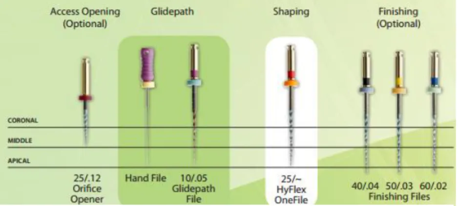
Provided as a modular system of sterile instruments, HyFlex EDM™ includes Glidepath, OneFile, Orifice Opener and Finishing files and may be used in combinat io n with HyFlex CM™ files (Figure 3) (Contene®, 2015).
The built-in shape memory of HyFlex EDM™ files prevents stress during canal preparation by changing their spiral shape. Like HyFlex CM™, a normal autoclaving process, following manufactures’ instructions, is enough to return the files to their original shape and fatigue resistance (Alazemi et al. 2015). Should the file fail to regain its shape after heat treatment, risk of fracture is increased, and the file should not be used after visual inspection (Contene®, 2015).
COM PARATIVE ANALYSIS OF ROOT CANAL ANATOM Y AFTER M ECHANICAL PREPARATION WITH HYFLEX CM™ AND HYFLEX EDM™
LUANA SILVA DE AMORIM |FMDUL 5
2 AIMS
One of the goals of this study was to compare the maintenance of original anatomy of prepared canals with an S-shaped curvature in clear resin blocks by the use of two different rotary files: HyFlex CM™ and HyFlex EDM™.
In a double curve canal, is important to understand whether shaping effect is bigger in the inner or outer portion of the curvature and whether shaping effect is more significant in the coronal or apical curvature.
The other goal was to verify if sterilization had influence on the cutting efficie nc y of both systems.
Specific goals:
1 - To compare the shaping effect of HyFlex CM™ and HyFlex EDM™.
H0 – the shaping effect is the same with HyFlex EDM™ and HyFlex CM™. H1 – the shaping effect is more significant with HyFlex EDM™.
H2 – the shaping effect is more significant with HyFlex CM™.
2 - To compare the shaping effect on inner of coronal curvature with HyFlex CM™ and HyFlex EDM™.
H0 – the shaping effect is the same with HyFlex EDM™ and HyFlex CM™. H1 – the shaping effect is more significant with HyFlex EDM ™.
H2 – the shaping effect is more significant with HyFlex CM ™.
3 - To compare the shaping effect on outer of coronal curvature with HyFlex CM™ and HyFlex EDM™.
H0 – the shaping effect is the same with HyFlex EDM™ and HyFlex CM™. H1 – the shaping effect is more significant with HyFlex EDM ™.
H2 – the shaping effect is more significant with HyFlex CM ™.
4 - To compare the shaping effect on inner of apical curvature with HyFlex CM™ and HyFlex EDM™.
H0 – the shaping effect is the same with HyFlex EDM™ and HyFlex CM™. H1 – the shaping effect is more significant with HyFlex EDM ™.
H2 – the shaping effect is more significant with HyFlex CM ™.
5 - To compare the shaping effect on outer of apical curvature with HyFlex CM™ and HyFlex EDM™.
H0 – the shaping effect is the same with HyFlex EDM™ and HyFlex CM™. H1 – the shaping effect is more significant with HyFlex EDM ™.
COM PARATIVE ANALYSIS OF ROOT CANAL ANATOM Y AFTER M ECHANICAL PREPARATION WITH HYFLEX CM™ AND HYFLEX EDM™
LUANA SILVA DE AMORIM |FMDUL 6
H2 – the shaping effect is more significant with HyFlex CM ™.
6 - To verify influence of sterilization on cutting efficiency of HyFlex CM™ and HyFlex EDM™:
H0 - The sterilization has influence on cutting efficiency of HyFlex CM™ and HyFlex EDM™.
H1 - The sterilization only has influence on cutting efficiency of HyFlex EDM™. H2 - The sterilization only has influence on cutting efficiency of HyFlex CM™. H3 - The sterilization has not any influence on cutting efficiency of HyFlex CM™ and HyFlex EDM™.
COM PARATIVE ANALYSIS OF ROOT CANAL ANATOM Y AFTER M ECHANICAL PREPARATION WITH HYFLEX CM™ AND HYFLEX EDM™
LUANA SILVA DE AMORIM |FMDUL 7
3 MATERIALS AND METHODS
3.1 Canal instrumentation
A total of 30 clear resin blocks with an S-shaped simulated canal (ISO 15, Endo-Training-Bloc-S .02 Taper; Dentsply-Maillefer, Ballaigues, Switzerlan) (Figure 4) were prepared by two different Ni-Ti rotary files system, using the technique recommended by the manufacturer: HyFlex CM™ (Coltene-Whaledent, Allstetten, Switzerland) and HyFlex EDM™ (Coltene-Whaledent, Allstetten, Switzerland).
Out of the 30 simulated canal resin blocks, two groups were defined, with 15 resin blocks each, prepared by one of the rotary system files above:
Group A – 15 simulated canal resin blocks prepared with HyFlex EDM™ (Coltene-Whaledent, Allstetten, Switzerland) (Figure 5);
Group B – 15 simulated canal resin blocks, prepared with HyFlex CM™ (Coltene-Whaledent, Allstetten, Switzerland) (Figure 6).
Each simulated canal was prepared to a working length of 16 millimeters. On Group A, the Glidepath file was used at 300 rpm and at a torque of 1.8 Ncm and the OneFile was used at 500 rpm and at a torque-control level of 2.5 Ncm, as the suggested
Figure 4 - S-shaped curvature in clear resin blocks (ISO 15, Endo-Training-Bloc-S .02).
Figure 6 - HyFlex CM™ kit used on Group B.
COM PARATIVE ANALYSIS OF ROOT CANAL ANATOM Y AFTER M ECHANICAL PREPARATION WITH HYFLEX CM™ AND HYFLEX EDM™
LUANA SILVA DE AMORIM |FMDUL 8
settings by manufacture, using a reduction hand-piece powered by an electric motor (Wave One®, Dentsply Maillefer) (Figure 7). The final apical preparation in Group A was set to HyFlex OneFile.
On Group B, the files were used at 500rpm and at a torque of 1.5Ncm, as the suggested setting by manufacture, using the same endo motor that Group A. The final apical preparation in Group B was set to file size 25, .04 taper.
Copious irrigation with water was performed after the use of each file, using a disposable syringe and 27G irrigation needle.
All canals were prepared by the same operator, who was not experience using these rotary files. After preparation of three canals, files were subjected to a steriliza t io n on autoclave, at approximately 134ºC (Table 1).
Blocks Sterilization
A1-A2-A3 / B1-B2-B3 Without sterilization
A4-A5-A6 / B4-B5-B6 After 1 sterilization
A7-A8-A9 / B7-B8-B9 After 2 sterilizations
A10-A11-A12 / B10-B11-B12 After 3 sterilizations
A13-A14-A15 / B13-B14-B15 After 4 sterilizations
The following preparation sequences were made, after all canal were scouted up to the working length with a #10 stainless-steal k-file (Dentsply Maillefer) (Figure 8).
Figure 7 - Wave One® endo motor used to prepare the simulated canals .
COM PARATIVE ANALYSIS OF ROOT CANAL ANATOM Y AFTER M ECHANICAL PREPARATION WITH HYFLEX CM™ AND HYFLEX EDM™
LUANA SILVA DE AMORIM |FMDUL 9
Group A
HyFlex EDM™files were set into rotation. Instrumentation followed the
sequence below, using shaping files up to the working length with brushing movements (Figure 9):
1º - Instrument size 10, 5% taper – Glidepath File 2º - Instrument size 25, variable taper – OneFile
Group B
HyFlex CM™ set into rotation. Instrumentation followed the sequence below, with in-and-out movements until reach the working length (Figure 10):
1º - Instrument size 20, 4% taper 2º - Instrument size 25, 4% taper
Figure 8 - #10 stainless-steal k-file used like permeability file (Image from Dentisply website).
Figure 9 - HyFlex EDM™ System used.
COM PARATIVE ANALYSIS OF ROOT CANAL ANATOM Y AFTER M ECHANICAL PREPARATION WITH HYFLEX CM™ AND HYFLEX EDM™
LUANA SILVA DE AMORIM |FMDUL 10
Were used copious irrigation and re-verify canal patency throughout the procedure. Upon completion of shaping, gauge the size of the foramen with an appropriate hand file.
3.2 Image analysis
Pre instrumentation and post instrumentation images of each block were recorded using a DSLR (Digita l Single- lens Reflex) camera (Olympus Digital Camera E500) with a 35mm macro lens, using a shutter speed of 1 second and a 22 F-stop. The footage was standardized: a landmark was made in each sample as a reference; the samples were all shot at the same distance and placed in the same position using a graph paper. To
accomplish this, a reproduction table was used (Kaiser Fototechnik GmbH & Co.KG) (Figure 11).
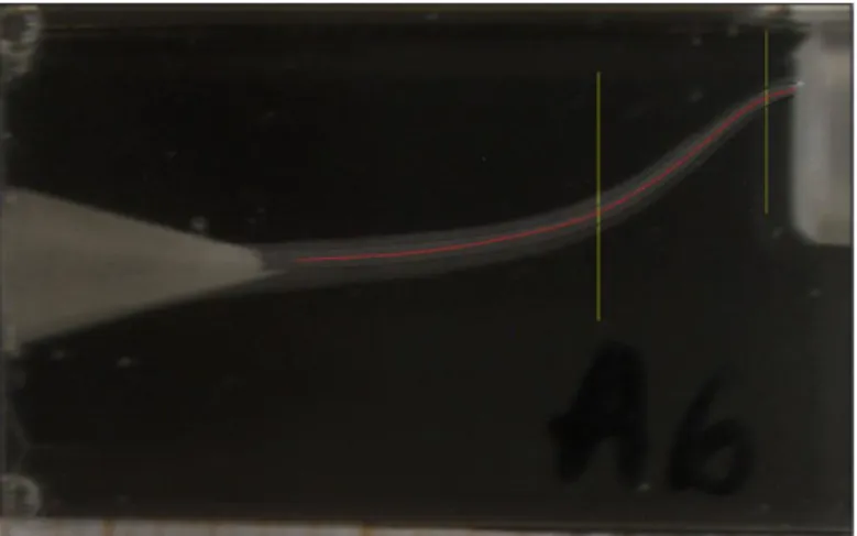
The Rhinoceros Software (version 5.0; Robert McNell & Associates, Seattle, WA) was used to identify the mean axis of the canal from the pre instrumentation images (Figure 12) and to identify the measure points, corresponding to the coronal and apical curvatures.
These measure points resulted from the interception of two tangent lines of each curve, drew by specific curve applications from the program as the sequence is shown below on Figure 13.
Figure 11 - Reproduction table (Kaiser Fototechnik GmbH & Co.KG).
Figure 12 – Identification of the mean axis of the canal using the Rhinoceros Software version 5.
COM PARATIVE ANALYSIS OF ROOT CANAL ANATOM Y AFTER M ECHANICAL PREPARATION WITH HYFLEX CM™ AND HYFLEX EDM™
LUANA SILVA DE AMORIM |FMDUL 11
The pre instrumentation digital images were superimposed over the post instrumentation images. This was accomplished by reducing the opacity of the post instrumentation images, done in a digital imaging software (Adobe Photoshop, version CS6; Adobe Systems Inc, San Jose, Ca) (Figure 14).
Figure 13 - This four pictures show the sequence made in the Rhinoceros Software to define the measure point of the coronal and apical curvature of the S-shape canal. Two tangent of each curve were trace and intercepted: first and second apical curvature tangents; interception of the two apical curve tangent – measure point; first and second coronal curvature tangent; interception of the two coronal curve tangent – measure point.
COM PARATIVE ANALYSIS OF ROOT CANAL ANATOM Y AFTER M ECHANICAL PREPARATION WITH HYFLEX CM™ AND HYFLEX EDM™
LUANA SILVA DE AMORIM |FMDUL 12
The canal width was assessed by measuring the distance from the centre of the canal to the inner and outer of pre and post prepared margins with specific dimension displays of the program. The distance between the margins were also registered. Rhinoceros Software allowed to get real measures. These paired images and the measures obtained give a quantitat i ve evaluation of the incidence of canal transportation after mechanical preparation.
3.3 Statistical analysis
The statistical analysis was obtained using the IBM SPSS® Statistics version 24.0.0 software. Descriptive statistical analysis was performed to each group (A and B). In each experimental group mean and standard deviation were calculated for the inner and outer of coronal and apical curvatures values. The Shapiro-Wilk test was used to evaluate the data normality. To analyse the homogeneity of the normality distributed sample was used Levene’s test. The Mann-Whitney U test was used to analyse the results with non-normal distribution or nonhomogeneous normal distribution and the student’s t-test was used to analyse the results with homogeneous normal distribution. Differences were considered statistically signific a nt when p<0,05.
COM PARATIVE ANALYSIS OF ROOT CANAL ANATOM Y AFTER M ECHANICAL PREPARATION WITH HYFLEX CM™ AND HYFLEX EDM™
LUANA SILVA DE AMORIM |FMDUL 13
4 RESULTS
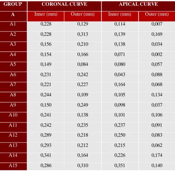
The distance from the mean axis of the canal to the inner and outer margins of the curvature was measured before and after the preparation of the canals, the tables with these results are in Appendix A. For comparison between systems files were used the distance between the pre and post instrumentation margins, inner and outer of both curvatures, because variations on the width of the canals between blocks were observed. In the following tables the values for groups A (Table 2) and B (Table 3) are presented:
GROUP CORONAL CURVE APICAL CURVE
A Inner (mm) Outer (mm) Inner (mm) Outer (mm)
A1 0,228 0,129 0,114 0,007 A2 0,228 0,313 0,139 0,169 A3 0,156 0,210 0,138 0,034 A4 0,154 0,166 0,071 0,002 A5 0,149 0,084 0,080 0,057 A6 0,231 0,242 0,043 0,088 A7 0,221 0,227 0,164 0,068 A8 0,244 0,109 0,105 0,134 A9 0,150 0,249 0,098 0,037 A10 0,241 0,138 0,101 0,106 A11 0,242 0,235 0,237 0,091 A12 0,289 0,218 0,250 0,083 A13 0,293 0,212 0,215 0,062 A14 0,341 0,164 0,226 0,174 A15 0,286 0,310 0,351 0,140
Table 2 - Group A – HyFlex EDM™. The difference of the measured distance margin before and after the preparation of the curve canal can be observed in each column.
COM PARATIVE ANALYSIS OF ROOT CANAL ANATOM Y AFTER M ECHANICAL PREPARATION WITH HYFLEX CM™ AND HYFLEX EDM™
LUANA SILVA DE AMORIM |FMDUL 14
Descriptive statistics of the four variables was done. The mean width and standard deviation for each experimenta l group are displayed in table 4.
Differences between the two systems files on the inner and outer side of the both curvatures (coronal and apical) are statistically significant, being HyFlex EDM™ system responsible for a bigger widening compared with HyFlex CM™.
On Group A, differences between the inner and outer of coronal curvature are not statistically significant. The differences between the inner of coronal curvature and the inner of apical curvature are statistically significant, with a bigger widening in the inner of coronal curvature. In the apical curvature, the differences between inner and outer of
GROUP CORONAL CURVE APICAL CURVE
B Inner (mm) Outer (mm) Inner (mm) Outer (mm)
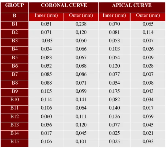
B1 0,051 0,238 0,070 0,065 B2 0,071 0,120 0,081 0,114 B3 0,033 0,050 0,053 0,007 B4 0,034 0,066 0,103 0,026 B5 0,083 0,067 0,054 0,009 B6 0,052 0,088 0,120 0,028 B7 0,085 0,086 0,077 0,007 B8 0,088 0,071 0,054 0,098 B9 0,105 0,059 0,175 0,043 B10 0,114 0,141 0,082 0,034 B11 0,106 0,064 0,140 0,017 B12 0,060 0,111 0,126 0,059 B13 0,056 0,120 0,077 0,045 B14 0,017 0,045 0,025 0,021 B15 0,106 0,101 0,025 0,093 Group Coronal Apical
Inner Outer Inner Outer
A – HyFlex EDM 0,23±0,06 0,20±0,07 0,16±0,08 0,08±0,05
B – HyFlex CM 0,07±0,03 0,10±0,05 0,08±0,04 0,04±0,03 Table 3 - Group B – HyFlex CM. The difference of the measured distance margin before and after
the preparation of the curve canal can be observed in each column.
Table 4 - Canal width in the measure points of the coronal and apical curvatures after instrumentation, mean values and their standard deviation.
COM PARATIVE ANALYSIS OF ROOT CANAL ANATOM Y AFTER M ECHANICAL PREPARATION WITH HYFLEX CM™ AND HYFLEX EDM™
LUANA SILVA DE AMORIM |FMDUL 15
curvature are statistically significant, showing a greater enlargement produced by HyFlex EDM™ in the inner part of curvature. The differences between outer of coronal curvature and outer of apical curvature are statistically significant, and the biggest widening is in the outer part of coronal curvature.
On Group B, differences between the inner and outer of coronal curvature are not statistically significant, as the differences between the inner of coronal and apical curvature. The difference between outer of coronal and apical curvatures are statistica l ly significant, showing that HyFlex CM™ cuts more on the coronal curvature.
In the apical curvature, the differences between inner and outer of curvature are statistically significant, with a bigger widening in the inner of curvature.
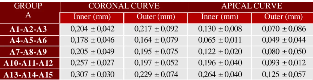
Regarding the influence of sterilization cycles in the inner of apical curvature on Group A, with HyFlex EDM™, the only statistically significant differences are between the files without sterilization and after first sterilization and between the files without sterilization and after four sterilizations (that has little statistical value because p≈0,05), being the biggest widening detected on blocks prepared with files without steriliza t io n and after four sterilizations, respectively. On the same group, in outer of coronal and apical curvatures, the differences are not statistically significant. About the inner of coronal curvature on the Group A, the only statistically significant differences are
GROUP A
CORONAL CURVE APICAL CURVE
Inner (mm) Outer (mm) Inner (mm) Outer (mm)
A1-A2-A3 0,204 ±0,042 0,217±0,092 0,130±0,008 0,070 ±0,086 A4-A5-A6 0,178 ±0,046 0,164±0,079 0,065±0,011 0,049 ±0,044 A7-A8-A9 0,205 ±0,049 0,195±0,075 0,122±0,020 0,080 ±0,050 A10-A11-A12 0,257 ±0,027 0,197±0,052 0,196±0,040 0,093 ±0,012 A13-A14-A15 0,307 ±0,030 0,229±0,074 0,264±0,040 0,125 ±0,057 GROUP B
CORONAL CURVE APICAL CURVE
Inner (mm) Outer (mm) Inner (mm) Outer (mm)
B1-B2-B3 0,052±0,019 0,136 ±0,095 0,068±0,014 0,062 ±0,054
B4-B5-B6 0,056±0,025 0,074 ±0,012 0,092±0,034 0,021 ±0,010
B7-B8-B9 0,093±0,011 0,072 ±0,014 0,102±0,064 0,049 ±0,046
B10-B11-B12 0,093±0,029 0,105 ±0,039 0,116±0,030 0,037 ±0,021
B13-B14-B15 0,060±0,045 0,089 ±0,039 0,042±0,030 0,053 ±0,037
Table 5 - Mean values and their standard deviation of canal width after each sterilization cycle, on Group A.
Table 6 - Mean values and their standard deviation of canal width after each sterilization cycle, on Group B.
COM PARATIVE ANALYSIS OF ROOT CANAL ANATOM Y AFTER M ECHANICAL PREPARATION WITH HYFLEX CM™ AND HYFLEX EDM™
LUANA SILVA DE AMORIM |FMDUL 16
between the files without sterilization and after three sterilizations and four sterilizatio ns, respectly, being the bigger widening measured on blocks prepared with files after three and four sterilizations.
On Group B, the differences are not statistically significant on outer of coronal curvature, inner and outer of apical curvature between sterilization cycles. In the inner of coronal curvature, statistically significant differences are observed only between the files without sterilization and after two sterilizations, being the blocks prepared with files after two sterilizations where the widening measured is bigger.
The HyFlex EDM™ system caused significantly greater widening of canals than HyFlex CM™, rejecting null hypothesis in the specific goals 1,2,3,4 and 5 and retaining hypothesis 1. More precisely, HyFlex EDM™ system removed more resin wall in coronal curvature compared to the apical curvature, and on the apical curvature this file removed more wall in inner than outer of curvature.
The HyFlex CM™ system removed more resin wall in outer of coronal curvature and in inner of apical curvature, compared to the inner of coronal curvature and the outer of apical curvature, respectively.
Regarding sterilizations cycles, on HyFlex EDM™ system the only differe nces statically significant are on inner of both curvatures, while on HyFlex CM™ system the only difference statically significant is on inner of coronal curvature, retaining null hypothesis on the specific goal 6.
COM PARATIVE ANALYSIS OF ROOT CANAL ANATOM Y AFTER M ECHANICAL PREPARATION WITH HYFLEX CM™ AND HYFLEX EDM™
LUANA SILVA DE AMORIM |FMDUL 17
5 DISCUSSION
Successful root canal treatment is dependent upon the effective debridement and shaping of the root canal system (Dhingra & Manchanda, 2014). Studies suggested that the analysis of modifications in canal curvature after instrumentation is a reliable method to evaluate the tendency of a shaping technique to maintain the original canal anatomy or to straighten the curves (Dhingra & Manchanda, 2014). Previous studies have shown that preserving the original canal shape with a less invasive approach minimizes the risk of canal transportation with a subsequently lower incidence of canal curvature straightening, the formation of ledges and irregular apical enlargement (Dhingra, Kochar, Banerjee, & Srivastava, 2014; Saber et al., 2014). Preservation of the original canal shape and the lack of canal aberrations are associated with increased antimicrobial and sealing efficie nc y and reduced weakening of the tooth structure (Dhingra et al., 2014; Saber et al., 2014).
In group A, the last file used was One File of HyFlex EDM™ system, which is a size 25 at the tip, with variable taper. To have a fair comparison, in group B the instrumentation was finished with the file size 25 (.04 taper) of HyFlex CM™ system. The literature suggested that sizes ISO 25-30 are a good target for apical preparation, minimizing the risk of transportation and extrusion of debris and irrigant (Darcey, Taylor, Roudsari, Jawad, & Hunter, 2015). Studies describe that larger preparations might result in canal straightening and undesirable weakening of the tooth structure, whereas smaller preparations may leave tissue remnants and infected dentin behind (Darcey et al., 2015; Saber et al., 2014). The results of this study shows that HyFlex EDM™ system causes a significant further widening of the canal than HyFlex CM™ system (p<0,05), inner and outer of both curvatures. This finding can be attributed to differences in instrument taper (variable for HyFlex EDM™ and 4% for HyFlex CM™) and differences in surface treatment which can affect their flexibility (HyFlex EDM™ files are produced using an innovative manufacturing process called Electrical Discharge Machining). Higher flexibility might be the predominant property responsible by the system’s ability to maintain the canal’s original anatomy.
The systems used in this study, Hyflex EDM™ and Hyflex CM™, remove more resin in the coronal curvature than the apical, contrary to the results obtained by another study (Neto, 2015), where the systems files used removed more resin in apical curvature than coronal. This finding can be attributed to the differences in flexibility of HyFlex
COM PARATIVE ANALYSIS OF ROOT CANAL ANATOM Y AFTER M ECHANICAL PREPARATION WITH HYFLEX CM™ AND HYFLEX EDM™
LUANA SILVA DE AMORIM |FMDUL 18
instruments compared to ProTaper instruments used by Neto 2015 (ProTaper Universal™, ProTaper Next™ and ProTaper Gold™).
To verify influence of sterilization on cutting efficiency of HyFlex CM™ and HyFlex EDM™, after preparation of three simulated canals the files of each system were sterilized in an autoclave. These three simulated canals intended to produce a molar with three canals, simulating a clinical act. After four sterilization cycles, all the instrume nts returned to the original shape.
On both systems, the obtained results are variable, verifying a reduction and increase in cutting efficiency of files between sterilizations. This fact can be explained by the small sample used in this study and the inability to control the standardization of instrumentation. This results of HyFlex CM™ system are not in harmony with the literature, as it is described that the number of sterilization cycles does not affect the cutting ability (Seago et al., 2015; Thompson, Sidow, Lindsey, Chuang, & McPherson, 2014). Regarding HyFlex EDM™ system, no literature was found. Therefore, a proper conclusion is difficult to obtain and further studies should contain a larger sample and more sterilization cycles.
In this study, analysis was performed through observation of differences between pre instrumentation and post instrumentation in simulated canal with an S-shaped curvature in clear resin blocks. Simulated root canals have been widely used to allow a direct analysis of post instrumentation changes in canal curvature and thus to evaluate the tendency of these techniques to maintain the original canal anatomy under standardized conditions, but always regarding the fact that this method only gives 2D dimens io ns (Dhingra et al., 2014). Care should be taken in the extrapolation of the results to the use of real roots because that resin blocks may not always reflect the action of the instrume nts in root canals of real teeth because of the many different configurations and differe nces between resin and dentin (Dhingra et al., 2014; Neto, 2015). To bypass this problem natural teeth can be used in future studies.
The experimental method used appeared to be reliable in representing changes in canal curvature and for extrapolating the results, however, this analysis may not be completely accurate taking in consideration that exists an uncertainty degree of the Rhinoceros Software (considered 0,006) and data dependent on operator’s skills and accuracy. During experimental procedure it is difficult to control instrumenta t io n technique and stabilization of the resin block.
COM PARATIVE ANALYSIS OF ROOT CANAL ANATOM Y AFTER M ECHANICAL PREPARATION WITH HYFLEX CM™ AND HYFLEX EDM™
LUANA SILVA DE AMORIM |FMDUL 19
6 CONCLUSION
Under the limitations of this study, HyFlex CM™ was the rotary file system that best maintained the original anatomy of the S-shaped canal with less modification of coronal and apical curvatures, revelling more flexibility compared to HyFlex EDM™ system.
The results of influence of sterilization on cutting efficiency of HyFlex CM™ and HyFlex EDM™ are inconclusive, being necessary further studies.
In clinical acts is important to choose an appropriate system files to each case, respecting as much as possible the original anatomy of the canal, to reduce the risk of ledging, transportation and perforation and contribute to a better prognosis of
COM PARATIVE ANALYSIS OF ROOT CANAL ANATOM Y AFTER M ECHANICAL PREPARATION WITH HYFLEX CM™ AND HYFLEX EDM™
x
REFERENCES
Alfoqom Alazemi, M., Bryant, S. T., & Dummer, P. M. H. (2015). Deformation of HyFlex CM instruments and their shape recovery following heat sterilization. International Endodontic Journal, 48(6), 593–601.
http://doi.org/10.1111/iej.12353
Burklein, S., Borjes, L., & Schafer, E. (2013). Comparison of preparation of curved root canals with Hyflex CM and Revo-S rotary nickel – titanium instruments.
Internacional Endodontic Journal, 1–7. http://doi.org/10.1111/iej.12171
Capar, I. D., Arslan, H., Akcay, M., & Uysal, B. (2014). Effects of ProTaper Universal, ProTaper Next, and HyFlex Instruments on Crack Formation in Dentin. Journal of Endodontics, 1–3. http://doi.org/10.1016/j.joen.2014.02.026
Capar, I. D., Ertas, H., & Arslan, H. (2014). Comparison of cyclic fatigue resistance of nickel-titanium coronal flaring instruments. Journal of Endodontics, 40(8), 1182– 1185. http://doi.org/10.1016/j.joen.2013.12.031
Coltene®. (n.d.). Changing the DNA of NiTi HyFlex® CMT M. Contene®. (2015). A nova geração de limas NiTi HyFlex®.
Darcey, J., Taylor, C., Roudsari, R. V., Jawad, S., & Hunter, M. (2015). Modern Endodontic Principles Part 3: Preparation. Dental Update, 42(9), 810–812,815– 818,821–822.
De Vasconcelos, R. A., Murphy, S., Carvalho, C. A. T., Govindjee, R. G., Govindjee, S., & Peters, O. A. (2016). Evidence for Reduced Fatigue Resistance of
Contemporary Rotary Instruments Exposed to Body Temperature. Journal of Endodontics, 42(5), 782–787. http://doi.org/10.1016/j.joen.2016.01.025
Dhingra, A., Kochar, R., Banerjee, S., & Srivastava, P. (2014). Comparative evaluation of the canal curvature modifications after instrumentation with One Shape rotary and Wave One reciprocating files. Journal of Conservative Dentistry : JCD, 17(2), 134–141.
Dhingra, A., & Manchanda, N. (2014). Modifications in Canal Anatomy of Curved Canals of Mandibular First Molars by two Glide Path Instruments using Cbct. Journal of Clinical and Diagnostic Research, 8(11), 13–18.
http://doi.org/10.7860/JCDR/2014/8702.5101
Iacono, F., Pirani, C., Generali, L., Bolelli, G., Sassatelli, P., Lusvarghi, L., … Prati, C. (2016). Structural analysis of HyFlex EDM instruments. International Endodontic Journal, 1–11. http://doi.org/10.1111/iej.12620
Kumar, R. V., & Shruthi, C. (2012). Evaluation of the sealing ability of resin cement used as a root canal sealer: An in vitro study. Journal of Conservative Dentistry : JCD. http://doi.org/10.4103/0972-0707.97958
Neto, F. (2015). Comparative analysis of root canal anatomy after different mechanical preparation. Universidade de Lisboa.
COM PARATIVE ANALYSIS OF ROOT CANAL ANATOM Y AFTER M ECHANICAL PREPARATION WITH HYFLEX CM™ AND HYFLEX EDM™
xi
Albuquerque, D. (2015). Apical Extrusion of Debris Produced during Continuous Rotating and Reciprocating Motion. Scientific World Journal, 2015(Cm).
http://doi.org/10.1155/2015/267264
Ninan, E., & Berzins, D. W. (2013). Torsion and bending properties of shape memory and superelastic nickel-titanium rotary instruments. Journal of Endodontics, 39(1), 101–104. http://doi.org/10.1016/j.joen.2012.08.010
Pedullà, E., Lo Savio, F., Boninelli, S., Plotino, G., Grande, N. M., La Rosa, G., & Rapisarda, E. (2016). Torsional and Cyclic Fatigue Resistance of a New Nickel-Titanium Instrument Manufactured by Electrical Discharge Machining. Journal of Endodontics, 42(1), 156–159. http://doi.org/10.1016/j.joen.2015.10.004
Pedullà, E., Lo Savio, F., Boninelli, S., Plotino, G., Grande, N. M., Rapisarda, E., & La Rosa, G. (2014). Influence of cyclic torsional preloading on cyclic fatigue
resistance of nickel - titanium instruments. International Endodontic Journal, 48(11), 1043–1050. http://doi.org/10.1111/iej.12400
Peters, O. A., Gluskin, A. K., Weiss, R. A., & Han, J. T. (2012). An in vitro assessment of the physical properties of novel Hyflex nickel-titanium rotary instruments. International Endodontic Journal, 45(11), 1027–1034.
http://doi.org/10.1111/j.1365-2591.2012.02067.x
Pirani, C., Iacono, F., Generali, L., Sassatelli, P., Nucci, C., Lusvarghi, L., … Prati, C. (2016). HyFlex EDM: Superficial features, metallurgical analysis and fatigue resistance of innovative electro discharge machined NiTi rotary instruments. International Endodontic Journal, 49(5), 483–493.
http://doi.org/10.1111/iej.12470
Plotino, G., Testarelli, L., Al-Sudani, D., Pongione, G., Grande, N. M., & Gambarini, G. (2014). Fatigue resistance of rotary instruments manufactured using different nickel-titanium alloys: A comparative study. Odontology, 102(1), 31–35. http://doi.org/10.1007/s10266-012-0088-8
Saber, S. E. D. M., Nagy, M. M., & Schafer, E. (2014). Comparative evaluation of the shaping ability of ProTaper Next, iRaCe and Hyflex CM rotary NiTi files in severely curved root canals. International Endodontic Journal, 48(2), 131–136. http://doi.org/10.1111/iej.12291
Savio, F. Lo, Boninelli, S., Plotino, G., Grande, N. M., Rosa, G. La, & Rapisarda, E. (2015). Torsional and Cyclic Fatigue Resistance of a New Nickel-Titanium Instrument Manufactured by Electrical Discharge Machining. Journal of Endodontics, 10–13. http://doi.org/10.1016/j.joen.2015.10.004
Seago, S. T., Bergeron, B. E., Kirkpatrick, T. C., Roberts, M. D., Roberts, H. W., Himel, V. T., & Sabey, K. A. (2015). Effect of Repeated Simulated Clinical Use and Sterilization on the Cutting Efficiency and Flexibility of Hyflex CM Nickel-Titanium Rotary Files. Journal of Endodontics, 41(5), 725–728.
http://doi.org/10.1016/j.joen.2015.01.011
Sharma, Shiv Aditya (Department of Conservative Dentistry and Endodontics, Kothiwal Dental College and Research centre, Moradabad, Uttar Pradesh, I., Tyagi, S. P., Sinha, D. J., Singh, U. P., Chandra, P., & Kaur, G. (2014). Influence of cervical preflaring using different rotary instruments on the accuracy of apical file size
COM PARATIVE ANALYSIS OF ROOT CANAL ANATOM Y AFTER M ECHANICAL PREPARATION WITH HYFLEX CM™ AND HYFLEX EDM™
xii
determination- A comparative in-vitro study. J Conserv Dent Nov-Dec, 17(6), 575– 578. http://doi.org/10.4103/0972-0707.144608
Shen, Y., Coil, J. M., Zhou, H., Zheng, Y., & Haapasalo, M. (2013). HyFlex nickel-titanium rotary instruments after clinical use: Metallurgical properties.
International Endodontic Journal, 46(8), 720–729. http://doi.org/10.1111/iej.12049
Testarelli, L., Plotino, G., Al-Sudani, D., Vincenzi, V., Giansiracusa, A., Grande, N. M., & Gambarini, G. (2011). Bending properties of a new nickel-titanium alloy with a lower percent by weight of nickel. Journal of Endodontics, 37(9), 1293–1295. http://doi.org/10.1016/j.joen.2011.05.023
Thompson, M., Sidow, S. J., Lindsey, K., Chuang, A., & McPherson, J. C. (2014). Evaluation of a new filing system’s ability to maintain canal morphology. Journal of Endodontics, 40(6), 867–870. http://doi.org/10.1016/j.joen.2013.10.016
Zhao, D., Shen, Y., Peng, B., & Haapasalo, M. (2013). Micro-computed tomography evaluation of the preparation of mesiobuccal root canals in maxillary first molars with Hyflex CM, twisted files, and K3 instruments. Journal of Endodontics, 39(3), 385–388. http://doi.org/10.1016/j.joen.2012.11.030
COM PARATIVE ANALYSIS OF ROOT CANAL ANATOM Y AFTER M ECHANICAL PREPARATION WITH HYFLEX CM™ AND HYFLEX EDM™
xiii
APENDIX A
GROUP CORONAL CURVE APICAL CURVE
A Inner (mm) Outer (mm) Inner (mm) Outer (mm) A1 0,223 0,451 0,223 0,352 0,179 0,293 0,156 0,163 A2 0,164 0,444 0,164 0,477 0,146 0,285 0,127 0,296 A3 0,211 0,367 0,230 0,440 0,192 0,330 0,192 0,226 A4 0,216 0,370 0,216 0,382 0,162 0,233 0,162 0,164 A5 0,254 0,403 0,254 0,338 0,169 0,249 0,169 0,226 A6 0,157 0,388 0,253 0,495 0,193 0,236 0,196 0,281 A7 0,184 0,405 0,184 0,411 0,130 0,294 0,138 0,206 A8 0,177 0,421 0,177 0,386 0,168 0,273 0,163 0,297 A9 0,228 0,378 0,228 0,477 0,186 0,284 0,186 0,223 A10 0,206 0,447 0,216 0,354 0,184 0,285 0,184 0,290 A11 0,165 0,407 0,165 0,400 0,156 0,393 0,146 0,237 A12 0,187 0,476 0,197 0,415 0,145 0,395 0,145 0,228 A13 0,148 0,441 0,159 0,371 0,126 0,341 0,126 0,188 A14 0,163 0,504 0,150 0,314 0,112 0,338 0,117 0,291 A15 0,208 0,494 0,208 0,518 0,143 0,494 0,143 0,283 Table A - HyFlex EDM™. Measures obtained with the Rhinoceros Software. Every left column of the inner and outer variables regards the distance from the center of the canal to the inner and outer margins of the pre instrumented curve canal and every right column regard the distance from the
COM PARATIVE ANALYSIS OF ROOT CANAL ANATOM Y AFTER M ECHANICAL PREPARATION WITH HYFLEX CM™ AND HYFLEX EDM™
xiv
GROUP CORONAL CURVE APICAL CURVE
B Inner (mm) Outer (mm) Inner (mm) Outer (mm) B1 0,161 0,212 0,179 0,417 0,125 0,195 0,125 0,190 B2 0,174 0,245 0,174 0,294 0,155 0,236 0,151 0,269 B3 0,204 0,237 0,204 0,254 0,167 0,220 0,167 0,170 B4 0,179 0,213 0,179 0,245 0,154 0,257 0,154 0,180 B5 0,154 0,237 0,205 0,272 0,179 0,233 0,179 0,188 B6 0,205 0,257 0,205 0,293 0,179 0,299 0,154 0,182 B7 0,186 0,271 0,185 0,271 0,167 0,244 0,167 0,174 B8 0,211 0,299 0,211 0,282 0,146 0,200 0,130 0,228 B9 0,186 0,291 0,189 0,248 0,151 0,326 0,151 0,194 B10 0,142 0,256 0,142 0,283 0,160 0,242 0,160 0,194 B11 0,160 0,266 0,196 0,260 0,161 0,301 0,142 0,159 B12 0,198 0,258 0,180 0,291 0,138 0,264 0,126 0,185 B13 0,169 0,225 0,169 0,289 0,148 0,225 0,148 0,193 B14 0,205 0,222 0,251 0,296 0,160 0,185 0,182 0,203 B15 0,144 0,250 0,160 0,261 0,175 0,200 0,170 0,263
Table B - HyFlex CM™. Measures obtained with the Rhinoceros Software. Every left column of the inner and outer variables regards the distance from the center of the canal to the inner and outer margins of the pre instrumented curve canal and every right column regard the distance from the
COM PARATIVE ANALYSIS OF ROOT CANAL ANATOM Y AFTER M ECHANICAL PREPARATION WITH HYFLEX CM™ AND HYFLEX EDM™
xv
APPENDIX B
Abbreviations
CM – Controlled Memory NiTi – Nickel-Titanium rpm – rotations per minute Wt – Weight Symbols % - Percentage p - Significance ® - Registered ™ - Trademark Units ºC - Degree Celsius G - Gauge mm - Millimeters Ncm – Newton centimeter