Effect of Acute Copper Sulfate
Exposure on Olfactory Responses to
Amino Acids and Pheromones in
Goldfish (Carassius auratus)
N I K O L A Y N . K O L M A K O V ,†P E T E R C . H U B B A R D ,† O R L A N D O L O P E S ,‡ A N D A D E L I N O V . M . C A N A R I O *, †
Centro de Cieˆncias do Mar, Universidade do Algarve, Campus de Gambelas, 8005-139 Faro, Portugal, and Centro de Ecologia e Ambiente, Universidade de E´vora, 7002-554 E´vora, Portugal
Received April 27, 2009. Revised manuscript received August 27, 2009. Accepted September 6, 2009.
Exposure of olfactory epithelium to environmentally relevant
concentrations of copper disrupts olfaction in fish. To examine
the dynamics of recovery at both functional and morphological
levels after acute copper exposure, unilateral exposure of
goldfish olfactory epithelia to 100 µM CuSO
4(10 min) was
followed by electro-olfactogram (EOG) recording and scanning
electron microscopy. Sensitivity to amino acids (
L-arginine
and
L-serine), generally considered food-related odorants,
recovered most rapidly (three days), followed by that to
catecholamines (3-O-methoxytyramine), bile acids (taurolithocholic
acid) and the steroid pheromone,
17,20β-dihydroxy-4-pregnen-3-one 20-sulfate, which took 28 days to reach full recovery.
Sensitivity to the postovulatory pheromone prostaglandin F
2Rhad
not fully recovered even at 28 days. These changes in
sensitivity were correlated with changes in the recovery of
ciliated and microvillous receptor cell types. Microvillous cells
appeared largely unaffected by CuSO
4treatment. Cilia in
ciliated receptor neurones, however, appeared damaged one
day post-treatment and were virtually absent after three days but
had begun to recover after 14 days. Together, these results
support the hypothesis that microvillous receptor neurones detect
amino acids whereas ciliated receptor neurones were not
functional and are responsible for detection of social stimuli
(bile acids and pheromones). Furthermore, differences in sensitivity
to copper may be due to different transduction pathways in
the different cell types.
Introduction
Olfaction mediates social behavior, reproduction, homing, schooling, food-search, and predator avoidance in teleost fishes (1-3). Olfactory neurons possess a unique capacity to regenerate, a property that has been used to reveal the cellular component of olfaction in fish (4), amphibians (5), snakes (6), birds (7), and mammals (8, 9). Furthermore, some chemicals, such as heavy metals and pesticides, even at sublethal concentrations, are able to disturb a broad range of olfaction-dependent behavior and cause long-lasting
damage to the sensory epithelium (10-12); this has been used to provoke degeneration and regeneration of olfactory neurons.
Copper is an essential metal, which in excess is toxic and which bioavailability vary widely from place to place ac-cording to potential sources and physicochemical water conditions. Highest concentrations are found in mines runoff but contributions from various sources, including industry, corrosion lixiviates, and pesticides, makes it an important metal in the urban water cycle (13). Values in the micromolar range are common at wastewater plant influents and acceptable levels in water for drinking vary between 15 and 45 µM (13). There are multiple reports on adverse effects of copper on the fish olfactory system even at concentrations below legally established levels (14).
Salmonid fishes tend to avoid relatively low concentrations of copper, whereas at high concentrations fish become anosmic (10). Chronic exposure to copper causes impaired avoidance of conspecific alarm cues in Iowa darters (Etheo-stoma exile) (15) and Colorado pike minnows (Ptychocheilus lucius) (16). Exposure during embryonic development renders fathead minnows (Pimephales promelas) insensitive to con-specific alarm cues (17). Chronic copper exposure also affects feeding behavior in rainbow trout (Onchorhyncus mykiss) (18) and olfactory sensitivity to amino acids in common carp (Cyprinus caprio) (19). Constant exposure to sublethal copper toxic pressure limits olfactory sensitivity and survival is affected (15). However, at subcritical chronic copper con-centrations, insufficient to suppress behavioral responses, the olfactory system may adapt: this was determined to be 0.25 µM for fathead minnow (17) and rainbow trout (18) and 1 µM for common carp (19).
Copper in the micromolar range causes immediate reduction of electro-olfactogram (EOG) amplitude evoked by amino and bile acids in salmonids (20-23). However, this inhibition is transient, and recovery is seen after one hour. Longer exposure, hours to days, does not affect olfaction (24, 25), as responses recover completely within one day. Thus, the EOG can be used to establish sublethal thresholds of toxicants that are apparently ineffective over short-term exposure.
Morphological changes after copper exposure are better studied. In the rainbow trout, 30 min exposure to 0.2 µM copper causes little change in the olfactory epithelium except increased mucus secretion. After 90 min, however, loss of cilia occurs and, after three hours, vacuoles and holes appear and neurons become shrunken and distorted (26). Similarly, a few hours of copper exposure (0.3-3 µM) causes necrosis of olfactory receptors in chinook salmon (Oncorhynchus tshawytscha). Yet some receptor cells survive and respond toL-serine (21). In brown trout (Salmo trutta), exposure to copper causes rapid apoptosis, when half the olfactory neurons die after one day of treatment and the epithelium takes six days to recover (27).
The timeline of olfactory receptor death in the constant presence of sublethal copper, and consequent recovery have been described in the rainbow trout (28-30). Mature neurons disappear from the surface during one week of copper exposure (0.1 µM), with a peak of death occurring after five days. During the second week, large numbers of prematurely differentiated ciliated neurons are seen, the dendrites of which fail to reach the epithelial surface. At day 15 there is a second wave of neuronal degeneration, consisting mainly of nearly mature neurons (29). No fully mature neurons are seen, even after two months of exposure, whereas receptor neurons with short cilia and goblet cells increase in number
* Corresponding author phone: 289800925; fax: +351-289800069; e-mail: [email protected].
†Universidade do Algarve. ‡Universidade de E´ vora.
Environ. Sci. Technol. 2009, 43, 8393–8399
(29). Adaptation of the olfactory system to toxic pressure takes about 5 months, with an increase in apoptosis and a decrease in the number of ciliated neurons (30). Transfer to clean water allows full recovery after six weeks (30). However, exposure to higher concentrations causes severe and pro-longed alterations. For example, 0.2 µM copper causes complete disorganization of stratification of the olfactory epithelium and hypertrophy of some goblet cells, which formed mucus-containing vacuoles within the epithelium. Structural recovery is delayed, with the epithelium recovering only after 14 weeks (30). Nevertheless, copper does not accumulate in the olfactory system (31).
The Cyprinidae is the largest family of fishes making 20% of the worlds freshwater fish species (32). The cyprinids include the model species zebrafish, carps used in aquac-ulture and the goldfish (Carassius auratus) a species with a well studied chemosensory system which is used in the current study (33). In cyprinids, information on morphologi-cal effects of copper on the olfactory epithelium, especially regeneration after exposure, is limited. In the Colorado pike-minnow, olfactory receptor cells are completely eliminated by four days’ copper exposure (1 µM). After two weeks, the number of olfactory receptor cells recovers (16). Acute exposure of big-scaled redfin, (Tribolodon hakonensis) to 4 µM copper causes cilia and microvilli to become stuck together and partially lost and, at the surface, excessive mucus excretion. At 40 µM copper, the olfactory epithelium becomes swollen with many holes; sensory neurons are reduced in number and size. At 80 and 160 µM the organization of the epithelium is distorted; all sensory and secretory cells are destroyed. The epithelium becomes dissociated from con-nective tissue (basal membrane); at the higher concentration, massive necrosis and a separation of fragments of epithelium into the olfactory cavity is seen. However, no data on recovery are available (34).
The objective of the current study was to investigate the physiological recovery of olfaction in goldfish after short exposure of the epithelium to high copper concentrations. The approach was to eliminate olfactory responses and to follow their recovery and compare with that after axotomy (35, 36). We determined the lowest copper concentration which caused long-term anosmia and used exposure of olfactory epithelium to this concentration to build a time-scale of response to known odorants for this fish: the amino acidsL-serine andL-arginine, the bile acid taurolithocholic acid (TLC) (37), the catecholamine 3-O-methoxytyramine (3-MT) (38), and pheromones prostaglandin F2R(PGF2R) and
17,20β-dihydroxy-4-pregnen-3-one 20-sulfate (17,20β-P-S) (39, 40).
Experimental Section
Animals. Normal body shaped goldfish (Carassius auratus
auratus) of both sexes, 10-15 cm and 20-25 g, from a naturally reproducing local stock were used. Fish were kept under natural photoperiod and temperature in shaded outdoor 1000 L tanks with aeration and fed once or twice a day (depending on temperature) on commercial food (Tet-raPond Pond Sticks, TetraWerke, Melle, Germany).
Reagents. All odorants except 17,20β-P-S (a gift from A.P.
Scott, CEFAS, Weymouth, UK) were bought from Sigma-Aldrich Chemical Co. (Madrid, Spain). Amino acids were dissolved in distilled water (10-3M); bile acid (10-2M), steroid (10-3M) and prostaglandin (2× 10⁻³M) were dissolved in ethanol and stored in aliquots at -20°C. All stimuli were diluted to working concentrations prior to use with dechlo-rinated, charcoal-filtered tap water (also used as background).
Surgery. Experiments were carried out in spring and
autumn as olfactory sensitivity to pheromones is not affected by reproductive stage (41). Goldfish were anaesthetized by immersion in 3-aminobenzoic acid ethyl ester (MS222; 100
mg/L), immobilized with intramuscular injection of gallamine triethiodide (1 mg/kg in 0.9% NaCl) and maintained with aerated water (containing 50 mg/L MS222) flowing over the gills. The flaps of skin overlying the nostrils were removed, and olfactory epithelia were continuously irrigated via glass tubes at a flow-rate of 6 mL/min with background water. Copper sulfate solutions were introduced into this flow via a computer-controlled three-way solenoid valve.
EOG Recording. The method used for recording EOGs
from goldfish has been previously described (42). Briefly, anaesthetized goldfish were maintained as described above with olfactory epithelia of both sides continuously irrigated with background water. Odorant containing solutions were introduced into this flow via a three-way solenoid valve for a period of 4 s. The recording electrode was placed near the raphe, between two adjacent olfactory lamellae. The following compounds were tested in each experiment: L-serine (10-7-10-4M),L-arginine (10-7-10-4M), 3-MT (10-7-10-4
M), TLC (10-8-10-5M), PGF
2R(10-9-10-6M) and 17, 20β-P-S
(10-9-10-6M).
Minimum Effective Copper Sulfate Concentration to Inactivate Olfactory Neurons. The sublethal copper
con-centration sufficient for suppression of olfactory responses, but not toxic to goldfish, was determined by exposure to copper and evaluation of the EOG response. The right nostril of each fish was exposed for 10 min with a given copper concentration followed by 10 min wash-out. The left nostril was superfused with background water during the whole procedure, serving as sham control. EOG responses from the right epithelium were recorded before and after exposure. The range of concentrations chosen, between 6 and 0.05 mM, were based on acute exposure in the cyprinid big-scaled redfin, the only species of this family for which there is a description of damage to the olfactory system after short-term exposure to copper (34), and considering that the subspecies Carassius carassius gibelium has a high copper tolerance (LC50for 96 h ) 22 µM) (43). Starting with the
higher concentration, if the EOG response was still absent 90 min after exposure, a copper concentration 5 times lower was tested on a new fish. This was repeated until there were clear signs of immediate recovery to copper exposure. The minimum effective copper sulfate required to fully inactivate olfactory responses was thus determined to be 0.1 mM.
Recovery of Olfactory Responses after Copper Exposure.
Having determined the minimum effective copper sulfate concentration required to fully inactivate olfactory responses, the temporal recovery of the olfactory epithelium was analyzed by recording EOG responses from right (treated) and left (control) epithelia at 1, 3, 7, 14, and 28 days after exposure to copper sulfate. The right epithelium was exposed to 0.1 mM copper sulfate for 10 min followed by 10 min wash-out. The left epithelium served as sham control as indicated above. The fish were then allowed to recover from anesthesia and placed in a tank similar to where they were kept prior to treatment (all fish survived).
Scanning Electron Microscopy. On completion of EOG
recording one fish for each time point was killed and used for scanning electron microscopy (SEM). Preparations for SEM were made according to Hansen et al. (44). The three most caudal olfactory lamelae from each side were isolated, processed for SEM - dehydrated in a graded series of acetone and isoamyl acetate, critical-point-dried in CO2, coated with
gold and examined at 5 or 10 kV with FEI Quanta 400FEG at the Centre of Materials, Porto University.
Statistics. Student’s paired t test was used to compare
EOG amplitudes from treated and control epithelia at each time-point. For clarity, only one concentration of each odorant is shown, corresponding to the concentration which evoked approximately 1-2 mV EOG amplitude in the control side; however, at all tested concentrations, each odorant
showed the same pattern of recovery, i.e., similar ratios of EOG the response between treated and control sides. For
L-arginine,L-serine and 3-MT it was 10-5M, for TLC 10-6M and for pheromones (17,20β-P-S and PGF2R) 10-7M. To test
whether handling-related procedures affected responses, EOGs from the control side were tested using one way repeated measures ANOVA with time as within-subjects factor, followed by the Holm-Sidak post hoc test. The statistical significance level was P < 0.05. All values are presented as mean ( SEM.
Results
Minimum Effective Copper Sulfate Concentration to In-activate Olfactory Neurons. Treatments with 0.2-6 mM
copper sulfate for 10 min caused complete elimination of EOG responses to any odorant (3-MT, 17,20β-PS and amino acids) for at least 90 min after the end of exposure (Table 1). A temporary EOG response 30 min after end of exposure to 0.1 mM copper sulfate, of between 80% of pretreatment levels forL-serine, 30% forL-arginine, and 50% for 3-MT was present, but by 90 min there was no response to any stimuli. At 0.05 mM copper sulfate it was possible to record responses (approximately 10% of pre-exposure amplitudes) immedi-ately after end of treatment. EOG responses 10 min after wash-out reached 25% of control, and there was 50% recovery within half an hour for amino acids and steroid. Further observation demonstrated nearly complete recovery of olfactory responses. Thus, 0.1 mM copper was sufficient to cause complete inactivation of the olfactory epithelium, leading to loss of sensitivity and was the concentration used to study olfactory recovery.
Recovery of Olfactory Responses after Copper Exposure.
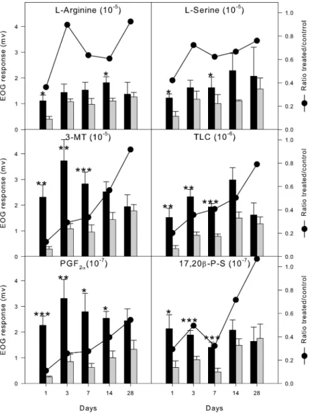
Amino Acids. One day after 10 min exposure to 0.1 mM copper sulfate, responses from the treated side toL-arginine and
L-serine were reduced to, respectively, 36 and 42% of the control side. After three days, sensitivity to amino acids recovered fully. Interestingly, differences in response between intact and exposed epithelia reappeared with statistical significance at day 14 forL-arginine (61% of control) and at day 7 forL-serine (62%). At 28 days, there was no significant difference in EOG amplitudes between treated and control epithelia in response toL-arginine (92%) and toL-serine (76%) (Figure 1, Table 2).
Bile Acid and 3-MT. The inhibition of responses to TLC and 3-MT lasted longer than to amino acids. At day one, EOG amplitudes were only 20% for TLC and 13% for 3-MT. The recovery followed a more linear path than amino acids (Figure 1, Table 2). The responses to both stimuli remained reduced after two weeks (50 and 57%, respectively). Only after four weeks did EOG amplitudes from control and treated sides become statistically similar, 92% for 3-MT and 79%. Steroid. Responses to the steroid pheromone were less affected than the bile acid but more than the amino acids.
After one day’s recovery, EOG amplitudes reached 29% of controls. After 3 days, responses to 17,20β-PS were already as much as half of those from the control side. Similar to the amino acids, there was a secondary decrease in sensitivity at day 7, when the treaded side only responded 32% of control. The later recovery pattern was similar to that of the amino acids, reaching 72% in two weeks and complete recovery after one month post exposure (Figure 1, Table 2).
Prostaglandin. Sensitivity to prostaglandin PGF2Rwas the
most affected by copper exposure and took the longest to recover. At day one, responses were as low as 11% of controls. Further slow linear recovery occurred, reaching 28% after 7 days and 40% after two weeks. One month after treatment, responses to PGF2Rwere still only about a half of those from
the control side (Figure 1, Table 2).
Control Epithelium Responses. In the control epithelium, repeated measures analysis of variance revealed no significant changes in olfactory response over time for any of the odorants (with P< 0.05), indicating the absence of damaging effects of the sham operation or any other form of handling stress on olfactory sensitivity.
Morphological Changes on Exposure to Copper. SEM of
the olfactory epithelium showed that after one day the treated side had a clear decrease in the number of ciliated neurons and an intensive release of content from goblet cells (Table 2, Figure 2). Microvillous cells were present on both sides and appeared unaffected. Olfactory knobs of the remaining ciliated neurons appeared damaged, with holes and loss of cilia (Figure 2a). After three days, the treated epithelium had hardly any ciliated neurons, whereas microvillous neurons were as abundant as on the control epithelium. Some increase in goblet cell activity was still evident on the treated side (Figure 2c). After one week, the treated side had virtually no gaps between nonsensory kinetocilia, which made it impos-sible to observe the sensory surface of the epithelium. In the spaces between less densely packed kinetocilia, microvilli were noticeable, but no typical sensory cilia radially arranged around olfactory knobs were seen (Figure 2e). After 14 days, sensory epithelia on both sides had a normal overall appearance with islands of kinetocilia and evenly distributed microvillous and ciliated olfactory receptor neurons. On both sides, patches of young neurons with developing olfactory knobs were seen. Also, goblet cell secretion seemed to have returned to normal. The morphology of the exposed epi-thelium had recovered completely by two weeks (Figure 2g).
Discussion
Exposure of goldfish to a 10 min “pulse” of copper concen-tration close to the reported 96 h LC50(43) does not affect
survival, but affects olfactory sensitivity to a broad range of odorants. Copper caused immediate inhibition of olfactory
TABLE 1
.Immediate Effect of 10 Minutes Exposure to Copper Sulfate on the Goldfish Olfactory Epithelium and EOG Response
[Cu2+] mM EOG response (%control) remarks
6 absent to any stimuli at any concentration white flakes precipitated on the surface of the chamber and rosette; olfactory chamber filled with mucus; fish did not survive
1 absent to any stimuli at any concentration white flakes; olfactory chamber filled with mucus; fish did not survive
0.2 absent to any stimuli at any concentration patches of white flakes; increased secretion of the mucus; fish survived
0.1 30-80% to amino acids after 30 min; no sensitivity to any stimuli after 90 min
small nodes of white flakes; increased secretion of the mucus; fish survived
0.05 10% after 10 min; 50% after 30 min to amino acids and steroid; 100% recovery after 90 min to amino acids and steroid; appearance of
responses to bile acid and 3-MT
No white flakes; increased secretion of the mucus; fish survived
FIGURE 1. EOG recordings at different time points after 10 min exposure to 0.1 mM copper sulfate. Treated (gray bars) correspond to recordings from the right olfactory epithelium. Control (black bars) correspond to recordings from the left (sham) epithelium. All bars represent mean ( SEM (mV). The line represents ratio of EOG amplitude mean values between copper-treated and the corresponding control at each time point. N ) 7 for each time-point except day 28 (N ) 4). * P< 0.05; ** P < 0.01, and *** P < 0.001 for a paired t test between amplitudes of EOG recorded from control and copper-treated sides at each time point.
TABLE 2. Recovery of the Goldfish Olfactory Epithelium and Eog Response Exposed for 10 Minutes to 0.1 mM Copper Sulfate
recovery
time (days) EOG response (% of control) SEM morphology of the treated epithelium
1 40% to amino acids; 30% to steroid, 20% to bile acid; below 10% to prostaglandin and catecholamine
microvillous cells appeared unaffected; ciliated neurons appeared damaged, with holes and loss of cilia; intensive release of content from goblet cells
3 70-90% to amino acids; 50% to steroid; below 40% to bile acid, prostaglandin and
catecholamine
microvillous cells appeared similar to control side; hardly any ciliated neurons visible; activity of goblet cells remains elevated 7 60% to amino acids; 30% to steroid, 40-45%
to bile acid and catecholamine; below 30% to prostaglandin
increase in the number of nonsensory kinetocilia, virtually no sensory surface visible; microvilli are present in the gaps between kinetocilia but no sensory cilia are visible
14 60% to amino acids; 70% to steroid; 50%-60% to bile acid and catecholamine; 40% to prostaglandin
overall appearance of the treated epithelium is similar to the control side; both microvillous and ciliated neurons are visible; goblet cells activity returned to normal
28 80-100% to amino acids, steroid, bile acid and 3-MT; 60% to prostaglandin
responses which, at low concentrations, was transient. This raises the possibility that copper may disturb the sense of smell via direct interaction with signal transduction pathways in addition to physical destruction of the sensory cells. The fact that the inhibitory effects of low copper concentrations on EOG responses in Atlantic salmon tended to weaken immediately after wash-out and that both mono- and divalent copper species were effective inhibitors of olfaction prior to any physical damage occurring within the epithelium (20), together with elimination of responses in olfactory bulb electro-encephalogram (EEG) in salmonids (21, 25) confirm that the effect is not caused by changes in the electrical conductance of the water due to copper. Sensitivity toL-serine
and 3-MT in our study was also partially reversible, as some recovery occurred shortly after wash-out. This could be a result of reversible locking of the cyclic nucleotide-gated channels by copper (45), preventing signal transduction by cAMP initiated by rhodopsin-type olfactory receptors. Sec-ondary loss of signaling to those odorants could be due to the entrance of copper into the cells and inhibition of the
adenylate cyclase transduction pathway by disturbing G-protein activation directly (46).
The inhibition of responses to odorants dependent on inositol-3-phosphate is more permanent and appears to be due to the oxidative ability of copper. Copper is known to directly sensitize vanilloid transient receptor channel 1 (TRPV1) by covalent modification of sulphydryl group of one of the cysteine residues on the extracellular side of the channel (47). Possible disturbance of other TRP channels, such as those expressed in fish olfactory epitheliumsTRPC2 (48), TRPM5 (49), and TRPV4 (50)scould cause an immediate and prolonged inhibition of responses to charged amino acids, as observed in our study, and to any other odorants relayed via phospholipase C (PLC) signaling.
Finally, oxidation of low density lipoproteins (LDLs) by copper may evoke apoptosis of neurones and glial cells (51, 52), triggering the death of ciliated cells and reduction of responses to functionally related odorants.
The strongest immediate effects of copper were on socially and reproductively important stimuli (steroids, prostaglan-dins, and bile acids), while food-related stimuli (amino acids) were less affected. Similar effects on EOG were described in axotomy experiments on goldfish (35, 36). However, several marked differences to axotomy were observed. First, sec-tioning the olfactory nerve caused no reduction in EOG amplitude during the first days (36), whereas copper exposure immediately abolished any response. Second, the develop-ment of sensory unresponsiveness in axotomized fish oc-curred two weeks after the operation while, in the present study, minimal sensitivity was present only one day after copper exposure, but responses were always detectable. Third, in axotomized fish, responses to amino acids took more than six weeks to recover (35) and responses to pheromones and bile acids were still reduced after 11 weeks post operation (36) while in copper-treated fish, full recovery of sensitivity (except to prostaglandin) was seen by the end of the [0][0]fourth week.
Overall, sensitivity to amino acids recovers much faster than that to any of the other stimuli tested. Surprisingly, responses to 17,20β-P-S recovered significantly faster than those to the other pheromone, PGF2R, and even faster than
those evoked by the bile acid or 3-MT. In addition, while the ratio treated/control sides of response amplitudes to 17,20β-P-S were smaller than those to amino acids, the dynamics of recovery demonstrated a similar secondary decrease at 7 days. This similarity could reflect the presence of common signaling mechanism for steroids and some amino acids. While receptors for these odorants are likely to be different, the neurons could belong to the same type and be regulated by the same differentiation and apoptotic factors. If this were the case, the timing of the decrease in EOG response should correspond to the previously reported wave of apoptosis seen in the olfactory epithelium after chronic copper exposure (29).
Responses to prostaglandin, bile acid, and monoamine demonstrated linear recovery, without any evidence of secondary inhibition. These results are also consistent with the presence of a common cellular type and mechanisms determining signal transduction for those odorants. It also corresponds to the segregation of recovery dynamics for those odorants observed in axotomized fish (36), even though the time-line of recovery differs.
In addition to alterations in olfactory sensitivity, there were significant alterations in olfactory epithelium morphol-ogy (Table 2). While the physiological effects were similar to those from chronic exposure experiments, morphological changes were more limited. Activation of mucus production in response to toxicant was the most immediate result. Exposure to copper also provoked immediate alterations in the subpopulation of ciliated olfactory neurons, whereas
FIGURE 2. Scanning electron micrograph images of recovery of the olfactory epithelium. On the left, epithelium from the right nostril exposed 10 min to 0.1 mM copper sulfate and side by side the corresponding left nostril exposed to vehicle water after 1 (a,b), 3 (c,d), 7 (e,f), and 14 (g,h) days of recovery after exposure. Arrows indicate ciliated neurons, arrowheads -microvillous neurons; gc, goblet cell. Scale bar is 5 µm except for b which is 10 µm.
microvillous neurons appeared to be morphologically unaf-fected. During the recovery period, ciliated neurons almost completely disappeared, recovering only after 14 days. This corresponds with the time of partial recovery of EOG responses to TLC and 3-MT, providing additional evidence for the proposed role of ciliated neurons in bile acid olfaction (53).
A possible correlation of ciliated neuron type to responses to monoamines has been first demonstrated in this study. The apparently unaltered number of microvillous ORNs correlates with the fast recovery of responses to amino acids, adding supporting evidence to the involvement of this cell type in amino acid reception (53). Surprisingly, it also provides some evidence for the possibility that microvillous receptor neurons could be signal transducers for steroid pheromones. This is supported by an apparent correlation between amino acid and steroid responses and a secondary decrease in sensitivity synchronized death of overpopulated microvillous neurons, as described above. An alternative explanation is that it could be related to the observed postexposure overgrowth of kinetocilia after one week (Figure 2c) which could hamper the access of odorants to sensory cells. The observed morphological changes differ from those described in goldfish after axotomy (44). The most noticeable difference was the complete elimination of sensory cells in axotomized fish at the second post operational week and, more impor-tantly, suppression of microvillous cells for most of the experimental time, whereas we observed disappearance only of ciliated neurons and persistence of microvillous cells. The limited literature available cannot resolve this discrepancy, but the observed loss of difference in EOG responses in control and treated epithelia to bile acid, prostaglandin, and catecholamine coincided with the reappearance of ciliated cells supports the proposed role of this cell type in sensing these odorants (53). The fact that the main markers of cAMP activatory olfactory pathway are strongly down regulated, together with rhodopsin-like olfactory receptor proteins, in zebrafish olfactory epithelium exposed to micromolar con-centrations of copper support our observation of a decrease in ciliated cells function/number. Simultaneously, microvil-lous neurons associated inhibitory G-protein were upregu-lated, together with neuronal proliferation and survival genes, demonstrating a possible activation of compensatory mech-anisms in surviving cells (54).
Concentrations of copper ions originating from multiple sources and often exceeding 0.1 mM quite commonly appear in water systems following rains (13). Our results suggest that acute exposures, such as those, with minutes to few hours duration, while not lethal, can still have a negative impact on fish populations by impairing the olfactory system and disturbing interactions between individuals and in-creasing their vulnerability toward predators and feeding ecology. Additionally, our results suggest that analysis of the olfactory responses can provide a sensitive method to determine the acute exposure to copper, and possibly other metals, since often this kind of exposure will not cause noticeable typical symptoms of chronic effects such as gill damage or elevated blood or tissue protein markers.
In conclusion, responses to amino acids recover within days of short-term exposure to sublethal concentrations of copper, whereas the recovery period for the bile acid and catecholamine took about a month. Surprisingly, preovu-latory and postovupreovu-latory pheromones demonstrated different recovery dynamics, with only partial recovery to prostag-landin even after a month. Our results allowed us to relate responses to steroid and charged amino acids to microvillous cells, whereas bile acid, catecholamine, and prostaglandin appear to signal through ciliated neurons, which corresponds to the segregation of these odorants in the olfactory bulb (55). Fast and partially selective anosmia with copper sulfate
exposure is a potential tool for molecular and histological studies of mechanisms of olfactory signal transduction.
Acknowledgments
We are grateful to Dr Eduardo N. Barata for help with the samples for microscopy. N.N.K. was in receipt of fellowship SFRH/BD/12736/20043 funded by the Portuguese National Science Foundation and European Social Funds.
Literature Cited
(1) Kotrschal, K. Taste(s) and olfaction(s) in fish: a review of specialized sub-systems and central integration. Pflu¨ gers Arch. 2000, 439 (7), r178–r180.
(2) Hara, T. J. Feeding behaviour in some teleosts is triggered by single amino acids primarily through olfaction. J. Fish Biol. 2006, 68 (3), 810–825.
(3) Dittman, A.; Quinn, T. Homing in Pacific salmon: mechanisms and ecological basis. J. Exp. Biol. 1996, 199 (Pt 1), 83–91. (4) Evans, R. E.; Hara, T. J. The characteristics of the
electro-olfactogram (EOG): Its loss and recovery following olfactory nerve section in rainbow trout (Salmo gairdneri). Brain Res.
1985, 330 (1), 65–75.
(5) Masukawa, L. M.; Hedlund, B.; Shepherd, G. M. Changes in the electrical properties of olfactory epithelial cells in the tiger salamander after olfactory nerve transection. J. Neurosci. 1985, 5 (1), 136–141.
(6) Wang, R. T.; Halpern, M. Neurogenesis in the vomeronasal epithelium of adult garter snakes. 2. Reconstitution of the bipolar neuron layer following experimental vomeronasal axotomy. Brain Res. 1982, 237 (1), 41–59.
(7) Jennings, R. A.; Keiger, C. J. H.; Walker, J. C. Time course of reinnervation of the olfactory bulb after transection of the primary olfactory nerve in the pigeon. Brain Res. 1995, 683 (2), 159–163.
(8) Graziadei, G. A.; Graziadei, P. P. Neurogenesis and neuron regeneration in the olfactory system of mammals. II Degenera-tion and reconstituDegenera-tion of the olfactory sensory neurons after axotomy. J. Neurocytol, 1979, 8 (2), 197–213.
(9) Konzelmann, S.; Saucier, D.; Strotmann, J.; Breer, H.; Astic, L. Decline and recovery of olfactory receptor expression following unilateral bulbectomy. Cell Tissue Res. 1998, 294 (3), 421–430. (10) Hansen, J. A.; Marr, J. C. A.; Lipton, J.; Cacela, D.; Bergman, H. L. Differences in neurobehavioral responses of chinook salmon (Oncorhynchus tshawytscha) and rainbow trout (Oncorhynchus mykiss) exposed to copper and cobalt: behavioral avoidance. Environ. Toxicol. Chem. 1999, 18 (9), 1972–1978.
(11) Sloman, K. A.; Scott, G. R.; Diao, Z.; Rouleau, C.; Wood, C. M.; McDonald, D. G. Cadmium affects the social behaviour of rainbow trout Oncorhynchus mykiss. Aquat. Toxicol. 2003, 65 (2), 171–185.
(12) Saglio, P.; Trijasse, S. Behavioral responses to atrazine and diuron in goldfish. Arch. Environ. Contam. Toxicol. 1998, 35 (3), 484– 491.
(13) Boulay, N.; Edwards, M. Copper in the urban water cycle. Crit. Rev. Environ. Sci. Technol. 2000, 30 (3), 297–326.
(14) Sandahl, J. F.; Baldwin, D. H.; Jenkins, J. J.; Scholz, N. L. A sensory system at the interface between urban stormwater runoff and salmon survival. Environ. Sci. Technol. 2007, 41, 2998. (15) Pyle, G. G.; Mirza, R. S. Copper-impaired chemosensory function
and behavior in aquatic animals. Hum. Ecol. Risk Assess. 2007, 13 (3), 492–505.
(16) Beyers, D. W.; Farmer, M. S. Effects of copper on olfaction of Colorado pikeminnow. Environ. Toxicol. Chem. 2001, 20 (4), 907–912.
(17) Carreau, N. D.; Pyle, G. G. Effect of copper exposure during embryonic development on chemosensory function of juvenile fathead minnows (Pimephales promelas). Ecotoxicol. Environ. Saf. 2005, 61 (1), 1–6.
(18) Niyogi, S.; Kamunde, C. N.; Wood, C. M. Food selection, growth and physiology in relation to dietary sodium chloride content in rainbow trout (Oncorhynchus mykiss) under chronic water-borne Cu exposure. Aquat. Toxicol. 2006, 77 (2), 210–221. (19) De Boeck, G.; Vlaeminck, A.; Blust, R. Effects of sublethal copper
exposure on copper accumulation, food consumption, growth, energy stores, and nucleic acid content in common carp. Arch. Environ. Contam. Toxicol. 1997, 33 (4), 415–422.
(20) Winberg, S.; Bjerselius, R.; Baatrup, E.; Doving, K. B. The effect of Cu (II) on the electro-olfactogram (EOG) of the Atlantic salmon (Salmo salar L) in artificial freshwater of varying inorganic
carbon concentrations. Ecotoxicol. Environ. Saf. 1992, 24 (2), 167–178.
(21) Hansen, J. A.; Rose, J. D.; Jenkins, R. A.; Gerow, K. G.; Bergman, H. L. Chinook salmon (Oncorhynchus tshawytscha) and rainbow trout (Oncorhynchus mykiss) exposed to copper: Neurophysi-ological and histNeurophysi-ological effects on the olfactory system. Environ. Toxicol. Chem. 1999, 18 (9), 1979–1991.
(22) Baldwin, D. H.; Sandahl, J. F.; Labenia, J. S.; Scholz, N. L. Sublethal effects of copper on coho salmon: impacts of nonoverlapping receptor pathways in the peripheral olfactory nervous system. Environ. Toxicol. Chem. 2003, 22 (10), 2266–2274.
(23) McIntyre, J. K.; Baldwin, D. H.; Meador, J. P.; Scholz, N. L. Chemosensory deprivation in juvenile coho salmon exposed to dissolved copper under varying water chemistry conditions. Environ. Sci. Technol. 2008, 42 (4), 1352–1358.
(24) Sandahl, J. F.; Miyasaka, G.; Koide, N.; Ueda, H. Olfactory inhibition and recovery in chum salmon (Oncorhynchus keta) following copper exposure. Can. J. Fish Aquat. Sci. 2006, 63 (8), 1840–1847.
(25) Sandahl, J. F.; Baldwin, D. H.; Jenkins, J. J.; Scholz, N. L. Odor-evoked field potentials as indicators of sublethal neurotoxicity in juvenile coho salmon (Oncorhynchus kisutch) exposed to copper, chlorpyrifos, or esfenvalerate. Can. J. Fish Aquat. Sci.
2004, 61 (3), 404–413.
(26) Starcevic, S. L.; Zielinski, B. S. Neuroprotective effects of glutathione on rainbow trout olfactory receptor neurons during exposure to copper sulfate. Comp. Biochem. Physiol., Part C: Pharmacol. Toxicol. Endocrinol. 1997, 117 (2), 211–219. (27) Moran, D. T.; Rowley, J. C.; Aiken, G. R.; Jafek, B. W.
Ultra-structural neurobiology of the olfactory mucosa of the brown trout Salmo trutta. Microsc. Res. Technol. 1992, 23 (1), 28–48. (28) Julliard, A. K.; Saucier, D.; Astic, L. Effects of chronic low-level copper exposure on ultrastructure of the olfactory system in rainbow trout (Oncorhynchus mykiss). Histol. Histopathol. 1993, 8 (4), 655–672.
(29) Julliard, A. K.; Saucier, D.; Astic, L. Time-course of apoptosis in the olfactory epithelium of rainbow trout exposed to a low copper level. Tissue Cell 1996, 28 (3), 367–377.
(30) Saucier, D.; Astic, L. Morphofunctional alterations in the olfactory system of rainbow trout (Oncorhynchus mykiss) and possible acclimation in response to long-lasting exposure to low copper levels. Comp. Biochem. Physiol., Part A: Physiol.
1995, 112 (2), 273–284.
(31) Julliard, A. K.; Saucier, D.; Astic, L. Metal X-Ray microanalysis in the olfactory system of rainbow trout exposed to low level of copper. Biol. Cell 1995, 83 (1), 77–86.
(32) NelsonJ. S. Fishes of the World, 4th ed.; John Wiley & Sons: New York, 2006; p 624.
(33) Stacey, N.; Chojnacki, A.; Narayanan, A.; Cole, T.; Murphy, C. Hormonally derived sex pheromones in fish: Exogenous cues and signals from gonad to brain. Can. J. Physiol. Pharmacol.
2003, 81 (4), 329–341.
(34) Byankin, A. G. Biomonitoring water environment using olfactory organ of fishes. 2003, 6, 1186-1208.
(35) Zippel, H. P.; Hansen, A.; Caprio, J. Renewing olfactory receptor neurons in goldfish do not require contact with the olfactory bulb to develop normal chemical responsiveness. J. Comp. Physiol., A 1997, 181 (5), 425–437.
(36) Zippel, H. P.; Sorensen, P. W.; Hansen, A. High correlation between microvillous olfactory receptor cell abundance and sensitivity to pheromones in olfactory nerve-sectioned goldfish. J. Comp. Physiol., A 1997, 180 (1), 39–52.
(37) Rolen, S. H.; Sorensen, P. W.; Mattson, D.; Caprio, J. Polyamines as olfactory stimuli in the goldfish Carassius auratus. J. Exp. Biol. 2003, 206 (Pt 10), 1683–1696.
(38) Hubbard, P. C.; Barata, E. N.; Canario, A. V. M. Olfactory sensitivity to catecholamines and their metabolites in the goldfish. Chem. Senses 2003, 28 (3), 207–218.
(39) Sorensen, P.; Hara, T.; Stacey, N.; Goetz, F. F prostaglandins function as potent olfactory stimulants that comprise the postovulatory female sex pheromone in goldfish. Biol. Reprod.
1988, 39 (5), 1039–1150.
(40) Sorensen, P. W.; Scott, A. P.; Stacey, N. E.; Bowdin, L. Sulfated 17,20β-dihydroxy-4-pregnen-3-one functions as a potent and specific olfactory stimulant with pheromonal actions in the goldfish. Gen. Comp. Endrocrinol. 1995, 100 (1), 128–142. (41) Sorensen, P. W.; Hara, T. J.; Stacey, N. E. Extreme olfactory
sensitivity of mature and gonadally-regressed goldfish to a potent steroidal pheromone, 17R,20β-dihydroxy-4-pregnen-3-one. J Comp. Physiol. 1987, 16A, 305–313.
(42) Hubbard, P. C.; Barata, E. N.; Canario, A. V. M. Possible disruption of pheromonal communication by humic acid in the goldfish Carassius auratus. Aquat. Toxicol. 2002, 60 (3-4), 169–183. (43) De Boeck, G.; Meeus, W.; De Coen, W.; Blust, R. Tissue-specific
Cu bioaccumulation patterns and differences in sensitivity to waterborne Cu in three freshwater fish: rainbow trout (Onco-rhynchus mykiss), common carp (Cyprinus carpio), and gibel carp (Carassius auratus gibelio). Aquat. Toxicol. 2004, 70 (3), 179–188.
(44) Hansen, A.; Zippel, H. P.; Sorensen, P. W.; Caprio, J. Ultra-structure of the olfactory epithelium in intact, axotomized, and bulbectomized goldfish Carassius auratus. Microsc. Res. Tech.
1999, 45 (4-5), 325–338.
(45) Nair, A. V.; Mazzolini, M.; Codega, P.; Giorgetti, A.; Torre, V. Locking CNGA1 channels in the open and closed state. Biophys. J. 2006, 90 (10), 3599–3607.
(46) Gao, X. L.; Du, Z. Y.; Patel, T. B. Copper and zinc inhibit G alpha(s) function. J. Biol. Chem. 2005, 280 (4), 2579–2586. (47) Susankova, K.; Tousova, K.; Vyklicky, L.; Teisinger, J.; Vlachova,
V. Reducing and oxidizing agents sensitize heat-activated vanilloid receptor (TRPV1) current. Mol. Pharmacol. 2006, 70 (1), 383–394.
(48) Sato, Y.; Miyasaka, N.; Yoshihara, Y. Mutually exclusive glom-erular innervation by two distinct types of olfactory sensory neurons revealed in transgenic zebrafish. J. Neurosci. 2005, 25 (20), 4889–4897.
(49) Kolmakov, N. N.; Kube, M.; Reinhardt, R.; Canario, A. V. Analysis of the goldfish Carassius auratus olfactory epithelium tran-scriptome reveals the presence of numerous non-olfactory GPCR and putative receptors for progestin pheromones. BMC Ge-nomics 2008, 9, 429.
(50) Mangos, S.; Liu, Y.; Drummond, I. A. Dynamic expression of the osmosensory channel trpv4 in multiple developing organs in zebrafish. Gene Expression Patterns 2007, 7 (4), 480–484. (51) Keller, J. N.; Hanni, K. B.; Markesbery, W. R. Oxidized
low-density lipoprotein induces neuronal death. Implications for calcium, reactive oxygen species, and caspases. J. Neurochem.
1999, 72 (6), 2601–2609.
(52) Chen, T. G.; Chen, T. L.; Chang, H. C.; Tai, Y. T.; Cherng, Y. G.; Chang, Y. T.; Chen, R. M. Oxidized low-density lipoprotein induces apoptotic insults to mouse cerebral endothelial cells via a Bax-mitochondria-caspase protease pathway. Toxicol. Appl. Pharmacol. 2007, 219 (1), 42–53.
(53) Hansen, A.; Rolen, S. H.; Anderson, K.; Morita, Y.; Caprio, J.; Finger, T. E. Correlation between olfactory receptor cell type and function in the channel catfish. J. Neurosci. 2003, 23 (28), 9328–9339.
(54) Tilton, F.; Tilton, S. C.; Bammler, T. K.; Beyer, R.; Farin, F.; Stapleton, P. L.; Gallagher, E. P. Transcriptional biomarkers and mechanisms of copper-induced olfactory injury in zebrafish. Environ. Sci. Technol. 2008, 42 (24), 9404–9411.
(55) Nikonov, A. A.; Caprio, J. Electrophysiological evidence for a chemotopy of biologically relevant odors in the olfactory bulb of the channel catfish. J. Neurophysiol. 2001, 86 (4), 1869–1876.