COMPOUNDS PRODUCED BY Clonostachys rosea DELETERIOUS TO Botrytis cinerea Rodrigo Moreira Saraiva1, Álefe Vitorino Borges1, Filipe Constantino Borel1, Luiz Antonio
Maffia1
1Federal University of Viçosa, E-mail: [email protected], [email protected],
[email protected], [email protected]
ABSTRACT
Clonostachys rosea is an antagonist to Botrytis cinerea, the causal agent of gray mold. It is known that C. rosea produces enzymes and secondary metabolism compounds deleterious to some pathogens, but its effect against B. cinerea has not been elucidated. In this paper, the activity of the compounds of C. rosea antagonistic to B. cinerea was assessed. We found that C. rosea does not produce volatile compounds that affect the pathogen. Compound(s) produced by the antagonist in culture medium inhibited the germination of conidia and the activities of sclerotia and mycelia. The effect was considered fungistatic because the activities of sclerotia and mycelia were resumed after placed in PDA medium without the compound(s). When the C. rosea culture filtrate was applied to tomato stem segments at different times, the size of the lesion caused by the pathogen reduced. The control efficiency was higher than 90% when the filtrate was applied either one day before or at the time of B. cinerea inoculation. In plants, the lesion length decreased by 50% when the filtrate was applied one day before the inoculation of the pathogen. According to the data, C. rosea isolates act by producing organic compound(s) that are deleterious, most likely through fungistasis, to B. cinerea.
Keywords: Gray mold, action mechanism, antifungal compound, biocontrol, antibiosis
COMPOSTOS PRODUZIDOS POR Clonostachys rosea DELETÉRIOS A Botrytis cinerea RESUMO
Clonostachys rosea é um antagonista de Botrytis cinerea, o agente causal do mofo-cinzento. Sabe-se que C. rosea produz enzimas e compostos do metabolismo secundário deletérios para alguns patógenos, mas seu efeito contra B. cinerea não foi elucidado. Neste trabalho, foi avaliada a atividade dos compostos de C. rosea antagônicos a B. cinerea. Descobrimos que C. rosea não produz compostos voláteis que afetam o patógeno. Os compostos produzidos pelo antagonista no meio de cultura inibiram a germinação de conídios e as atividades de escleródios e micélio. O efeito
foi considerado fungistático, porque as atividades de esclerócios e micélios foram retomadas após colocadas em meio PDA sem o(s) composto(s). Quando o filtrado da cultura de C. rosea foi aplicado aos segmentos do caule de tomateiros em diferentes momentos, o tamanho da lesão causada pelo patógeno diminuiu. A eficiência do controle foi superior a 90% quando o filtrado foi aplicado um dia antes ou no momento da inoculação com B. cinerea. Nas plantas, o comprimento da lesão diminuiu em 50% quando o filtrado foi aplicado um dia antes da inoculação do patógeno. De acordo com os dados, os isolados de C. rosea atuam produzindo composto(s) orgânico(s) que são prejudiciais, provavelmente por fungistase, para B. cinerea.
Palavras-chave: Mofo-cinzento, mecanismo de ação, composto antifúngico, biocontrole, antibiose INTRODUCTION
Clonostachys rosea has different antagonism mechanisms against plant pathogens (LAHOZ et al., 2004; ROBERTI et al., 2008; RODRÍGUEZ et al., 2011 SUTTON et al., 2002), including Botrytis cinerea the agent of gray mold. This antagonist is known to act mainly by mycoparasitism and/or space and nutrient competition with B. cinerea. Clonostachys rosea parasitizes conidia, sclerotia and hyphae of B. cinerea (SUTTON et al., 1997). Through microscopy, the antagonist hyphae were visualized inside the B. cinerea hyphae (MORANDI et al., 2001), and disruption and degradation of conidia and the germinative tube of the pathogen were also observed (LI et al., 2002). Through competition, the antagonist inhibited B. cinerea sporulation in various plant hosts, such as begonia, tomato, geranium, raspberry, strawberry, rose, and eucalyptus (NOBRE et al., 2005; SUTTON et al., 1997). The successful biocontrol of B. cinerea in different hosts is due to C. rosea abilities to compete with the pathogen in absence of leaf wetness and during prolonged periods of leaf wetness, to be effective disregard the developmental stage of the host tissue, to be unaffected by solar radiation, and to have temperature requirements similar to the pathogen’s (COTA et al., 2008; MORANDI et al., 2003; MORANDI et al., 2008; SUTTON et al., 2002).
For an antagonist to be an effective biocontrol agent, it is advantageous to have more than one mechanism of action because it is expected to act against various pathogens in different hosts under variable environmental conditions. It is known that C. rosea produces enzymes and secondary metabolic compounds deleterious to other pathogens (KARLSON et al., 2015; LI et al., 2006; MAMARABADI et al., 2009; RODRÍGUEZ et al., 2011). A gene, cr -nag1, in the IK726
strain of C. rosea was isolated and identified as the responsible for the production of the N-acetyl-β-D-glucosaminidase enzyme, which hydrolyzes chitin. During interaction with IK726, it was observed both the inhibition of the mycelium of Fusarium culmorum and high expression of the gene cr-nag1 (MAMARABADI et al., 2009). An extracellular protease of C. rosea was purified and degraded the cuticle of the nematode Panagrellus redivivus (LI et al., 2006). Additionally, secondary metabolism compounds produced by C. rosea also have fungicide activity: the BAF3874 isolate produced a peptaibiotic peptide belonging that was deleterious to Sclerotinia sclerotiorum (RODRÍGUEZ et al., 2011).
Four Brazilian isolates of the antagonist, NCR19/F, NCR60/F, NCR61/F, and NCR62/F, reduced B. cinerea sporulation in strawberry leaves, rose, eucalyptus, and tomato (NOBRE et al., 2005). In field experiments, all four isolates were as effective as fungicides in controlling the pathogen on strawberries and were reported to act mainly by competition (COTA et al., 2008), although they also can act by hyperparasitism (unpublished data). These isolates were also effective in controlling gray mold in tomatoes under greenhouse conditions (BORGES et al., 2015). However, it is not known whether the C. rosea isolates can or cannot produce any antifungal substances. Therefore, we aimed to evaluate the production of compound(s) as an antagonistic mechanism of C. rosea to B. cinerea.
MATERIAL AND METHODS General procedures
The four C. rosea isolates, NCR19/F, NCR60/F, NCR61/F, and NCR62/F (NOBRE et al., 2005) as well as the isolate T1 of B. cinerea, re-isolated from petiole of tomato with abundant sporulation (BORGES et al., 2014), were used. All isolates were kept in potato-dextrose-agar (PDA) medium at 4ºC.
Each antagonist isolate was grown on PDA in Petri plates at 25 ± 2 ºC under 12 h photoperiod, for 10 days. One 1 cm-diameter mycelial disc of each isolate and four mycelial discs (one of each isolate) were placed in respective erlenmeyers with potato-dextrose-broth (PDB). The erlenmeyers were kept under agitation at 120 rpm at the dark; after 4 days, the filtrate was taken through a syringe and sterile millipore membranes (pore size 0.45 µm, Millex®).
In a greenhouse at 25±2 °C, 'Santa Clara' tomatoes were seeded in polystyrene trays (Styrofoam) with 128 cells containing a commercial organic substrate, Tropstrato®. After 25 -30
days, the seedlings were transplanted to plastic pots 17.5 cm in diameter (2 L) with the same substrate.
Each experiment described below was run twice, except the experiment with the whole plants, which was run three times, each in a completely randomized design. Statistical procedures were conducted using General Linear Model Procedure (PROC GLM) in SAS (Statistical Analysis System, version 9.2), the data of the runs were pooled together.
Inhibition of conidia germination of Botrytis cinerea by the filtrate of Clonostachys rosea In a preliminary experiment, the filtrate from the growth of the mixture of isolates was mixed with a suspension of B. cinerea at 2 x 10⁴ conidia mL-1, in a 1:1 ratio. As a control, sterile PDB medium was added to the pathogen suspension. A 15 µL drop of the filtrate was poured on a slide set over a nylon screen with a moistened foam in a 11x11x5 cm-plastic box with a lid (Gerbox). The Gerboxes were kept at 25 °C with a 12 h photoperiod. After 12 h, 100 conidia were examined under a light microscope at 400x magnification; a conidium was considered germinated when the germ tube was at least twice its width. Each experimental run included 11 replicates (one experimental unit = one slide).
A second experiment, with procedures similar to the preliminary, included six treatments: the filtrates from the growth of each isolate, the filtrate from the mixture and the control. Each experimental run included ten replicates (one experimental unit = one slide).
Inhibition of Botrytis cinerea by compounds produced by Clonostachys rosea Non-volatile compounds
Following Rodríguez et al. (2011), a disc of sterilized cellophane (90 mm diameter) was laid on the PDA surface in Petri plates; 0.2 mL of a 106 conidia mL-1 suspension of each C. rosea isolate was dropped on the cellophane and smeared by a Drigalski glass. As a control, sterilized distilled water was applied on the cellophane. The Petri plates were kept at 25°C, with a 12 h photoperiod. After 3 days, the cellophane with the antagonist colony was removed. To ensure the inactivity of possible antagonist propagules, the Petri plates were kept under ultraviolet light (UV) for 50 min. Subsequently, mycelial discs (9 mm diameter) or sclerotia of B. cinerea were set in the center of each plate, incubating at 25 °C, 12 h photoperiod (culture 1). The diameter of B. cinerea
colonies were measured daily up to the first colony reached the plate edge. The mycelial growth rate was calculated.
The mycelial discs and sclerotia that did not grow on PDA with the compound(s) were transferred to new Petri plates with PDA (culture 2).
Each experimental run included five replicates (one Petri plate = one experimental unit). A descriptive statistic was used to study the rate of growth of sclerotia or mycelial discs in the culture 1 and the percent development of mycelium and sclerotia in both cultures 1 and 2.
Volatile compounds
A 1 cm-diameter mycelial disc of each isolate grown on PDA was cut and laid on PDA in a Petri plate, incubating at 25°C, 12 h photoperiod. After 24 h, a 1 cm diameter mycelial disc of B. cinerea was laid on the PDA in the Petri plate. After removing the lids of all plates, the bottom plates with either an isolate of C. rosea or B. cinerea were joined, facing each other, the antagonist on the bottom, and wrapped with a PVC film. A control included a bottom plate with plain PDA. The plate sets were kept at 25°C, 12 h photoperiod. After 5 days, the radial colony growth of the pathogen was measured.
Each experimental run included with five replicates (one Petri plate = one experimental unit).
Effect of the filtrate of Clonostachys rosea on the intensity of tomato gray mold
Two experiments were conducted, one with stem segments and one with whole tomato plants. Autoclaved (120 ºC for 40 min) and non-autoclaved filtrates were used in both experiments.
Tomato stem segments
A modified methodology of O'Neill et al. (1997) was used. Segments were cut (6 cm long) in bevel with a pruning shear from the middle third of 65-70 days plants (BORGES et al., 2014). Two segments were set over a nylon screen with moistened foam in a Gerbox and kept at 22 °C, 12 h photoperiod. At the apex of each segment a 30 μL drop of a suspension of 105 conidia.mL-1 of B. cinerea or/and 30 μL drop filtrate of C. rosea was/were deposited. The filtrate was applied: one day before, simultaneously, 1 day after or 5 days after B. cinerea inoculation. Controls included PDB and pathogen conidia suspension. Daily, up to 8 days after B. cinerea inoculation, lesion
length was measured. To calculate the efficiency in the reduction of disease intensity, lesion length with filtrate application was divided by the lesion length in the pathogen-inoculated control. Each run had three replicates (one experimental unit = one Gerbox).
Whole tomato plants
Following Borges et al. (2014), a sprout in the upper third of a 65-70 day-old plant was removed with a pruning shear. At the injury site, a 30 μL drop of a suspension of 105 conidia.mL -1 of B. cinerea or/and 30 μL of C. rosea filtrate was/were deposited. The plants were kept in a growth chamber at 18 °C, 12 h photoperiod. The treatments and the evaluation proceeded as in the previous experiment. There were four replicates in the first and second run and five in the third run (one experimental unit = one plant).
RESULTS
Inhibition of conidia germination of Botrytis cinerea by the filtrate of Clonostachys rosea In the preliminary experiment, conidia germination was 5.9 % with the filtrate of mixture of isolates and 94 % in the control.
In the second experiment, the filtrates of each isolate and of the mixture significantly reduced the germination. The germination was significantly less with the mixture filtrate (Figure 1).
Figure 1. Germination of conidia B. cinerea poured in culture filtrates of isolates NCR19/F, NCR60/F, NCR61/F and NCR62/F of C. rosea individually or in a mixture (second experiment). Each value is the averages of ten replicates. Means followed by the same letter do not differ significantly (Duncan, α = 0,05).
Inhibition of Botrytis cinerea by compounds produced by Clonostachys rosea Non-volatile compounds
Clonostachys rosea did not grow and sclerotia did not germinate after removing the cellophane from the PDA. The growth of pathogen was reduced at least 3.5 times and germination of sclerotia was reduced at least 6.25 times (Figure 2). In the first run, in culture 1, 40 % mycelial discs and 15 % sclerotia developed; in culture 2, 90 % mycelial discs and 85 % sclerotia developed (Figure 3A). In the second run, 70 % mycelial discs and 50 % sclerotia developed in culture 1, whereas 100 % developed in culture 2 (Figure 3B). In the controls, 100 % of either mycelial discs or sclerotia developed in both cultures 1 and 2.
Volatile compounds
The radial growth of B. cinerea colonies in the presence of C. rosea did not differ from the pathogen growth on the control plates (p = 0.2929).
Figure 2. Mycelial growth of B. cinerea from mycelial discs and sclerotia on PDA amended with filtrates of C. rosea isolates. Each value is the average of ten replicate. The bars represent the standard error of the mean.
Figure 3. Development of B. cinerea in potato dextrose agar (PDA) in Petri plates. The fungus was set to grow in PDA amended with filtrates of C. rosea isolates (culture) and was transferred to plain PDA (subculture). Means of two experiments.
Effect of the filtrate of Clonostachys rosea on the intensity of tomato gray mold
There was no effect of autoclaving the filtrate in reducing the intensity of gray mold. Therefore, the averages of autoclaved and non-autoclaved extracts were pooled for the statistical analysis.
Tomato stem segments
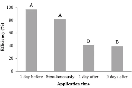
The lesion length caused by B. cinerea on the stems reduced at all application times of the filtrate, the efficiency in reduction varied from 40 - 90 % depending on the application time. The efficiency was higher when the filtrate was applied one day before or at the time of pathogen inoculation (Figure 4).
Figure 4. Efficiency (%) in reducing the lesion length in tomato stem segments treated with filtrates of C. rosea isolates at four application times based on B. cinerea inoculation. The values are the averages of 12 replicates. The bars represent the standard error of the mean.
Whole tomato plants
The lesion length on plants only reduced when the filtrate was applied one day before B. cinerea inoculation. At the other application times, the efficiency was less than 9 % (Figure 5).
Figure 5. Efficiency (%) in reducing the lesion length in tomato plants following application of filtrates of C. rosea filtrate at four application times related to B. cinerea inoculation. The values are averages of 13 replicates. The bars represent the standard error of the mean.
DISCUSSION
The efficiency of C. rosea in controlling pathogenic fungi, including B. cinerea, is attributed to different mechanisms of antagonism: competition, mycoparasitism and induction of resistance (MOUEKOUBA et al., 2014; NOBRE et al., 2005; ROBERTI et al., 2008; SUTTON et al., 2002). It is known that C. rosea produces enzymes (LI et al., 2006; MAMARABADI et al., 2009) and secondary metabolism compounds that are peptide and polyketides antibiotics with antifungal action (FATEMA et al., 2018; KARLSON et al., 2015; PACHANARI & DIX, 1980; RODRÍGUEZ et al., 2011). In this paper, the effect of compound(s) anti-B. cinerea was evaluated in vitro and in vivo.
It was observed that when it was mixed B. cinerea conidia to a C. rosea filtrate, the germination of the pathogen conidia were reduced. Similarly, when conidia of B. allii were added to a filtrate of C. rosea, a reduction in conidia germination of over 90 % was observed (PACHENARI & DIX, 1980). Therefore, we concluded that the four C. rosea isolates produce antifungal compound(s). We found variability in the production of C. rosea compound(s) between the experimental runs. The amount of time C. rosea was grown slightly varied and the compound(s) concentration was not quantified. The best conditions for production of the compound(s) by C. rosea are not yet defined, but we noticed differences in colony and on culture medium color depending on temperature and light intensity. According to Rodríguez et al. (2011), the formation of the inhibition zone, a variable used to assess C. rosea antibiosis against S. sclerotiorum, was dependent on the culture medium. In addition, genes that are related to some compounds produced by fungi cannot be expressed, or are under expressed, under laboratory conditions (BRAKHAGE & SCHROECKH, 2011), possibly because the secondary metabolic compounds are produced only when the fungus needs to communicate or defend against other organisms (CHIANG et al., 2011; O'BRIEN & WRIGHT, 2011). Indeed, the polyketides genes pks22 and pks29, were highly induced at different culture media, especially PDB, and during C. rosea-B. cinerea interaction. Two of four polyketides encoded by pks22, named Clonorosein A and B, showed high inhibition of conidial germination and germ tube growth of B. cinerea, suggesting an important role in the antagonism and biocontrol (FATEMA et al., 2018).
The compound(s) produced by C. rosea on PDA also reduced the mycelial and sclerotia development. When B. cinerea has grown in PDA in the presence of C. rosea compound(s) (first subculture), the mycelial growth was thinner and less cottonous than in the control. Therefore, it is
confirmed that C. rosea is producing compound(s) deleterious to B. cinerea. When both mycelium and sclerotia that stopped growing in the first subculture were transferred to sterile PDA (second subculture), more than 90% of both resumed the development. Therefore, it is possible that C. rosea compound(s) have a fungistatic effect on B. cinerea. It is important to report that peptide antibiotics, compounds of the secondary metabolism of C. rosea (RODRÍGUEZ et al., 2011), are also produced by species of Trichoderma (MUKHERJEE et al., 2011; VEY et al., 2001), a genus taxonomically related to Clonostachys. These peptides stop mycelial growth and lyse hyphae cells (DANIEL & RODRIGUES FILHO, 2007; VITERBO et al., 2007). Both reduction of mycelial growth and resuming growth of S. sclerotiorum by C. rosea compounds, when pathogen mycelial discs was transferred from culture medium with C. rosea compounds to a medium without the compounds, took place (RODRÍGUEZ et al., 2011). A reduction on appressoria formation by Magnaporthe grisea was also observed (THINES et al., 1998). Therefore, we conclude that C. rosea produces compound(s) with similar effects to a substance produced by Trichoderma, which can inhibit germination and paralyze hyphal growth, with a fungistatic effect. Probably the compound(s) produced by the isolates we tested is(are) non-volatile, as found by Rodríguez et al. (2011) that tested C. rosea against S. sclerotiorum.
The filtrate of growth of C. rosea in PDB was also tested against B. cinerea in tomato stem segments and tomato plants. In the segments, the filtrate reduced the severity of gray mold at all application times we evaluated, although it was most effective when applied one day before or simultaneously with the pathogen inoculation. Less efficiency was achieved with application one day after B. cinerea inoculation, most likely because the infection had already started and the filtrate did not reach the pathogen mycelium. In plants, the filtrate was efficient only when applied one day before pathogen inoculation, but the effect was less than in the cut stem. The filtrate, either autoclaved or not, was efficient. There is evidence of the instability of compounds produced by C. rosea against pathogens (PACHENARI & DIX, 1997). We did not test the stability of the compound(s), but it is likely that the temperature does not affect the compound(s) stability. It seems that this(se) compound(s) has(ve) a fungistatic effect on B. cinerea. If it is the case, the compound could give a competitive advantage to C. rosea: the antagonist can grow over a substrate and occupy it, and is no longer effective after the antagonist colonizes the substrate. This mechanism would increase the competitive ability of the antagonist. As reported above, C. rosea seems to produce antifungal compound(s) with non-specific action, which can affect different species and
different fungi structures. As a saprophytic, the production of fungistatic compound(s) could help C. rosea colonize the substrate more quickly and efficiently, without necessarily killing other species of microorganism.
As we mentioned, the compound(s) was(were) not identified and quantified. Both identification and quantification will help to determine whether the compound(s) can be used as a sustainable commercial product. The four isolates of C. rosea were as effective as fungicides in suppressing B. cinerea in both laboratory and field (COTA et al., 2008; NOBRE et al., 2005). It is known that these isolates act by competition and hyperparasitism, multiple mechanisms would increase their potential as biocontrol agents. The production of anti-B. cinerea compound(s) increases the potential of isolates to be used either individually or mixed. This is the first report indicating that C. rosea produces compound(s) with a fungistatic effect against B. cinerea. More studies are underway to identify these compounds and to develop strategies to use them in the management of gray mold on plants.
ACKNOWLEDGEMENTS
To National Council for Scientific and Technological Development (CNPq) and Foundation for Research of the State of Minas Gerais (FAPEMIG).
REFERENCES
BORGES, A. V.; SARAIVA, R. M.; MAFFIA, L. A., 2014. Key factors to inoculate Botrytis cinerea in tomato plants. Summa Phytopathologica, v.40, p.221-225, http://dx.doi.org/10.1590/0100-5405/1929
BORGES, A.V.; SARAIVA, R. M.; MAFFIA, L. A. 2015. Biocontrol of gray mold in tomato plants by Clonostachys rosea. Tropical Plant Pathology, v.40, p.71-76, https://doi.org/10.1007/s40858-015-0010-3
BRAKHAGE, A. A.; SCHROECKH, V. 2011. Fungal secondary metabolites – strategies to activate silent gene clusters. Fungal Genetics and Biology, v.48, p.15-22, http://dx.doi.org/10.1016/j.fgb.2010.04.004
CHIANG, Y. M.; CHANG, S. L.; OAKLEY, B.R.; WANG, C. C. 2011. Recent advances in awakening silent biosynthetic gene cluster and linking orphan clusters to natural products in microorganisms. Current Opinion in Chemical Biology, v.15, p.137-143, http://dx.doi.org/10.1016/j.cbpa.2010.10.011
COTA, L. V.; MAFFIA, L. A; MIZUBUTI, E. S. G.; MACEDO, P. E. F.; ANTUNES, R. F., 2008. Biological control of strawberry gray mold by Clonostachys rosea under field conditions. Biological Control, v.46, p.515-522, https://doi.org/10.1016/j.biocontrol.2008.04.023
DANIEL, J. F.; RODRIGUES FILHO, E. 2007. Peptaibols of Trichoderma. Natural Product Reports, v.24, p.1128-1141, https://doi.org/10.1039/b618086h
FATEMA, U.; BROBERG, A.; JENSEN, D. F.; KARLSON, M.; DUBEY, M. 2018. Functional analysis of polyketide synthase genes in the biocontrol fungus Clonostachys rosea. Scientific Reports, v.8, p.15009, https://doi.org/ 0.1038/s41598-018-33391-1
KARLSON, M.; DURLING, M. B.; CHOI, J.; et al., 2015. Insights on the evolution of mycoparasitism from the genome of Clonostachys rosea. Genome Biology and Evolution 7:465-80, https://doi.org/ 10.1093/gbe/evu292
LAHOZ, E.; CONTILLO, R.; PORRONE, F. 2004. Induction of systemic resistance to Erysiphe orontii cast in tobacco by application on roots of an isolate of Gliocladium roseum Bainier. Journal of Phytopathology, v.152, p.465-470, https://doi.org/ 10.1111/j.1439-0434.2004.00876.x
LI, G. Q.; HUANG, H. C.; KOKKO, E. G.; ACHARYA, S. N., 2002. Ultrastructural study of mycoparasitism of Gliocladium roseum on Botrytis cinerea. Botanical Bulletin of Academia Sinica, v. 43, p.211-218, https://doi.org/10.7016/BBAS.200207.0211
LI, J.; YANG, J. K.; HUANG, X. W.; ZHANG, K. Q. 2006. Purification and characterization of an extracellular serine protease from Clonostachys rosea and its potential as a pathogenic factor. Process Biochemistry, v.41, p.925-929, https://doi.org/10.1016/j.procbio.2005.10.006 MAMARABADI, M.; JENSEN, D. F.; LUBECK, M. 2009. An N-acetyl-b-D-glucosaminidase gene, cr-nag1, from the biocontrol agent Clonostachys rosea is up-regulated in antagonistic interactions with Fusarium culmorum. Mycological Research, v.113, p.33-43, https://doi.org/10.1016/j.mycres.2008.07.005
MORANDI, M. A. B.; MAFFIA, L. A.; MIZUBUTI, E. S. G.; ALFENAS, A. C.; BARBOSA, J. G. 2003. Suppression of Botrytis cinerea sporulation by Clonostachys rosea on rose debris: a valuable component in Botrytis blight management in commercial greenhouses. Biological Control, v.26, p.311–317, https://doi.org/10.1016/S1049-9644(02)00134-2
MORANDI, M. A. B.; MAFFIA, L. A.; SUTTON, J. C., 2001. Development of Clonostachys rosea and interactions with Botrytis cinerea in rose leaves and residues. Phytoparasitica, v.29, p.1-11, https://doi.org/0.1007/BF02983954
MORANDI, M. A. B.; MATTOS, L. P. V.; SANTOS, E. R.; BONUGLI, R. C. 2008. Influence of application time on the establishment, survival, and ability of Clonostachys rosea to suppress Botrytis cinerea sporulation on rose debris. Crop Protection, v.27, p.77-83, https://doi.org/10.1016/j.cropro.2007.04.008
MUKHERJEE, P. K.; HORWITZ, B. A.; KENERLEY, C. M. 2011. Secondary metabolism in Trichoderma - a genomic perspective. Microbiology-SGM, v.158, p.35-45, https://doi.org/10.1099/mic.0.053629-0
MOUEKOUBA, L. D. O.; ZHANG, L.; GUAN, X.; CHEN, X.; CHEN, H.; ZHANG, J.; ZHANG, J.; LI, J.; YANG, Y.; WANG, A. 2014. Analysis of Clonostachys rosea-induced resistance to
tomato gray mold disease in tomato leaves. Plos One, v.7, p.e102690,
https://doi.org/10.1371/journal.pone.0102690
NOBRE, S. A. M.; MAFFIA, L. A.; MIZUBUTI, E. S. G.; COTA, L. V.; DIAS, A. P. S., 2005. Selection of Clonostachys rosea isolates from Brazilian ecosystems effective in controlling Botrytis cinerea. Biological Control, v.34, p.132-143, https://doi.org/10.1016/j.biocontrol.2005.04.011
O’BRIEN, J.; WRIGHT, G. D. 2011. An ecological perspective of microbial secondary metabolism. Current Opinion in Biotechnology, v.22, p.552-558, https://doi.org/10.1016/j.copbio.2011.03.010
O'NEILL, T. M.; SHTIENBERG, D.; ELAD, Y., 1997. Effect of some host and microclimate factors on infection of tomato stems by Botrytis cinerea. Plant Disease, v.81, p.36-40, https://doi.org/10.1094/PDIS.1997.81.1.36
PACHENARI, A.; DIX, N. J. 1980. Production of toxins and wall degrading enzymes by Gliocladium roseum. Transactions of the British Mycological Society, v.74, p.561-566, https://doi.org/10.1016/S0007-1536(80)80057-X
PENG, G.; SUTTON, J. C. 1991. Evaluation of microorganisms for biocontrol of Botrytis cinerea in strawberry. Canadian Journal of Plant Pathology, v.13, p.247-257, https://doi.org/10.1080/07060669109500938
ROBERTI, R.; VERONESI, A.; CESARI, A.; CASCONE, A.; DI BERARDINO, I.; BERTINI, L.; CARUSO, C. 2008. Induction of PR proteins and resistance by the biocontrol agent Clonostachys rosea in wheat plants infected with Fusarium culmorum. Plant Science, v.175, p.339-347, https://doi.org/10.1016/j.plantsci.2008.05.003
RODRÍGUEZ, M. A.; CABRERA, G.; GOZZO, F. C.; EBERLIN, M. N.; GODEAS, A. 2011. Clonostachys rosea BAFC3874 as a Sclerotinia sclerotiorum antagonist: mechanisms involved and potential as a biocontrol agent. Journal of Applied Microbiology, v.110, p.1177-1186, https://doi.org/10.1111/j.1365-2672.2011.04970.x
SUTTON, J. C.; LI, D. W.; PENG, G.; YU, H.; ZHANG, P.; VALDEBENITO-SANHUENZA, R. M., 1997 Gliocladium roseum: A versatile adversary of Botrytis cinerea in crops. Plant Disease, v.81, p.316-328, http://dx.doi.org/10.1094/PDIS.1997.81.4.316
SUTTON, J. C.; LIU, W.; HUANG, R.; OWEN-GOING, N., 2002. Ability of Clonostachys rosea to establish and suppress sporulation potential of Botrytis cinerea in deleafed stems of hydroponic greenhouse tomatoes. Biocontrol Science and Technology, v.12, p.413-425, https://doi.org/10.1080/09583150220146004
THINES, E.; EILBERT, F.; ANKE, H.; STERNER, O. 1998. Glisoprenis C; D and E; new inhibitors of appressorium formation in Magnaporthe grisea, from cultures of Gliocladium roseum. Production and biological activities. Journal of Antibiotics, v.51, p.117-22, https://doi.org/10.7164/antibiotics.51.228
VEY, A.; HOAG, I. R. E.; BUT, T. M. 2001. Toxic metabolites of fungal biocontrol agents. In: BUT, T. M.; JACKSON, C.; MAGAN, N. (Eds) Fungi as biocontrol agents: progress, problems and potential. USA, CaB International Publishing, 311p.
VITERBO, A.; WIEST, A.; BROTMAN, Y.; CHET, I.; KENERLEY, C. 2007. The 18mer peptaibols from Trichoderma virens elicit plant defence responses. Molecular Plant Pathology, v.8, p.737-746, https://doi.org/10.1111/j.1364-3703.2007.00430.x
Received in: November 26 2019 Accepted in: February 26, 2020