C.H. Santiago et al.
ABSTRACT: Cassava root rot diseases such as dry and black root rot are listed among the major threats, since its affect the main product
(tuberous roots), causing a progressive decline in yield and affecting
subsequent crop cycles, being the use of resistant varieties the most
reliable control measure on field level. The objective of this study was
to identify inoculation methods for the early evaluation of genotypes,
considering the level of resistance to dry (DRR) and black (BRR) root
rot diseases. Different methodologies and plant tissues were evaluated,
based on the immersion of cassava tissues (roots and stem cuttings),
soil infestation, and inoculation of detached tissues (leaves, tuberous
roots and stem cuttings). The following parameters were evaluated
for inoculations based on tissue immersion: disease index (ω); aerial
part weight (g); fresh weight of the roots (g); and volume (cm3). For
BASIC AREA - Article
Methodologies for selecting cassava with
resistance to dry and black root rot under
controlled conditions
Camila Hohenfeld Santiago1, Mariana Pereira Santana1, Luiz Rodriguez Cairo Junior1,
Saulo Alves Santos de Oliveira2*, Eder Jorge de Oliveira2
1.Universidade Federal do Recôncavo da Bahia - Centro de Ciências Agrárias, Ambientais e Biológicas - Cruz das Almas (BA), Brazil. 2.Embrapa Mandioca e Fruticultura - Núcleo de Recursos Genéticos e Desenvolvimento de Variedades - Cruz das Almas (BA), Brazil.
*Corresponding author: [email protected]
Received: Mar. 8, 2017 – Accepted: Nov. 14, 2017
the inoculations on detached tissues, the percentage of lesioned area
was determined. Immersion methods for roots and lesioned stems did
not show typical symptoms of DRR and BRR during the two-month
evaluation period. The soil infestation method did not differ from the
stem immersion method as to the reduction of aerial part weight and
the disease index, whereas both can be recommended for resistance
selection trials. There was a positive correlation between the BRR and
DRR data for the stem inoculation (r = 0.94, p = 0.001) and for DRR in
the peel and root pulp (r = 0.73, p = 0.05). Therefore, the resistance
within each tissue is apparently independent and should be compared
with the behavior of the genotypes in the field.
INTRODUCTION
Dry (DRR) and black (BRR) root rot are two of the most destructive pathologies in cassava (Manihot esculenta
Crantz), whose economic importance is increasing in major producing countries (Onyeka et al. 2005a; Vilas Boas et al. 2017). The increase in DRR and BRR is causing a progressive decline in yield and affecting subsequent crop cycles (Vilas Boas et al. 2016). DRR and BRR are caused by pathogens of different fungi genera, and their symptoms differ depending on the associated species. Dry root rot (DRR) is characterized by the dry appearance of lesions, usually with a brownish halo, and the main species from the genus Fusarium spp. (e.g., F. solani and F. oxysporum) (Bandyopadhyay et al. 2006).
In contrast, black root rot (BRR) presents lesions with brownish or black colors, which could be associated with the softening of the infected tissue. The pathogens that cause black root rot include those from the genus Lasiodiplodia spp. as well as Neoscytalidium hyalinum (Machado et al. 2014).
The use of resistant varieties is the most economic and feasible approach for DRR and BRR control (Onyeka et al. 2005a). However, development of resistant varieties is difficult, since root rot is caused by soil-borne pathogens and the onset of symptoms can be influenced by climate and soil conditions. Currently, the screening for resistance has been based on the evaluation of genotypes with a history of the disease under field conditions (Okechukwu et al. 2009). However, this type of evaluation requires several experimental trials and can still be inaccurate, due to the presence of different species that cause the disease and the heterogeneous distribution of pathogens in the soil (Onyeka et al. 2005b).
The most frequent species causing DRR and BRR in the North and Northeast regions of Brazil are those associated with dry root rot and black root rot (Vilas Boas et al. 2017). Based on these results, it is necessary to develop strategies for the identification of multiple sources of resistance against the major pathogens associated with DRR and BRR, taking into consideration the different resistance mechanisms in plant tissues.
Therefore, this study aimed to identify inoculation methods for different plant tissues, for evaluating the levels of dry and black root rot resistance in cassava genotypes.
MATERIAL AND METHODS
All evaluated methodologies used an isolate of Fusarium
solani (CBPPR0057) and Neoscytalidium hyalinum
(CBPPR1011), which causes DRR and BRR, respectively, previously characterized both as pathogenic to cassava and the most aggressive of the two groups of pathogens studied (Vilas Boas et al. 2017). These fungi belong to the cassava root rot pathogen collection at the Plant Pathology Laboratory in Embrapa Mandioca e Fruticultura.
Isolates were grown in Petri dishes containing potato dextrose agar (PDA) medium at 25 ± 2 °C and 12 h of light, for seven days. The spore suspensions for the methods based on immersion of cassava stem cuttings and roots and for inoculation of detached tuberous roots were prepared from the addition of 10 mL of sterile distilled water in petri dishes containing colonies of each isolate, and the conidia mass released with a soft toothbrush. The suspension was filtered with a double layer of cheesecloth and the concentration of spores determined using a hemocytometer (Neubauer chamber) under a light microscope adjusted to 1 × 10⁵ conidia·mL-1. Sterile distilled water was used as a
control treatment (mock).
The inoculum for the trials involving soil infestation was produced in a sand and cornmeal mixture in the ratio of 3:1 w/w. The mixture was stored in transparent plastic bags and autoclaved twice at 120 °C for 1 h. After 24 h of cooling, 150 mL of the spore suspension of each isolate was added. To promote the uniform growth of fungi, the bags containing the sand and cornmeal infested substrate were homogenized every two days and incubated in a growth chamber at 25 ± 2 °C for 20 days.
Different cassava varieties were used across the experiments, and they were choose based on previous works aiming the selection of resistant varieties, and the behavior of the cassava genotypes in the field (Oliveira et al. 2013; Vilas Boas et al. 2016; Vilas Boas et al. 2017).
Tissue immersion and soil infestation
The varieties Fécula Branca and Olho Junto were used in the evaluations with the following methods:
a. Immersion of cassava stems cuttings
planted in plastic bags containing 3 kg of sterilized soil. The experiment was set in a 2·2 factorial completely randomized, with 2 varieties and 3 inoculation procedures. There were three replicates with six plants each.
The inoculation procedures were as follows:
1. Uninfested soil with healthy cassava cuttings (control);
2. Uninfested soil × cassava stem cuttings inoculated with dry root rot pathogen;
3. Uninfested soil × cassava stem cuttings inoculated with black root rot pathogen.
The evaluations were performed at 60 days after inoculation, based on the following parameters:
I. Fresh weight of the aerial part, in grams (g);
II. Fresh weight of the roots, in grams (g);
III. Root system volume (cm3), determined based on
the measurement of the displacement of the water column in a graduated cylinder;
IV. Internal colonization of stem cuttings, based on following rating scale:
0 = not colonized;
1 = colonization of < 1/3 of the interior of the cutting; 2 = colonization of ≥ 1/3 and < 2/3 of the interior of the cutting;
3 = colonization of ≥ 2/3 of the interior of the cutting
b. Root immersion
Stem cuttings of 12 cm of each cassava variety were germinated in a sand bed for 15 days. The roots of the stem cuttings were immersed in a spore suspension of each pathogen for 1 h and then planted in plastic bags containing 3 kg of sterilized soil. The control treatment was represented by immersing plant root systems in sterile distilled water only. The experiment was set as 2 × 2 factorial in completely randomized design, containing 2 varieties and 2 pathogens. There were four replicates with six plants each. An additional treatment was used for both varieties, composed by non-inoculated plants (control). The evaluation was performed from five days after inoculation up to two months.
c. Soil infestation
Plastic bags were filled with a with a mixture composed of 3 kg of sterilized soil and 200 g of the inoculum produced in the sand–cornmeal (ratio 3:1) substrate, which was infested with each pathogen. As a control (non-inoculated), 100 g of mixing sand and pure cornmeal were distributed. The inoculation procedure was performed by planting
the cassava stem cuttings of each variety in the infested soil, as described above. The experiment was set as 2 × 2 in completely randomized design, with 2 varieties and 2 inoculation procedures.
The inoculation procedures were as follows:
1. Soil infested with DRR pathogen × healthy stem cassava cuttings;
2. Soil infested with BRR pathogen × healthy cassava stem cuttings
An additional treatment was used for both varieties, composed by uninfested soil and healthy cassava stem cuttings (control). The evaluations were performed according to the methodology described above on see section (a) Immersion of cassava stem cuttings.
Inoculation of detached tissue
Varieties BRS Verdinha, BRS Dourada, BRS Aramaris, BRS Tapioqueira, BRS Poti Branca, BRS Formosa, Olho Junto, and Salangó Preta, were used in the inoculation experiments with detached tissue, in addition to the varieties BRS Kiriris (resistant to root rot under field conditions) and Fécula Branca (highly susceptible under field conditions).
a. Inoculation of tuberous roots
The experiments were carried out following the methodology for inoculating whole roots, as proposed by Onyeka et al. (2005c) and adapted by Oliveira et al. (2013). The roots were washed and disinfested with sodium hypochlorite (0.5%), punched at three equidistant points (6 mm in diameter), and then a 40 µL drop of each pathogen spore suspension was added. As a control, 40 µL of sterile distilled water was added to the central perforations. The experiment was set as 10 × 2 factorial in completely randomized design with 10 varieties and 2 pathogens, with 12 replicates.
b. Detached leaves and stem segments
as 10 × 2 factorial in completely randomized design with 10 varieties and 2 pathogens, with 6 replicates (3 lobes of each leaf).
Stem segments were harvested from each two-month old variety, cut into 10 cm pieces, disinfested with 0.5% sodium hypochlorite, and washed in sterile distilled water. The ends of the segments were covered with wet cotton to avoid desiccation. With a single needle, three holes were punched in a triangular arrangement on the surface of the stem, and a 5 mm mycelial plug wounds. As a control, a sterile PDA plug was used. The experiment was set as 10 × 2 factorial in completely randomized design with 10 varieties and 2 pathogens, with 3 replicates (5 stem each).
The inoculated leaves and stems were maintained in a transparent gerbox, with the base covered with sterile filter paper. The development of the lesions was assessed seven days after inoculation, and the digital images analyzed using the ASSESS 2.0 program (APS Press®, 2002–2008).
c. Inoculation on wounded cuttings
Stem cuttings of 10 cm from mature plants (approximately 12 months old) were punched with 6 mm holes between the first and the second nodes. Discs of the culture medium (5 mm in diameter) containing fungal structures from each pathogen were then deposited. The inoculation sites were covered with moistened cotton and wrapped with PVC film to prevent external contamination and ensure moisture at the site of inoculation. A PDA plug without fungal growth was used as control.
The inoculated stem cuttings and controls were planted in plastic bags containing 3 kg of sterilized soil, mixed with equal parts of washed sand, coconut fiber, and vermiculite. The experimental design was completely randomized, with 10 varieties and 2 pathogens, with 4 replicates (6 plants each). The evaluation was performed from five days after inoculation up to two months. The parameters evaluated were mortality of accessions and incidence and external symptoms by a rating scale, as follows:
(0) no symptoms;
(1) leaf chlorosis and/or wilt of the lower third; (2) leaf chlorosis and/or wilt of the middle third; (3) leaf chlorosis and/or wilt of the whole plant; (4) complete defoliation and/or death of the plant. The inoculated tuberous roots were kept in plastic trays and covered with transparent plastic bags to keep the moisture in. Evaluations were performed 10 days after
inoculation, measuring the size of the lesioned area through digital analysis of the images using the software ImageTool (University of Texas Health Science Center, San Antonio, TX, USA) (Oliveira et al. 2013).
Statistical Analysis
The data obtained from arbitrary scales were converted into the disease index (ω) according to Czermainsky (1999). Afterwards, the quantitative data were submitted to an analysis of variance as a double factorial design, and means were grouped using the Scott-Knott test (p < 0.05) (Chapola et al. 2016). In order to verify the relationship between the different methods tested, the Pearson correlation test (p ≤ 0.05) (Koerner and Zhang, 2017) and a hierarchical clustering analysis were conducted based on Euclidean distance using the R program (R core team, 2015). All assays were conducted in growth chambers at a controlled temperature (26 ± 2 °C) in the dark and with relative humidity greater than 85%.
RESULTS AND DISCUSSION
According to the ANOVA analysis for all treatments, there were significant effects regarding the variety and pathogen species and for the interaction between variety and the two pathogen species. Among the eight methods tested, the immersion of the roots and wounding of stem cuttings did not present the typical symptoms of root rot during the evaluation period of two months. However, the other methodologies developed symptoms similar to what was previously reported for each pathogen, enabling the re-isolation thereof and in such, completing Koch’s postulates (Oliveira et al. 2013), and were effective in determining varieties of cassava that are resistant to BRR and DRR diseases under controlled conditions.
Tissue immersion and soil infestation
of the plant at 60 DAI. The same results were obtained in treatments with soil infested with dry root rot isolates (SI-DRR).
The control treatment for all methodologies showed higher aerial part weight, root weight and root volume, which differed from the inoculated treatments. Moreover, root rot disease significantly reduced the growth of the aerial
parts of both varieties. There was no significant difference in the severity of symptoms between the varieties Fécula Branca and Olho Junto, considering the different methods of inoculation. There was also no significant difference between the immersion methods and the infestation of the soil within the same variety considering the weight of the aerial part (Fig. 1a and 1b).
Means followed by the same capital letter (comparing varieties within the same treatment) and lower case letters (treatments within varieties) belong to the same group by the Scott-Knott test (p < 0.05)
IM-BRR = Immersion of stem segment in black root rot pathogen suspension SI-BRR = Soil infestation by black root rot pathogen
IM-DRR = Immersion of stem segment in dry root rot pathogen suspension SI-DRR = Soil infestation by dry root rot pathogen.
Figure 1. (a) Fresh weight of the aerial part of varieties Fécula Branca and Olho Junto when inoculated with black root rot pathogens; (b) Fresh weight of the aerial part of varieties Fécula Branca and Olho Junto when inoculated with dry root rot pathogens; (c) Disease Index when inoculated with black root rot pathogens; (d) Disease Index when inoculated with dry root rot pathogens; (e) Root volume when inoculated with black root rot pathogens; (f) Root volume when inoculated with dry root rot pathogens; (g) Fresh weight of the roots when inoculated with black root rot pathogens and (h) Fresh weight of the roots when inoculated with dry root rot pathogens.
0 10 20 30 40
IM - DRR SI-DRR Control 0
10 20 30 40
IM - BRR SI - BRR Control
FW A
erial P
art (g)
0 10 20 30 40
IM - DRR SI-DRR
0 10 20 30 40 50
IM - BRR SI - BRR
Dise
a
se Index (
ω
)
0 0,5 1 1,5 2 2,5 3 3,5
IM - DRR SI-DRR Control 0
0,5 1 1,5 2 2,5 3 3,5
IM - BRR SI - BRR Control
R
oot V
olume (cm³)
0 1 2 3 4
IM - BRR SI - BRR Control
0 1 2 3 4
IM - DRR SI-DRR Control
bA bA bA bA
aB aA
bA bA bA bA
aB aA
bB aB
aA aA aA aA
aB
aB
aA
bA
bA aA
bB
aA aA
bA
bA
bA bA
aA
bA
bA
cA cA
aA aA
bA bA
cA
aA aA
bB
Fécula Branca Olho Junto
FW of the r
oots (g)
FW A
erial P
art (g)
Dise
a
se Index (
ω
)
R
oot V
olume (cm³)
FW of the r
oots (g)
(a)
(c)
(e)
(g)
(b)
(d)
(f)
The weight of the aerial part of the varieties Fécula Branca and Olho Junto for each treatment, respectively, were IM-DRR (3.08 and 4.89 g), IM-BRR (0.6 and 2.54 g), SI-DRR (0.7 and 3.59 g), and SI-BRR (4.7 and 2.02 g), while the controls for the two varieties were 23.18 and 35.49 g, respectively. The disease index (ω) values for the Fécula Branca and Olho Junto varieties were 17.96 and 42.02, respectively, for the four methods of inoculation (Fig. 1c and 1d).
The methodologies based on tissue immersion and soil infestation were also effective in reducing the volume (Fig. 1e and 1f) and the fresh weight of roots (Fig. 1g and 1h). Varietal differences were observed only in the IM-DRR method, with the highest values being for the Olho Junto variety when compared with Fécula Branca for both parameters. No significant difference was observed in the development of the root system with the SI-DRR method. When considering the possibility of simultaneous evaluation for different pathogens, soil infestation was more effective in reducing the weight and volume of roots for the two diseases (DRR and BRR), not differing from the stem cuttings immersion method. Therefore, the soil infestation method can be recommended for future evaluations of the reaction of cassava genotypes to DRR and BRR in greenhouse experiments. Similar results were obtained with inoculations of Fusarium solani on rosewood trees, in which soil infestation enabled the appearance of typical symptoms of root rot, with greater reduction of fresh weight, root volume, and aerial part length compared to direct inoculation on the stem (Rajput et al. 2008).
Although using pathogens of the Botryosphaeriaceae family and Fusarium spp. to inoculate detached cassava stem cuttings has been reported (Onyeka et al. 2005c; Msikita et al. 2005), and although similar reports have been made for other pathosystems (Linaldeddu et al. 2011), the results of planting the stem cuttings inoculated by wounding did not result in typical external DRR and BRR symptoms. Therefore, after two months of evaluation, this methodology was adapted, and stems that sprouted from the infected stem cuttings were wounded again and used in the second trial.
The inoculation method based on the immersion of roots has also been commonly and successfully used in resistance tests using Fusarium species on other crops (Li et al. 2008; Aguiar et al. 2013). However, the inefficiency may be related to the regeneration capacity of the cassava root system. This inoculation did not prevent the development of new roots by the cassava stem cuttings (manivas) or the recovery of
the inoculated plant, even if there was death of adventitious roots present at the time of inoculation.
Inoculation of detached tissue
In general, there was a difference between how the inoculated tissues of the different varieties responded to the pathogens. For example, in assessing the resistance to BRR in the root peel, the variety with the largest lesioned area was BRS Aramaris (421.44 mm²), while the varieties Salangó Preta, BRS Tapioqueira, and Olho Junto presented the smallest lesioned areas (34.89 mm², 69.05 mm², and 78.52 mm², respectively) (Fig. 2a). Furthermore, in assessing the inoculation of the root pulp, the varieties BRS Dourada (86.70 mm²), Salangó Preta (64.46 mm²), and BRS Tapioqueira (130.69 mm²) presented the smallest lesions (Fig. 2b). When inoculated with pathogens causing DRR, BRS Formosa showed a larger lesioned area on the root peel (266.72 mm²) (Fig. 2c), while for the pulp, the major lesioned areas were observed in the BRS Kiriris (208.44 mm²), BRS Formosa (193.80 mm²), and BRS Verdinha (173.75 mm²) varieties (Fig. 2d).
The inoculation of DRR and BRR pathogens on detached leaves was also effective in the expression of symptoms in all cassava varieties analyzed. These symptoms appeared in the form of lesions on the leaf surface 48 h after inoculation. The method of inoculating detached stems was also very efficient in terms of the onset of symptoms associated with cassava root rot pathogens and varietal behavior.
As a result, the inoculation of detached leaves revealed significant differences in the percentage of lesioned areas on different varieties, once the BRS Tapioqueira (70.06%) and BRS Kiriris (68.03%) were quite susceptible to the pathogens causing BRR (Fig. 3a) differing from the inoculation on detached stem, where the variety BRS Aramaris was the most susceptible, with a percentage of 34.29% of the lesioned area (Fig. 3b).
Figure 3. Percentage of lesioned area by pathogens that cause black root rot (BRR) when inoculated on leaves (a) and stems (b) from different varieties of cassava; Percentage of lesioned area by pathogens that cause dry root rot (DRR) when inoculated on leaves (c) and stems (d) from different varieties of cassava. Means followed by the same letter belong to the same group based on Scott-Knott test (p < 0.05).
Figure 2. Lesioned area (mm 2 by pathogens that cause black root rot (BRR) when inoculated in the peel (a) and pulp (b) of the root in different varieties of cassava; Lesioned area by pathogens that cause dry root rot (DRR), in different varieties of cassava, when inoculated in the peel (c) and pulp (d) of the root. Means followed by the same letter belong to the same group based on Scott-Knott test (p < 0.05).
50 150 250 350 450 a a a a a a a a a a a b b b b b b b b b BRS Ar amaris BRS Dour ada BRS F ormosa BRS Kiriris BRS P oti Br anc a BRS T apioqueir a BRS V er dinha Fécula Br anc a Olho J unt o S alangó BRS Ar amaris BRS Dour ada BRS F ormosa BRS Kiriris BRS P oti Br anc a BRS T apioqueir a BRS V er dinha Fécula Br anc a Olho J unt o S alangó BRS Ar amaris BRS Dour ada BRS F ormosa BRS Kiriris BRS P oti Br anc a BRS T apioqueir a BRS V er dinha Fécula Br anc a Olho J unt o S alangó BRS Ar amaris BRS Dour ada BRS F ormosa BRS Kiriris BRS P oti Br anc a BRS T apioqueir a BRS V er dinha Fécula Br anc a Olho J unt o S alangó 0 50 100 150 200 250 300 0 50 100 150 200 250 300 0 50 100 150 200 250 Pulp Peel Pulp Peel a
b b b b
b
b b b
b
a a
a
b
b b b
Although all of the evaluated methods for screening germplasm in the search for sources of resistance have presented typical lesions in the tested varieties, there was discrepancy in the ranking of varieties by different methods and diseases. However, there was a positive correlation between BRR and DRR on the stem (r = 0.94, p = 0.001) and between inoculation by DRR on the root pulp and peel (r = 0.73, p = 0.05). There was also a moderate correlation between the BRR on the stem and on the root peel (r = 0.56), but was not significant (Fig. 4).
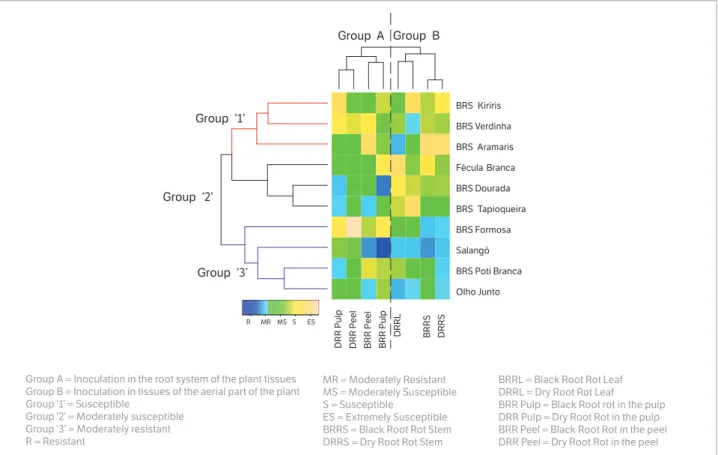
The clustering of the varieties based on the reaction when inoculated on different tissues revealed the formation of three groups (Fig. 5). In general, the varieties of higher susceptibility (Group 1) were BRS Kiriris, BRS Verdinha, and BRS Aramaris. Within Group 1, only the variety BRS Kiriris was not classified as being resistant or moderately resistant for any inoculated tissue, while BRS Verdinha and BRS Aramaris proved moderately resistant to the BRR and DRR pathogens on the detached leaf.
The varieties Fécula Branca, BRS Dourada, and BRS Tapioqueira were allocated in Group 2 (Fig. 5). Among these varieties, only Fécula Branca did not show any resistance in the inoculated tissue. In contrast, BRS Dourada was classified as being resistant in the root pulp to both diseases (DRR and BRR), and BRS Tapioqueira had moderate resistance to BRR pathogens in the peel and to DRR pathogens in the root pulp.
Group 3 comprised the varieties with higher resistance to DRR and BRR, and included BRS Formosa, Salangó Preta, BRS Poti Branca, and Olho Junto (Fig. 5). Among these varieties, Salangó Preta presented the highest percentage of agreement between the different methods and was classified as resistant or moderately resistant in 75% of the evaluated methodologies (6 out of 8). Additionally, based on the different methodologies and disease groups (DRR and BRR), the variety Salangó Preta was the most resistant genotype. However, new experiments must be undertaken in order to confirm the field resistance of this variety.
Figure 4. Correlogram displays of correlation matrices for methods of black and dry root rot inoculation in different cassava varieties. Above diagonal are listed the Pearson’s coefficients, below diagonal the scatter plot and tendency line for each pairwise comparison.
BRRS 10 30
0.94*** 0.24
15 30 45
0.23 0.56
.
100 200
-0.26 0.22
50 150
5
15
2
5
3
5
-0.02
10
3
0 DRRS
0.43 0.11 0.47 -0.19 0.12 0.12
BRRL
0.45 -0.26 -0.26 -0.049
2
0
4
0
6
0
-0.049
15
3
0
4
5 DRRL
-0.11 -0.26 0.047 -0.26
BRR Peel
0.32 0.49
10
0
30
0
0.23
10
0
20
0 DRR Peel
0.27 0.73*
BRR Pulp
10
0
20
0
0.36
5 15 25 35
5
0
15
0
20 40 60 100 300 100 200
DRR Pulp Moderately Resistante
DRR Peel = Dry Root Rot in the peel *Significant at p < 0.05
***Significant at p < 0.001
Mean values for different inoculation methods are presented as a histogram (blue) with the distribution of values for the treatments as a trend line DRRL = Dry Root Rot leaf
BRR Pulp = Black Root Rot in the pulp DRR Pulp = Dry Root Rot in the pulp
Figure 5. Heatmap and hierarchical clustering of cassava varieties in behavior when inoculated by Black and Dry Root Rot in different plant tissues.
BRRL = Black Root Rot Leaf DRRL = Dry Root Rot Leaf
BRR Pulp = Black Root rot in the pulp DRR Pulp = Dry Root Rot in the pulp BRR Peel = Black Root Rot in the peel DRR Peel = Dry Root Rot in the peel Group A = Inoculation in the root system of the plant tissues
Group B = Inoculation in tissues of the aerial part of the plant Group ‘1’ = Susceptible
Group ‘2’ = Moderately susceptible Group ‘3’ = Moderately resistant R = Resistant
MR = Moderately Resistant MS = Moderately Susceptible S = Susceptible
ES = Extremely Susceptible BRRS = Black Root Rot Stem DRRS = Dry Root Rot Stem
Olho Junto BRS Poti Branca Salangó BRS Formosa BRS Tapioqueira BRS Dourada Fécula Branca BRS Aramaris BRS Verdinha BRS Kiriris
DR
R P
u
lp
DR
R P
ee
l
B
RR
P
ee
l
B
RR P
u
lp
DRR
L
B
RR
S
DRR
S
R MR MS S ES
Group ‘1’
Group ‘2’
Group ‘3’
Group A Group B
In relation to the different methods of inoculation (Fig. 5), two distinct groups were formed in which there was greater agreement between the reactions expected and the inoculations of the root system (Group A) as well as the inoculations of the aerial part (Group B).
A moderate correlation (r = 0.73) between the symptoms of root rot in the peel and pulp of the root when inoculated with F. solani was also observed by Vilas Boas et al. (2016), suggesting that the selection of resistant varieties can be made based on the symptoms in both root tissues. The differences of resistance to root rot disease among varieties of cassava may be related to their structural and biochemical mechanisms, which can hinder or entirely block the entry or subsequent activity of pathogens in their tissues.
Regarding the performance of the evaluated methods of inoculation, although screening based on the methodology of detached root inoculation is currently used in the screening, one method that is widely used to determine the resistance to root rot disease requires the programming of the harvest of the tuberous roots (minimum of 10 months of planting time) and a rigorous selection of healthy roots, meaning no injuries, wounds, or symptoms of infections from the
field (Onyeka et al. 2005a; Oliveira et al. 2013; Villas-Boas et al. 2016).
Senthil et al. (2013) compared inoculations of cassava stems, roots, and leaves and observed a positive correlation (0.88) between the symptoms in leaves and roots. Negative correlations for leaves and stems (-0.66) and for roots and stems (-0.68) were also observed by these authors. They also reported that the inoculation of detached leaves is a fast and reliable method with a smaller variation in the response of cassava cultivars to the soft root rot disease caused by Phytophthora palmivora. This method can replace the detached root inoculation method.
Similar results were reported for Phytophthora tropicalis, which also causes soft root rot in cassava, with a moderate positive correlation (0.37) between the symptoms in the leaves and roots (Loke et al. 2004)3. However, the literature
still lacks works presenting the correlation of this kind of selection, with results of field experiments.
3Loke, J., Álvarez, E., Corredor, J. A., Folgueras, M., Jaramillo G. and
Ceballos, H. (2004). Preliminary evidence of correlation between foliar and root resistance to root rot caused by Phytophthora tropicalis
Methodologies for inoculating detached leaves have been used successfully in other pathosystems using species from the genus Fusarium. They have also been used for the red root rot of soybeans, which is caused by Fusarium tucumaniae (Franco et al. 2009), and for the rot of the ‘‘Egusi’’ melon (Colocynthis citrullus L.), which is caused by Fusarium oxysporum (Ntui et al. 2010). However, in this work, the correlation between the symptoms of DRR and BRR when the leaves and stems were inoculated was positive, but not significant. Furthermore, no correlation was observed between inoculations of leaves and roots. For these reasons, the correlation between inoculation methods in different plant tissues still needs to be investigated in order to reflect more accurately the varietal resistance in field conditions.
Regardless of the pathogens, the inoculation methods were grouped separately for root peels and pulp and for stems and leaves. The absence of significant correlations between inoculations of detached tissues, as well their grouping according to tissue (aerial parts or the root system), indicate that root rot resistance mechanisms are different in each tissue evaluated.
A moderate correlation (r = 0.73) between the symptoms of root rot in the peel and pulp of the root when inoculated with F. solani was also observed by Vilas Boas et al. (2016), suggesting that the selection of resistant varieties can be made based on the symptoms in both root tissues. The differences of resistance to root rot disease among varieties of cassava may be related to their structural and biochemical mechanisms, which can hinder or block the entry or subsequent activity of pathogens in their tissues.
As to the behavior of the 10 cassava varieties studied in this work, it was not possible to observe any relationship between the phenotypes of BRS Kiriris and BRS Aramaris, both of which were considered resistant to DRR and BRR under field conditions (Fukuda et al. 2002). In addition, these varieties showed no uniformity in the ranking when evaluated by different inoculation methods.
The variety BRS Aramaris presented moderate resistance to pathogens, showing DRR only when the leaves were inoculated. Oliveira et al. (2013) evaluated the behavior of seven varieties of cassavas when inoculated with Fusarium spp., also rating BRS Aramaris and Fécula
Branca as being susceptible to root rot, while BRS Kiriris was moderately resistant. These results show that the resistance to DRR and BRR may have a quantitative trait, controlled by minor genes whose inheritance is characterized by the absence of complete resistance and by the influence of the environment on the incidence and severity of disease (Villas Boas et al. 2016). Moreover, as in the previous experiment, the variety Fécula Branca was classified as being highly susceptible to root rot in field conditions, and also as being susceptible for all of the tissues evaluated.
Studying the infection of the peel (pre-penetration) and pulp (post-penetration), Loke et al. (2004) attributed the different levels of resistance of 22 cassava genotypes to the different levels of iron (essential in phytoalexin synthesis), fungitoxic substances, and manganese in the plant. Thus, it is necessary to investigate which mechanisms may be related to structural and biochemical factors that are pre or post-formed in cases of infection by DRR and BRR pathogens, aiming to understand the resistance levels within each genotype.
CONCLUSION
The soil infestation and stem immersion methodologies can be recommended for the evaluation of the reaction of cassava genotypes to root rot disease under greenhouse environment. The low correlation between inoculation methods in detached tissue is an indication that the types of tissue may have different levels of resistance.
ORCID IDs
C. H. Santiago
https://orcid.org/0000-0003-2518-7716
M. P. Santana
https://orcid.org/0000-0002-0096-1966
L. R. Cairo-Junior
https://orcid.org/0000-0002-0521-9429
S. A. S. Oliveira
https://orcid.org/0000-0002-2992-1570
E. J. Oliveira
Aguiar, F. M., Michereff, S. J., Boiteux, L. S. and Reis, A. (2013).
Search for sources of resistance to Fusarium wilt (Fusarium
oxysporum f. sp. vasinfectum) in okra germplasm. Crop Breeding
and Applied Biotechnology, 13, 33-40. http://dx.doi.org/10.1590/
S1984-70332013000100004.
Bandyopadhyay, R., Mawangi, M., Aigbe, S. O. and Leslie, J.
(2006). Fusarium species from the cassava root rot complex
in West Africa. Phytopathology, 96, 673-676. http://dx.doi.
org/10.1094/PHYTO-96-0673.
Chapola, R. G., Hoffmann, H. P. and Massola, N. S. (2016).
Reaction of sugarcane varieties to orange rust (Puccinia kuehnii)
and methods for rapid identification of resistant genotypes.
Tropical Plant Pathology, 41, 139-146. https://doi.org/10.1007/
s40858-016-0076-6.
Czermainski, A. B. C. (1999). Generalização de um índice de
intensidade de infecção em experimentos de avaliação de
doenças em plantas. Pesquisa Agropecuária Brasileira, 34,
1545-1555. http://dx.doi.org/10.1590/S0100-204X1999000900004.
Franco, H. B. J., Centurion, M. P. P. C. and Barbosa. J. C. (2009).
Inoculation methods study to evaluation soybean cultivars to
Fusarium tucumaniae. Summa Phytopathologica,35, 32-38.
http://dx.doi.org/10.1590/S0100-54052009000100005.
Fukuda, W. M. G., Silva, S. O. and Iglesias, C. (2002). Cassava
breeding. Crop Breeding and Applied Biotechnology, 2, 617-638.
Koerner, T. K. and Zhang, Y. (2017). Application of Linear
Mixed-Effects Models in Human Neuroscience Research: A Comparison
with Pearson Correlation in Two Auditory Electrophysiology
Studies. Brain Sciences. 7, 26. http://dx.doi.org/10.3390/
brainsci7030026.
Li, X. M. and Liu, C. J., Chakraborty, S., Manners, J. M. and Kazan,
K. (2008). A simple method for the assessment of crown rot
disease severity in wheat seedlings inoculated with Fusarium
pseudograminearum. Journal of Phytopathology, 156, 751-754.
http://dx.doi.org/10.1111/j.1439-0434.2008.01425.x.
Linaldeddu, B. T., Scanu, B., Maddau, L. and Franceschini,
A. (2011). Diplodia africana causing dieback on Juniperus
phoenicea: a new host and first report in the northern hemisphere.
Phytopathologia Mediterranea, 50, 473-477.
REFERENCES
Machado, A. R., Pinho, D. B., Oliveira, S. A. S. and Pereira, O. L.
(2014). New occurrences of Botryosphaeriaceae causing black
root rot of cassava in Brazil. Tropical Plant Pathology, 39,
464-470. http://dx.doi.org/10.1590/S1982-56762014000600008.
Msikita, W., Bissang, B., James, B. D., Baimey, H., Wilkinson,
H. T., Ahounou, M. and Fagbemissi, R. (2005). Prevalence and
severity of Nattrassia mangiferae root and stem rot pathogen
of cassava in Bénin. Plant Disease, 89, 12-16. http://dx.doi.
org/10.1094/PD-89-0012.
Ntui, V. O., Thirukkumaran, G., Azadi, P., Khan, R. S. and Nakamura,
I., Mii, M. (2010). Stable integration and expression of wasabi
defensin gene in ‘‘Egusi’’ melon (Colocynthis citrullus L.)
confers resistance to Fusarium wilt and Alternaria leaf spot.
Plant Cell Reports, 29, 943-954. http://dx.doi.org/10.1007/
s00299-010-0880-2.
Okechukwu, R. U., Dixon, A. G. O., Akoroda, M. O., Mwangi,
M. and Bandyopadhyay, R. (2009). Root rot resistance in
new cassava varieties introduced to farmers in Nigeria.
Experimental Agriculture, 45, 15-24. https://doi.org/10.1017/
S0014479708006777.
Oliveira, S. A. S., Hohenfeld, C. S., Santos, V. S., Haddad, F. and
Oliveira, E. J. (2013) Resistance to Fusarium dry root rot disease
in cassava accessions. Pesquisa Agropecuária Brasileira, 48,
1414-1417. http://dx.doi.org/10.1590/S0100-204X2013001000014.
Onyeka, T. J., Dixon A. G. O. and Ekpo E. J. A. (2005a). Identification
of levels of resistance to cassava root rot disease (Botryodiplodia
theobromae) in African landraces and improved germplasm
using in vitro inoculation method. Euphytica,145, 281-288.
http://dx.doi.org/10.1007/s10681-005-1646-8.
Onyeka, T. J., Dixon, A. G. O. and Ekpo, E. J. A. (2005b). Field
evaluation of root rot disease and relationship between disease
severity and yield in cassava. Experimental Agriculture, 41,
357-363. http://dx.doi.org/10.1017/S0014479705002668.
Onyeka, T. J., Dixon, A. G. O. and Ekpo, E. J. A. (2005c). Assessment
of laboratory methods for evaluating cassava genotypes for
resistance to root rot disease. Mycopathologia, 159, 461-467.
http://dx.doi.org/10.1007/s11046-004-6156-z.
R Core Team. (2015). R: A language and environment for statistical
computing. R Foundation for Statistical Computing, Vienna,
Rajput, N. A., Pathan, M. A., Jiskani, M. M., Rajput, A. Q. and
Arain, R. R. (2008). Pathogenicity and host range of Fusarium
solani (Mart.) Sacc., causing dieback of Sisham (Dalbergia sissoo
Roxb.). Pakistan Journal of Botany, 40, 2631-2639.
Senthil, M., Nath, V. S., Lajapathyjeeva, M., Hegde, V. M. and
Misra, R. S. (2013). In vitro standardization of resistance screening
methods in cassava against tuber rot disease. Archives of
Phytopathology and Plant Protection, 46, 1255-1261. http://
dx.doi.org/10.1080/03235408.2013.763618.
Vilas Boas, S. A., Hohenfeld, C. S., Oliveira, S. A. S., Silva, S. V.
and Oliveira, E. J. (2016). Sources of resistance to cassava root
rot caused by Fusarium spp.: a genotypic approach. Euphytica,
209, 237-251. http://dx.doi.org/10.1007/s10681-016-1676-4.
Vilas Boas, S. A., Oliveira, S. A. S., Bragança, C. A. D., Ramos, J. B.
and Oliveira, E. J. (2017). Survey of fungi associated with cassava
root rot from different producing regions in Brazil. Scientia Agricola,