Yusuf Kenan DagliogluI, Ozgul DuzgunII, Inanc Samil SariciIII,Kemal Turker UlutasIV
Comparison of platelet rich plasma
versus
fibrin glue on
colonic anastomoses in rats
1Acta Cir Bras. 2018;33(4):333-340
Abstract
Purpose: To compare platelet rich plasma (PRP) and fibrin glue about the effect of anastomotic healing.
Methods: Thirty six Wistar-Albino male rats diveded into 3 groups according to control(Group1), PRP (Group 2) and fibrin glue(Tisseel VH) (Group 3). The colon was transected with scissor and subsequently an end to end anastomosis was performed using continuous one layer 6/0 vicryl sutures. Postoperative 7th day effect of anastomotic healing measuring with tissue hydroxyproline(TH) level and anastomotic bursting pressure(ABP); moreover comparison of cytokine (IL-6 and IL-10) and procalcitonin levels on 1st,3rd and 7th
days.
Results: There was no statistically significant difference of the ABP and hydroxyproline levels between PRP and fibrin glue on the 7th day. There was no statistically significant difference between levels of proinflammatory cytokine 6) (P=0.41), anti-inflammatory cytokine (IL-10) (P=0.35), and procalcitonin levels (P=0.63) on 1, 3 and 7 days.
Conclusion: Fibrin glue and platelet rich plasma are shown to be effective in healing intestinal anastomoses without superior to each other.
Key words: Anastomosis, Surgical. Fibrin Tissue Adhesive. Platelet-Rich Plasma. Rats.
DOI: http://dx.doi.org/10.1590/s0102-865020180040000005
IAssociate Professor, Department of Experimental Medical Research and Application Center, Faculty of Medicine,
Cukurova University, Adana, Turkey. Critical revision.
IIMD, Department of Surgical Oncology, Umraniye Training and Research Hospital, Istanbul, Turkey. Conception, design,
scientific, and intellectual content of the study; technical procedures.
IIIMD, Department of General Surgery, Kanuni Sultan Suleyman Training and Research Hospital, Istanbul, Turkey. Statistical
analysis, manuscript writing, final approval.
IVMD, Department of Clinical Biochemistry, Kadirli State Hospital, Osmaniye, Turkey. Interpretation of data, critical
are released and accelerate wound healing
process. Fibrin glue is natural hemostatic agents which contain thrombin,fibrinogen, calcium, aprotinin and fibrin stabilizing factor6. It is indicated that fibrin glue is useful in maintaining anastomosis safety by support for
intestinal anastomosis.
The aim of this experimental study
in the rats was to compare PRP an fibrin glue about the effect of anastomotic healing by
measuring microcirculatory parameters with
tissue hydroxyproline(TH) level and quantifying anastomotic bursting pressure(ABP); moreover
comparison of cytokine (IL-6 and IL-10) and
procalcitonin levels.
■
Methods
This study was established at the
Experimental Research Center after obtaining the ethical committee approval of Cukurova University, Faculty of Medicine (Approval number: 2016/12).
Thirty six Wistar-Albino male rats, weighting 250–300 g were used in the present study. The animals were maintained at 2°C, humidity at 40–60% with a 12 hr light/dark
cycle and allowed free access to water and
standard chow during the study. All animals were observed closely and weighed on days 7 after surgery. This research was carried out in accordance with the Guide for the Care and Use of Laboratory Animals.
Preperation of anastomosis of colon and follow-up
Rats were anesthetized by intramuscular injection of ketamine 40 mg/kg (Ketalar;Parke Davis, Eczacibasi, Istanbul,Turkey) and %2 xylazinebio 8 mg/kg (Rometar; Bioveta ,Chech). A heating lamp was used to maintain the body temperature at 37.2°C. Every efforts was made
by the same surgeon with surgical technique
■
Introductıon
Gastrointestinal anastomotic leaks
continue to be the nightmare of surgeons
with increasing morbidity and mortality, leading to an increase in the length of hospital
stay and increased hospital costs. Although technological developments and production of advance devices (laparoscopic and robotic surgery) have become widespread throughout the world however intestinal leaks still remain between 5-10%1,2.
The causes of intestinal anastomotic
leaks are examined in two groups as local
and systemic factors. Hypoalbuminemia, hypovolemia, severe anemia, acidosis, sepsis,
immunosuppression, diabetes mellitus,
malignancy, cachexia due to malnutrition are systemic factors; surgical technique and suture materials, tension and inadequate blood flow in anastomosis, presence of healty tissue tips, bacterial contamination, distal obstruction, hyperthermia, radiation damage, mechanical trauma and antibiotic use are local factors3,4.
Most local factors are associated with surgical
factors, associated with surgeon, his/her technique and suture materials with or without local applications which facilitate anastomotic strength.
Developing leaks from anastomosis
one of the most important causes of surgical
morbidity and mortality. However many materials have been used for the potency of anastomotisis, no ideal therapy has yet been described. PRP (Platelet-rich plasma) and fibrin
glue are frequently used in studies because they are easily accessible in clinical practice, simple to administer and easily available everywhere. However, the comparison of
these two materials on colon anastomoses has
not yet been made.
Platelet-rich plasma (PRP) is known as an autologous platelet concentrate overhead
due to ensure the standardization. After asepsi a 3 cm midline incision was made and the descending colon was mobilized. The colon was transected with scissor and subsequently
an end to end anastomosis was performed
using continuous one layer 6/0 vicryl sutures (Figure 1). In order to compare the effects; of PRP and fibrin glue applied on anastomotic
line and rats diveded into 3 groups( 12 rats in
each group) according to control(Group1), PRP (Group 2) and fibrin glue(Tisseel VH) (Group 3). The median incision was closed with a 3/0 polypropilen sutures. In the post-operative period, no antibiotics were given to the rats.
Oral food was started on the post-operative
first day. On postoperative day 7 all animals were euthanized by intraperitoneal overdose of 2 ml pentobarbital sodium (175mg/ml, KU Life, Copenhagen, Denmark). After the sacrification, the rat fascia was opened and the anastomosis was achieved by carefully dissecting the adhesions.
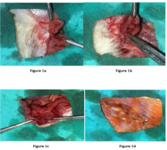
Figure 1 – a. The descending colon was transected
with scissor. b. End to end anastomosis was
performed using continuous one layer sutures. c.
Final of colonic anastomosis. d. Median incision
was closed with a 3/0 polypropilen sutures.
PRP preparation
Blood taken from each rat was used to prepare their own PRP during 3 consecutive days. No morbidity and mortality was seen in any rats. The tail of each animal in the PRP group was shaved and cleaned with antiseptic solution. The tail veins were placed in the water at 37°C for 5 minutes to allow them to more appearance. Friction with alcohol was performed and 2.5 ml blood was taken from each rat with 21 G cannulae. Blood samples taken from each rats buffered with citrate phosphate dextrose (0.5 ml citrate phosphate dextrose buffer (CPD)) was added in the ratio of 0.5 ml of CPD buffer to 2.5 ml of blood. The blood was then centrifuged at 1000 rpm for 5 min using the manufacturer’s
recommendation (Joan Lab Medical, China). The material containing PRP was removed and
recentrifuged at 3000 rpm for 10 minutes. The
PRP was applied anastomotic area. Each 0.5ml of PRP was administered locally.
Anastomotic bursting pressure measurement
The anastomosis was resected with a margin of at least 4 cm on each side with
regard to tissue hydroxyproline level followed by measuring the anastomotic bursting pressure (ABP). A 16-gauage silicon catheter
was inserted from both sides and feces in the resected column segment were cleared with
0.9% NaCl, then the both part of colon segment was ocluded with 3/0 silk. Isotonic saline was
administered at 5ml/min rate with an infusion pump (perfusor secura FT-Braun) while the
pressure within the lumen was monitored
Tissue hydroxyproline level measurement
After measuring ABP, 1 cm of the anastomosis including 0.5 cm proximal and
distal from the anastomosis were excised
and blunt dissection was performed to clear the anastomotic line from adherent tissues.
The tissues were cooled in the room heat
and neutralized with potassium hydroxide
in the phenolphthalein solution indicator. The volumes of all of them were equalized and centrifuged. Alanine and potassium borate buffer were added for saturation oxidation after saturation with a certain volume of potassium chloride. Chloramine-T solution, thiosulfate and toluene were added sequentially after standing for twenty minutes in the room temperature. Since the oxyde form of hydroxyproline was not dissolved
with toluene, the samples were heated to the soluble form and the hydroxypyrrole
organic phase was removed by addition of toluene again. P-dimethylaminobenzaldehyde was added to provide coloring. The resulting color and intensity were evaluated spectrophotometrically at 560 nm.
Measurement of cytokines and procalcitonin
Blood samples were collected at the 24th postoperative hour, and on the 3rd and 7th( just before sacrifice) postoperative days. We performed enzyme-linked immunosorbent assays (ELISAs) (Eclectica, Germany) to measure levels of IL-6 ( reference range : < 17.4 pg/mL), IL-10( reference range : < 2 pg/mL) and procalcitonin levels( reference range : < 0.15 ng/mL) in collected blood samples of the rats. Histopathological analysis
The sample was taken containing
the whole anastomosis line and fixed in 10% formaldehyde then embedded in paraffin. Tissues were sectioned at 4 μm and stain
hematoxylin-eosin for evaluating inflammation parameters and Masson trichrome for collagen. Healing parameters fibroblastic infiltration, capillary vascularisation and inflammatory infiltrations in each specimen were assessed with a score of 1–5 for each parameter.
Masson’s trichrome stain was assessed for the presence or absence of regular collagen
fibers. The degree of collagen deposition was assessed by pathologists.
Statistical analysis
Statistical analyzes were done using
(SPSS version 20.0 for Windows; SPSS Inc, Chicago, IL) Kruskal Wallis test was used in
the comparison of the groups. Chi-square test
was used to compare the three groups in the
parameters where significant differences were found. P<0.05 was accepted as statistically significant.
■
Results
One rat died in each Group 2 and Group
3 and 2 rats died in each Group 1 during the follow-up. These cases were excluded from the study. There is no significant difference in baseline weight values between the three
groups. Although the mean body weights
were reduced in all groups during the study
period, there was no statistical difference
among the groups in terms of body weight
changes. No local or systemic complications related to PRP and fibrin glue application were observed. There was no statistically significant difference of the ABP between Group 2 and
Group 3 (Group1:110.1±35.65mmHg, Group
2: 146.3±44.55 mmHg, Group 3:149.1±72.29 mmHg) of the rats after sacrification on the
7th day (Table 1). There was no statistically
day of rats (Table 1). There was no statistically
significant difference between levels of proinflammatory cytokine (IL-6) (P=0.41),
anti-inflammatory cytokine (IL-10) (P=0.35),
and procalcitonin levels (P=0.63) on 1, 3 and
7 days (Table 2). There was no statistically
significant difference indicators of wound
healing parameters (inflammatory infiltrations, capillary vascularisation, fibroblastic infiltration)
between Group 2 and Group 3 (p=0.316, Figure 2). Additionally, there was no statistically significant difference in terms of collagen formation between the groups. No significant differences were observed in the comparisons for inflammatory cell (P > 0.05). Fibroblast density and neovascularization showed no differences in the PRP compared to fibrin glue group.
Table 1 - AnastomoticBurst Pressures (ABP) and hydroxyproline levels in all groups.
Parameters
Group 1 (Mean±SD)
Group 2 (Mean±SD)
Group 3 (Mean±SD)
P value (Group1-2)
P value (Group1-3)
P value (Group2-3)
Anastomotic
Burst Pressure (mm/hg)
110.1±35.65 146.3±44.55 149.1±72.29 0.026 0.018 0.896
Hydroxyproline
Level (μg/mg) 96.2±29.22 120.1±51.50 118.71±42.18 0.023 0.022 0.745 SD: Standart Deviation
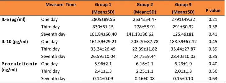
Table 2 - Cytokine and procalcitonin levels at 1st, 3rd and 6th day of each group.
Measure Time Group 1 Group 2 Group 3
P value
(Mean±SD) (Mean±SD) (Mean±SD)
IL-6 (pg/ml) One day 2805±89.56 2534±54.47 2791±49.32 0.21
Third day 330±61.15 278±58.91 291±30.32 0.38
Seventh day 101.84±66.40 141.13±36.62 125.49±81 0.41
IL-10 (pg/ml) One day 161.59±29.21 203.70±87.78 188.59±67.12 0.45
Third day 33.24±26.45 22.39±11.82 35.44±27.87 0.39
Seventh day 26.59±10.04 24.75±9.44 28.40±10.03 0.35
P r o c a l c i t o n i n (ng/ml)
One day 5.96±2.1 6.16±2.1 6.23±1.9 0.40
Third day 2.41±1.3 2.25±1.1 2.01±1.3 0.56
Seventh day 0.14±0.09 0.16±0.08 0.15±0.10 0.63
SD: Standart Deviation
Figure 2 - a. Control (Group1): moderate mononuclear cell infiltration with fibroblastic activity. b.
PRP (Group 2): moderate infiltration of inflammatory cells with moderate collagen synthesis. c. Fib
■
Dıscussıon
Leakage of anastomoses is a challenging problem causing morbidity and mortality in
colorectal surgery in several clinical studies.
Many local and systemic factors play a role in the healing of colon anastomoses7,8. These
factors are directly or indirectly influential in the healing process. Numerous procedure and methods are being used to applied intestinal anastomoses that all have the same aim to constitute the safest anastomosis9,10. Therefore,
local tissue materials have been used for years
and the most important examples of these
materials are fibrin glue and PRP.
Fibrin glue was initially intended for
hemostasis and control bleeding during surgical procedures11. Due to the biological adhesive
material made from concentrated fibrinogen,
it started to be used in wound healing. It is a
water-resistant cover and can thus constitute a physical barrier around the anastomosis. Clinical reports indicate that it might prevent anastomotic leakage after colonic operations.
Kanellos et al.10 reported that the application
of fibrin glue around a sutured anastomosis reduced the rate of anastomotic leaks and strengthened the anastomosis. In a study by
Akgün et al.12, sutured colocolic anastomosis
has been compared to the application of fibrin glue over the sutures. When subjects were
compared in terms of anastomosis safety on
the 72nd hour postoperatively, they have detected higher bursting pressures of the group that was applied with fibrin glue and have defended safer anastomosis12.
PRP is generraly applied locally in clinical and experimental studies but systemic use is rare13,14. PRP application to
gastrointestinal anastomosis is one of the
most useful methods to deliver concentrated amounts of growth factors throughout the surgical site, and expected to accelerate13,14.
Many studies showed positive effects of PRP on intestinal anastomoses. In a study conducted
open-abdominal rat groups compaired that PRP
was effective in intestinal anastomoses in open
abdomen cases. In another study conducted by Serdar et al.16 compared PRP and bioglue
and they found that PRP is superior to control
and bioglue group in the recovery of intestinal
anastomosis. Another experimental study by Alper et al.17 demonstrated that PRP has a
positive recovery on intestinal anastomosis in the sespis model.
The most important structure in the healing of the anastomosis is obtaining the
tensile strength in the submucosal connective tissue and the collagen contained. In the study of Irvin et al.18on rats, level of collagen tissue
decreased after the 3rd day and their value getting to normal approximately on the seventh day. In the study of Croin et al.19anastomotic
bursting pressure gradually increased from the third day after the anastomosis and reaching maximum in 7th day. At the same time they found 40% of decrease the concentration of hydroxyproline levels in the first 3 days on the anastomotic region and normalized 5th day.
Thus, although the debate continues about the postoperative day for measuring the ABP, we evaluated the bursting pressure on postoperative day 7 since anastomotic leakage is often diagnosed in the first postoperative week in clinical practice. In this study, there
were no difference in ABP and hydroxyproline levels between the groups of PRP and fibrin glue on the 7th day .
Cytokine levels are frequently used to assess intestinal anastomoses as well as indicators of inflammatory parameters20-23. In
many studies in the literature, cytokine levels have important role for assessing tissue healing degree with numerical data. The cytokine levels of proinflammatory (IL-1, IL-6, TNF-α) and anti-inflammatory (IL-4, IL-10) cytokines are in continuous relationship with each other during the tissue regeneration process24.
of early postoperative complications and these
parameters related to anastomosis evaluation in literature is often done on similar days as our
study25. IL-10 is the most important of the
anti-inflammatory cytokines and is responsible for suppressing the pro-inflammatory cytokines22. In a study conducted by Herwig et al.20, early
elevation of IL-6 and TNF-α levels and higher levels of IL-1 on 3rd day were shown as a marker of intestinal anastomosis leaks. Also procalcitonin levels are considered to be an important indicator of acute inflammation (especially in bacterial infection). It has important role in predicting intestinal anastomotic leaks in clinical practice26,27. In our study we found no difference between
groups of the cytokine (IL-6 and IL-10) and procalcitonin levels at 1,3and 7 days. Although
the anastomotic power and hydroxyproline levels are more effective than Group 1, they are not reflected in the inflammatory parameters. We interpreted that local application of the anastomosis healing systematically did not affect the laboratory values.
■
Conclusıon
Fibrin glue and platelet rich plasma
are shown to be effective in healing colon
anastomoses we found no differences
between fibrin glue and PRP effects of colonic anastomosis significantly superior to each other.
■
References
1. Walker KG, Bell SW, Rickard MJ. Mehanna D, Dent OF, Chapuis PH, Bokey EL.Anastomotic
leakage is predictive of diminished survival after potentially curative resection for colorectal cancer. Ann Surg. 2004;240(2):255-9. PMID: 15273549.
2. Law WL, Choi HK, Lee YM, Ho JW, Seto CL.
Anastomotic leakage is associated with
poor long-term outcome in patients after
curative colorectal resection for malignancy. J Gastrointest Surg. 2007;11(1):8-15.
PMID:17390180.
3. Kingham TP, Pachter HL. Colonic anastomotic leak: risk factors, diagnosis, and treatment. J Am Coll Surg. 2009;208(2):269-78. PMID: 19228539.
4. Buchs NC, Gervaz P, Secic M, Bucher P, Mugnier-Konrad B, Moerl P. Incidence, consequences, and risk factors for anastomotic dehiscence after colorectal surgery: a prospective monocentric study. Int J Colorectal Dis. 2008;23(3):265-70. PMID: 18034250.
5. Yamaguchi R, Terashima H, Yoneyama S, Tadano S, Ohkohchi N. Effects of plateletrichplasma on intestinal anastomotic healing in rats: PRP concentration is a key factor. J SurgRes. 2012;173(2):258-66. PMID: 21074782.
6. Bonanomi G, Prince JM, McSteen F, Schauer PR, Hamad GG. Sealing effect of fibrin glue on
the healing of gastrointestinal anastomoses: implications for the endoscopic treatment of leaks. Surg Endosc. 2004 Nov;18(11):1620-4. PMID: 15931477.
7. Schrock TR, Deveney CW, Dunphy JE. Factor contributing to leakage of colonic anastomoses. Ann Surg. 1973;177(5):513– 8. PMID: 4540874.
8. Strunden MS, Heckel K, Goetz AE, Reuter DA. Perioperative fluid and volume management:physiological basis, tools and
strategies.Ann Intensive Care. 2011;1(1):2. PMID: 21906324.
9. Zhou B, Ren J, Ding C, Wu Y, Chen J, Wang G, Gu G, Li J.Protection of colonic anastomosis with platelet-rich plasma gel in the open abdomen. Injury. 2014;45(5):864-8. PMID: 24552769.
10. Kanellos D, Mantzoros L, Goulirnaris,
Zacharakis E, Zavitsanakis A, Betsis D. Effects
of the use of fibrin glue around the colonic anastomosis of the rat. Tech Coloproctol. 2003;7(2):82-4. PMID: 14605925.
11. Martinowitz U, Schulman S. Fibrin sealent in surgery of patients with hemorrhagic diathesis. Tromb Haemost. 1995;74:486-92. PMID: 8578511.
12. Akgün A, Kuru S, Uraldi C, Tekin O, Karip B, Tug T, Ongören AU. Early effects of fibrin sealant on colonic anastomosis in rats: an
experimental and case-control study. Tech Coloproctol. 2006;10(3):208-14. PMID: 16969615.
Nurden AT.Autologous platelets as a source of proteins for healing and tissue regeneration. Thromb Haemost. 2004;91(1):4–15. PMID: 14691563.
14. Yamaguchi R, Terashima H, Yoneyama S,
Tadano S, Ohkohchi N. Effects of platelet-rich
plasma on intestinal anastomotic healing in rats: PRP concentration is a key factor. J Surg Res. 2012;173(2):258-66. PMID: 21074782. 15. Zhou B, Ren J, Ding C, Wu Y, Chen J, Wang G,
Gu G, Li J. Protection of colonic anastomosis
with platelet-rich plasma gel in the open abdomen. Injury. 2014;45(5):864-8. PMID: 24552769.
16. Yol S, Tekin A, YilmazH, Küçükkartallar T, Esen H, Caglayan O, Tatkan Y. Effects of
platelet rich plasma on colonic anastomosis. J Surg Res. 2008;146(2):190-4. PMID: 18028949.
17. Sozutek A, Colak T, Cetinkunar S, Reyhan E, Irkorucu O, Polat G, Cennet A. The effect
of platelet-rich-plasma on the healing of left colonic anastomosis in a rat model of ıntra-abdominal sepsis. J Invest Surg. 2016;29(5):294-301. PMID: 26822265. 18. İrvin TT.Collagen metabolism in infected
colonic anastamoses. Surg Gynecol Obstet. 1976;143(2):220-4. PMID: 941076.
19. Croinin K, Jackson DS, Dunphy JE. Specific activity of hidroxyproline tritium in the healing colon. Surg Gynecol Obstet. 1968;126(5):1061-5. PMID: 5652652.
20. Herwig R, Glodny B, Kuhle C, Schluter B, Brinkmann OA, Strasser H, Senninger N, Winde G. Early identification of peritonitis by peritoneal cytokine measurement. Dis Colon Rectum. 2002;45(4):514-21. PMID: 12006934.
21. Marques e Silva S, Jerônimo MS, Silva-Pereira Id, Tavares AH, Bocca AL, Sousa JB. Effects of metoclopramide on the expression
of metalloproteinases and interleukins in
left colonic anastomoses. An experimental study. Acta Cir Bras. 2015;30(11):762-9. PMID: 26647796.
22. Ishimura K, Moroguchi A, Okano K, Maeba T, Maeta H. Local expression of tumor necrosis factor-alpha and interleukin-10 on wound healing of intestinal anastomosis during endotoxemia in mice. J Surg Res. 2002;108(1):91-7. PMID: 12443720.
23. Mateo RB, Reichner JS, Albina JE. Interleukin-6 activity in wounds. Am J Physiol. 1994;266(6):1840-4. PMID: 8024036.
24. Sousa JB, Soares EG, Aprilli F. Effects of diclofenac sodium on intestinal anastomotic healing. Experimental study on the small intestine of rabbits. Dis Colon Rectum.1991;34(7):613-7. PMID: 2055147. 25. DeCherney AH, diZerega GS. Clinical problem
of intraperitoneal postsurgical adhesion
formation following general surgery and the use of adhesion prevention barriers. Surg Clin North Am. 1997;77(3):671-88. PMID: 9194886
26. Rongione AJ, Kusske AM, Ashley SW, Reber HA, McFadden DW. Interleukin-10 prevents early cytokine release in severe intraabdominal infection and sepsis. J Surg Res. 1997;70(2):107-12. PMID: 9237883. 27. Novotny AR, Emmanuel K, Hueser N, Knebel
C, Kriner M, Ulm K, Bartels H, Siewert JR,
Holzmann B. Procalcitonin ratio indicates
successful surgical treatment of abdominal
sepsis. Surgery. 2009;145:20-6. PMID: 19081471.
Correspondence: Inanc Samil Sarici
Bahçeşehir 1. Kısım Mahallesi Ispartakule Caddesi Vaditepe Bahçeşehir 5. Bölge Sitesi K01 Blok No:33D İç kapı No:16 Başakşehir İstanbul Turkey
Phone: +905532271140 issarici2015@gmail.com
Received: Dec 26, 2017 Review: Feb 22, 2018 Accepted: Mar 23, 2018
Conflict of interest: none
Financial source: none
1Research performed at Experimental